EP0696201B2 - Supplemented tissue sealants, methods of their production and use - Google Patents
Supplemented tissue sealants, methods of their production and use Download PDFInfo
- Publication number
- EP0696201B2 EP0696201B2 EP94910927A EP94910927A EP0696201B2 EP 0696201 B2 EP0696201 B2 EP 0696201B2 EP 94910927 A EP94910927 A EP 94910927A EP 94910927 A EP94910927 A EP 94910927A EP 0696201 B2 EP0696201 B2 EP 0696201B2
- Authority
- EP
- European Patent Office
- Prior art keywords
- supplemented
- tet
- hbgf
- cells
- fgf
- Prior art date
- Legal status (The legal status is an assumption and is not a legal conclusion. Google has not performed a legal analysis and makes no representation as to the accuracy of the status listed.)
- Expired - Lifetime
Links
Images


Classifications
-
- A—HUMAN NECESSITIES
- A61—MEDICAL OR VETERINARY SCIENCE; HYGIENE
- A61L—METHODS OR APPARATUS FOR STERILISING MATERIALS OR OBJECTS IN GENERAL; DISINFECTION, STERILISATION OR DEODORISATION OF AIR; CHEMICAL ASPECTS OF BANDAGES, DRESSINGS, ABSORBENT PADS OR SURGICAL ARTICLES; MATERIALS FOR BANDAGES, DRESSINGS, ABSORBENT PADS OR SURGICAL ARTICLES
- A61L27/00—Materials for grafts or prostheses or for coating grafts or prostheses
- A61L27/50—Materials characterised by their function or physical properties, e.g. injectable or lubricating compositions, shape-memory materials, surface modified materials
- A61L27/507—Materials characterised by their function or physical properties, e.g. injectable or lubricating compositions, shape-memory materials, surface modified materials for artificial blood vessels
-
- A—HUMAN NECESSITIES
- A61—MEDICAL OR VETERINARY SCIENCE; HYGIENE
- A61K—PREPARATIONS FOR MEDICAL, DENTAL OR TOILETRY PURPOSES
- A61K38/00—Medicinal preparations containing peptides
- A61K38/16—Peptides having more than 20 amino acids; Gastrins; Somatostatins; Melanotropins; Derivatives thereof
- A61K38/17—Peptides having more than 20 amino acids; Gastrins; Somatostatins; Melanotropins; Derivatives thereof from animals; from humans
- A61K38/18—Growth factors; Growth regulators
- A61K38/1825—Fibroblast growth factor [FGF]
-
- A—HUMAN NECESSITIES
- A61—MEDICAL OR VETERINARY SCIENCE; HYGIENE
- A61K—PREPARATIONS FOR MEDICAL, DENTAL OR TOILETRY PURPOSES
- A61K38/00—Medicinal preparations containing peptides
- A61K38/16—Peptides having more than 20 amino acids; Gastrins; Somatostatins; Melanotropins; Derivatives thereof
- A61K38/17—Peptides having more than 20 amino acids; Gastrins; Somatostatins; Melanotropins; Derivatives thereof from animals; from humans
- A61K38/18—Growth factors; Growth regulators
- A61K38/1875—Bone morphogenic factor; Osteogenins; Osteogenic factor; Bone-inducing factor
-
- A—HUMAN NECESSITIES
- A61—MEDICAL OR VETERINARY SCIENCE; HYGIENE
- A61K—PREPARATIONS FOR MEDICAL, DENTAL OR TOILETRY PURPOSES
- A61K38/00—Medicinal preparations containing peptides
- A61K38/16—Peptides having more than 20 amino acids; Gastrins; Somatostatins; Melanotropins; Derivatives thereof
- A61K38/17—Peptides having more than 20 amino acids; Gastrins; Somatostatins; Melanotropins; Derivatives thereof from animals; from humans
- A61K38/36—Blood coagulation or fibrinolysis factors
- A61K38/363—Fibrinogen
-
- A—HUMAN NECESSITIES
- A61—MEDICAL OR VETERINARY SCIENCE; HYGIENE
- A61K—PREPARATIONS FOR MEDICAL, DENTAL OR TOILETRY PURPOSES
- A61K47/00—Medicinal preparations characterised by the non-active ingredients used, e.g. carriers or inert additives; Targeting or modifying agents chemically bound to the active ingredient
- A61K47/30—Macromolecular organic or inorganic compounds, e.g. inorganic polyphosphates
- A61K47/42—Proteins; Polypeptides; Degradation products thereof; Derivatives thereof, e.g. albumin, gelatin or zein
-
- A—HUMAN NECESSITIES
- A61—MEDICAL OR VETERINARY SCIENCE; HYGIENE
- A61L—METHODS OR APPARATUS FOR STERILISING MATERIALS OR OBJECTS IN GENERAL; DISINFECTION, STERILISATION OR DEODORISATION OF AIR; CHEMICAL ASPECTS OF BANDAGES, DRESSINGS, ABSORBENT PADS OR SURGICAL ARTICLES; MATERIALS FOR BANDAGES, DRESSINGS, ABSORBENT PADS OR SURGICAL ARTICLES
- A61L15/00—Chemical aspects of, or use of materials for, bandages, dressings or absorbent pads
- A61L15/16—Bandages, dressings or absorbent pads for physiological fluids such as urine or blood, e.g. sanitary towels, tampons
- A61L15/22—Bandages, dressings or absorbent pads for physiological fluids such as urine or blood, e.g. sanitary towels, tampons containing macromolecular materials
- A61L15/32—Proteins, polypeptides; Degradation products or derivatives thereof, e.g. albumin, collagen, fibrin, gelatin
-
- A—HUMAN NECESSITIES
- A61—MEDICAL OR VETERINARY SCIENCE; HYGIENE
- A61L—METHODS OR APPARATUS FOR STERILISING MATERIALS OR OBJECTS IN GENERAL; DISINFECTION, STERILISATION OR DEODORISATION OF AIR; CHEMICAL ASPECTS OF BANDAGES, DRESSINGS, ABSORBENT PADS OR SURGICAL ARTICLES; MATERIALS FOR BANDAGES, DRESSINGS, ABSORBENT PADS OR SURGICAL ARTICLES
- A61L24/00—Surgical adhesives or cements; Adhesives for colostomy devices
- A61L24/001—Use of materials characterised by their function or physical properties
- A61L24/0015—Medicaments; Biocides
-
- A—HUMAN NECESSITIES
- A61—MEDICAL OR VETERINARY SCIENCE; HYGIENE
- A61L—METHODS OR APPARATUS FOR STERILISING MATERIALS OR OBJECTS IN GENERAL; DISINFECTION, STERILISATION OR DEODORISATION OF AIR; CHEMICAL ASPECTS OF BANDAGES, DRESSINGS, ABSORBENT PADS OR SURGICAL ARTICLES; MATERIALS FOR BANDAGES, DRESSINGS, ABSORBENT PADS OR SURGICAL ARTICLES
- A61L26/00—Chemical aspects of, or use of materials for, wound dressings or bandages in liquid, gel or powder form
- A61L26/0009—Chemical aspects of, or use of materials for, wound dressings or bandages in liquid, gel or powder form containing macromolecular materials
- A61L26/0028—Polypeptides; Proteins; Degradation products thereof
- A61L26/0042—Fibrin; Fibrinogen
-
- A—HUMAN NECESSITIES
- A61—MEDICAL OR VETERINARY SCIENCE; HYGIENE
- A61L—METHODS OR APPARATUS FOR STERILISING MATERIALS OR OBJECTS IN GENERAL; DISINFECTION, STERILISATION OR DEODORISATION OF AIR; CHEMICAL ASPECTS OF BANDAGES, DRESSINGS, ABSORBENT PADS OR SURGICAL ARTICLES; MATERIALS FOR BANDAGES, DRESSINGS, ABSORBENT PADS OR SURGICAL ARTICLES
- A61L26/00—Chemical aspects of, or use of materials for, wound dressings or bandages in liquid, gel or powder form
- A61L26/0061—Use of materials characterised by their function or physical properties
- A61L26/0066—Medicaments; Biocides
-
- A—HUMAN NECESSITIES
- A61—MEDICAL OR VETERINARY SCIENCE; HYGIENE
- A61L—METHODS OR APPARATUS FOR STERILISING MATERIALS OR OBJECTS IN GENERAL; DISINFECTION, STERILISATION OR DEODORISATION OF AIR; CHEMICAL ASPECTS OF BANDAGES, DRESSINGS, ABSORBENT PADS OR SURGICAL ARTICLES; MATERIALS FOR BANDAGES, DRESSINGS, ABSORBENT PADS OR SURGICAL ARTICLES
- A61L26/00—Chemical aspects of, or use of materials for, wound dressings or bandages in liquid, gel or powder form
- A61L26/0061—Use of materials characterised by their function or physical properties
- A61L26/0085—Porous materials, e.g. foams or sponges
-
- A—HUMAN NECESSITIES
- A61—MEDICAL OR VETERINARY SCIENCE; HYGIENE
- A61L—METHODS OR APPARATUS FOR STERILISING MATERIALS OR OBJECTS IN GENERAL; DISINFECTION, STERILISATION OR DEODORISATION OF AIR; CHEMICAL ASPECTS OF BANDAGES, DRESSINGS, ABSORBENT PADS OR SURGICAL ARTICLES; MATERIALS FOR BANDAGES, DRESSINGS, ABSORBENT PADS OR SURGICAL ARTICLES
- A61L27/00—Materials for grafts or prostheses or for coating grafts or prostheses
- A61L27/28—Materials for coating prostheses
- A61L27/34—Macromolecular materials
-
- A—HUMAN NECESSITIES
- A61—MEDICAL OR VETERINARY SCIENCE; HYGIENE
- A61P—SPECIFIC THERAPEUTIC ACTIVITY OF CHEMICAL COMPOUNDS OR MEDICINAL PREPARATIONS
- A61P17/00—Drugs for dermatological disorders
-
- A—HUMAN NECESSITIES
- A61—MEDICAL OR VETERINARY SCIENCE; HYGIENE
- A61P—SPECIFIC THERAPEUTIC ACTIVITY OF CHEMICAL COMPOUNDS OR MEDICINAL PREPARATIONS
- A61P17/00—Drugs for dermatological disorders
- A61P17/02—Drugs for dermatological disorders for treating wounds, ulcers, burns, scars, keloids, or the like
-
- A—HUMAN NECESSITIES
- A61—MEDICAL OR VETERINARY SCIENCE; HYGIENE
- A61P—SPECIFIC THERAPEUTIC ACTIVITY OF CHEMICAL COMPOUNDS OR MEDICINAL PREPARATIONS
- A61P25/00—Drugs for disorders of the nervous system
- A61P25/04—Centrally acting analgesics, e.g. opioids
-
- A—HUMAN NECESSITIES
- A61—MEDICAL OR VETERINARY SCIENCE; HYGIENE
- A61P—SPECIFIC THERAPEUTIC ACTIVITY OF CHEMICAL COMPOUNDS OR MEDICINAL PREPARATIONS
- A61P31/00—Antiinfectives, i.e. antibiotics, antiseptics, chemotherapeutics
-
- A—HUMAN NECESSITIES
- A61—MEDICAL OR VETERINARY SCIENCE; HYGIENE
- A61P—SPECIFIC THERAPEUTIC ACTIVITY OF CHEMICAL COMPOUNDS OR MEDICINAL PREPARATIONS
- A61P31/00—Antiinfectives, i.e. antibiotics, antiseptics, chemotherapeutics
- A61P31/04—Antibacterial agents
-
- A—HUMAN NECESSITIES
- A61—MEDICAL OR VETERINARY SCIENCE; HYGIENE
- A61P—SPECIFIC THERAPEUTIC ACTIVITY OF CHEMICAL COMPOUNDS OR MEDICINAL PREPARATIONS
- A61P35/00—Antineoplastic agents
-
- A—HUMAN NECESSITIES
- A61—MEDICAL OR VETERINARY SCIENCE; HYGIENE
- A61P—SPECIFIC THERAPEUTIC ACTIVITY OF CHEMICAL COMPOUNDS OR MEDICINAL PREPARATIONS
- A61P9/00—Drugs for disorders of the cardiovascular system
-
- A—HUMAN NECESSITIES
- A61—MEDICAL OR VETERINARY SCIENCE; HYGIENE
- A61L—METHODS OR APPARATUS FOR STERILISING MATERIALS OR OBJECTS IN GENERAL; DISINFECTION, STERILISATION OR DEODORISATION OF AIR; CHEMICAL ASPECTS OF BANDAGES, DRESSINGS, ABSORBENT PADS OR SURGICAL ARTICLES; MATERIALS FOR BANDAGES, DRESSINGS, ABSORBENT PADS OR SURGICAL ARTICLES
- A61L2300/00—Biologically active materials used in bandages, wound dressings, absorbent pads or medical devices
- A61L2300/40—Biologically active materials used in bandages, wound dressings, absorbent pads or medical devices characterised by a specific therapeutic activity or mode of action
- A61L2300/402—Anaestetics, analgesics, e.g. lidocaine
-
- A—HUMAN NECESSITIES
- A61—MEDICAL OR VETERINARY SCIENCE; HYGIENE
- A61L—METHODS OR APPARATUS FOR STERILISING MATERIALS OR OBJECTS IN GENERAL; DISINFECTION, STERILISATION OR DEODORISATION OF AIR; CHEMICAL ASPECTS OF BANDAGES, DRESSINGS, ABSORBENT PADS OR SURGICAL ARTICLES; MATERIALS FOR BANDAGES, DRESSINGS, ABSORBENT PADS OR SURGICAL ARTICLES
- A61L2300/00—Biologically active materials used in bandages, wound dressings, absorbent pads or medical devices
- A61L2300/40—Biologically active materials used in bandages, wound dressings, absorbent pads or medical devices characterised by a specific therapeutic activity or mode of action
- A61L2300/404—Biocides, antimicrobial agents, antiseptic agents
- A61L2300/406—Antibiotics
-
- A—HUMAN NECESSITIES
- A61—MEDICAL OR VETERINARY SCIENCE; HYGIENE
- A61L—METHODS OR APPARATUS FOR STERILISING MATERIALS OR OBJECTS IN GENERAL; DISINFECTION, STERILISATION OR DEODORISATION OF AIR; CHEMICAL ASPECTS OF BANDAGES, DRESSINGS, ABSORBENT PADS OR SURGICAL ARTICLES; MATERIALS FOR BANDAGES, DRESSINGS, ABSORBENT PADS OR SURGICAL ARTICLES
- A61L2300/00—Biologically active materials used in bandages, wound dressings, absorbent pads or medical devices
- A61L2300/40—Biologically active materials used in bandages, wound dressings, absorbent pads or medical devices characterised by a specific therapeutic activity or mode of action
- A61L2300/412—Tissue-regenerating or healing or proliferative agents
- A61L2300/414—Growth factors
-
- A—HUMAN NECESSITIES
- A61—MEDICAL OR VETERINARY SCIENCE; HYGIENE
- A61L—METHODS OR APPARATUS FOR STERILISING MATERIALS OR OBJECTS IN GENERAL; DISINFECTION, STERILISATION OR DEODORISATION OF AIR; CHEMICAL ASPECTS OF BANDAGES, DRESSINGS, ABSORBENT PADS OR SURGICAL ARTICLES; MATERIALS FOR BANDAGES, DRESSINGS, ABSORBENT PADS OR SURGICAL ARTICLES
- A61L2300/00—Biologically active materials used in bandages, wound dressings, absorbent pads or medical devices
- A61L2300/40—Biologically active materials used in bandages, wound dressings, absorbent pads or medical devices characterised by a specific therapeutic activity or mode of action
- A61L2300/416—Anti-neoplastic or anti-proliferative or anti-restenosis or anti-angiogenic agents, e.g. paclitaxel, sirolimus
Definitions
- This invention is directed to supplemented TSs (TS), such as fibrin glue (FG), as well as to methods of their production and use.
- TS TS
- FG fibrin glue
- this invention is directed to TSs which do not inhibit full thickness skin wound healing.
- the invention is directed to TSs which have been supplemented with a drug which is ciprofloxacin HCl, as well as to methods of their production and use.
- Wound healing the repair of lesions, begins almost instantly after injury. It requires the successive coordinated function of a variety of cells and the close regulation of degradative and regenerative steps.
- the proliferation, differentiation and migration of cells are important biological processes which underlie wound healing, which also involves fibrin clot formation, resorption of the clot, tissue remodeling, such as fibrosis, endothelialization and epithelialization.
- Wound healing involves the formation of highly vascularized tissue that contains numerous capillaries, many active fibroblasts, and abundant collagen fibrils, but not the formation of specialized skin structures.
- thromboplastin which flows out of injured cells.
- Thromboplastin contacts plasma factor VII to form factor X activator, which then, with factor V and in a complex with phospholipids and calcium, converts prothrombin into thrombin.
- factor X activator which then, with factor V and in a complex with phospholipids and calcium, converts prothrombin into thrombin.
- Thrombin catalyzes the release of fibrinopeptides A and B from fibrinogen to produce fibrin monomers, which aggregate to form fibrin filaments.
- Thrombin also activates the transglutaminase, factor XIIIa, which catalyzes the formation of isopeptide bonds to covalently cross-link the fibrin filaments.
- Alpha 2 -antiplasmin is then bound by factor XIII onto the fibrin filaments to thereby protect the filaments from degradation by plasmin (see, for example, Doolittle et al., Ann. Rev. Biochem. 53:195-229 (1984) ).
- polypeptide growth factors which exhibit an array of biological activities, are released into the wound where they play a crucial role in healing (see, e.g ., Hormonal Proteins and Peptides (Li, C.H., ed.) Volume 7, Academic Press, Inc., New York, N.Y. pp. 231-277 (1979 ) and Brunt et al., Biotechnology 6:25-30 (1988) ).
- These activities include recruiting cells, such as leukocytes and fibroblasts, into the injured area, and inducing cell proliferation and differentiation.
- Growth factors that may participate in wound healing include, but are not limited to: platelet-derived growth factors (PDGFs); insulin-binding growth factor-1 (IGF-1); insulin-binding growth factor-2 (IGF-2); epidermal growth factor (EGF); transforming growth factor- ⁇ (TGF- ⁇ ); transforming growth factor- ⁇ (TGF- ⁇ ); platelet factor 4 (PF-4); and heparin binding growth factors one and two (HBGF-1 and HBGF-2, respectively).
- PDGFs platelet-derived growth factors
- IGF-1 insulin-binding growth factor-1
- IGF-2 insulin-binding growth factor-2
- EGF epidermal growth factor
- TGF- ⁇ transforming growth factor- ⁇
- TGF- ⁇ transforming growth factor- ⁇
- PF-4 platelet factor 4
- HBGF-1 and HBGF-2 heparin binding growth factors one and two
- PDGFs are stored in the alpha granules of circulating platelets and are released at wound sites during blood clotting (see, e . g ., Lynch et al., J. Clin. Invest. 84:640-646 (1989) ).
- PDGFs include: PDGF; platelet derived angiogenesis factor (PDAF); TGF- ⁇ ; and PF-4, which is a chemoattractant for neutrophils ( Knighton et al., in Growth Factors and Other Aspects of Wound Healing: Biological and Clinical Implications, Alan R. Liss, Inc., New York, New York, pp. 319-329 (1988 )).
- PDGF is a mitogen, chemoattractant and a stimulator of protein synthesis in cells of mesenchymal origin, including fibroblasts and smooth muscle cells. PDGF is also a nonmitogenic chemoattractant for endothelial cells (see, for example, Adelmann-Grill et al., Eur. J. Cell Biol. 51:322-326 ( 1990) ).
- IGF-1 acts in combination with PDGF to promote mitogenesis and protein synthesis in mesenchymal cells in culture.
- Application of either PDGF or IGF-1 alone to skin wounds does not enhance healing, but application of both factors together appears to promote connective tissue and epithelial tissue growth ( Lynch et al., Proc. Natl. Acad. Sci. 76:1279-1283 (1987) ).
- TGF- ⁇ is a chemoattractant for macrophages and monocytes. Depending upon the presence or absence of other growth factors, TGF- ⁇ may stimulate or inhibit the growth of many cell types. For example, when applied in vivo , TGF- ⁇ increases the tensile strength of healing dermal wounds. TGF- ⁇ also inhibits endothelial cell mitosis, and stimulates collagen and glycosaminoglycan synthesis by fibroblasts.
- EGF epithelial growth factor
- TGF- ⁇ the HBGFs and osteogenin
- Other growth factors are also important in wound healing
- EGF which is found in gastric secretions and saliva
- TGF- ⁇ which is made by both normal and transformed cells
- EGF EGF
- TGF- ⁇ which is made by both normal and transformed cells
- These receptors mediate proliferation of epithelial cells. Both factors accelerate reepithelialization of skin wounds.
- Exogenous EGF promotes wound healing by stimulating the proliferation of keratinocytes and dermal fibroblasts ( Nanney et al., J. Invest. Dermatol. 83:385-393 (1984) and Coffey et al., Nature 328:817-820 (1987) ).
- HBGFs also known as Fibroblast Growth Factors (FGFs), which include acidic HBGF (aHBGF also known as HBFG-1 or FGF-1) and basic HBGF (bHBGF also known as HBGF-2), are potent mitogens for cells of mesodermal and neuroectodermal lineages, including endothelial cells (see, e . g ., Burgess et al., Ann. Rev. Biochem. 58:575-606 (1989) ).
- HBGF-1 is chemotactic for endothelial cells and astroglial cells. Both HBGF-1 and HBGF-2 bind to heparin, which protects them from proteolytic degradation.
- the array of biological activities exhibited by the HBGFs suggests that they play an important role in wound healing.
- Basic fibroblast growth factor (FGF-2) is a potent stimulator of angiogenesis and the migration and proliferation of fibroblasts (see, for example, Gospodarowicz et al., Mol. Cell. Endocinol. 46:187-204 (1986) and Gospodarowicz et al., Endo. Rev. 8:95-114 (1985) ).
- Acidic fibroblast growth factor (FGF-1) has been shown to be a potent angiogenic factor for endothelial cells (Burgess et al ., supra , 1989). However, it has not been established if any FGF growth factor is chemotactic for fibroblasts.
- Growth factors are, therefore, potentially useful for specifically promoting wound healing and tissue repair.
- their use to promote wound healing has yielded inconsistent results (see, e.g., Carter et al., in Growth Factors and Other Aspects of Wound Healing: Biological and Clinical Implications, Alan R. Liss, Inc., New York, New York, pp. 303-317 (1988 )).
- PDGF, IGF-1, EGF, TGF- ⁇ , TGF- ⁇ and FGF also known as HBGF
- applied separately to standardized skin wounds in swine had little effect on the regeneration of connective tissue or epithelium in the wounds ( Lynch et al., J. Clin. Invest. 84:640-646 (1989) ).
- TGF- ⁇ stimulated the greatest response alone.
- factors such as PDGF-bb homodimer and IGF-1 or TGF- ⁇ produced a dramatic increase in connective tissue regeneration and epithelialization.
- Tsuboi et al . have reported that the daily application of bFGF to an open wound stimulated wound healing in healing-impaired mice but not in normal mice (J. Exp. Med. 172:245-251 (1990) ).
- Surgical adhesives and TSs which contain plasma proteins are used for sealing internal and external wounds, such as in bones and skin, to reduce blood loss and maintain hemostasis.
- TSs contain blood clotting factors and other blood proteins.
- FG also called fibrin sealant, is a gel similar to a natural clot which is prepared from plasma. The precise components of each FG are a function of the particular plasma fraction which is used as a starting material. Fractionation of plasma components can be effected by standard protein purification methods, such as ethanol, polyethylene glycol, and ammonium sulfate precipitation, ion exchange, and gel filtration chromatography.
- FG contains a mixture of proteins including traces of albumin, fibronectin and plasminogen. In Canada, Europe and possibly elsewhere, commercially available FG typically also contains aprotinin as a stabilizer.
- FGs generally are prepared from: (1) a fibrinogen concentrate, which contains fibronectin, Factor XIII, and von Willebrand factor; (2) dried human or bovine thrombin; and (3) calcium ions.
- Commercially prepared FGs generally contain bovine components.
- the fibrinogen concentrate can be prepared from plasma by cryoprecipitation followed by fractionation, to yield a composition that forms a sealant or clot upon mixture with thrombin and an activator of thrombin such as calcium ions.
- the fibrinogen and thrombin concentrates are prepared in lyophilized form and are mixed with a solution of calcium chloride immediately prior to use. Upon mixing, the components are applied to a tissue where they coagulate on the tissue surface and form a clot that includes cross-linked fibrin.
- Factor XIII which is present in the fibrinogen concentrate, catalyzes the cross-linking.
- Australian Patent 75097/87 describes a one-component adhesive, which contains an aqueous solution of fibrinogen, factor XIII, a thrombin inhibitor, such as antithrombin III, prothrombin factors, calcium ions, and, if necessary, a plasmin inhibitor.
- Stroetmann U.S. Patent Nos. 4,427,650 and 4,427,651 , describes the preparation of an enriched plasma derivative in the form of a powder or sprayable preparation for enhanced wound closure and healing that contains fibrinogen, thrombin and/or prothrombin, and a fibrinolysis inhibitor, and may also contain other ingredients, such as a platelet extract. Rose et al ., U.S. Patent Nos.
- IMMUNO AG (Vienna, Austria) and BEHRINGWERKE AG (Germany) ( Gibble et al., Transfusion 30:741-747 (1990) ) presently have FGs on the market in Europe and elsewhere (see, e.g. , U.S. Patent Nos. 4,377,572 and 4,298,598 , which are owned by IMMUNO AG).
- TSs are not commercially available in the U.S.
- the American National Red Cross and BAXTER/HYLAND (Los Angeles, CA) have recently co-developed a FG (ARC/BH FG) which is now in clinical studies.
- the TSs available in Europe contain proteins of non-human origin such as aprotinin and bovine thrombin. Since these proteins are of non-human origin, people may develop allergic reactions to them.
- heat inactivation is used to inactivate viruses which may be present in the components of the FG. However, this heat inactivation method may produce denatured proteins in the FG which may also be allergenic.
- the ARC/BH FG has advantages over the TSs available in Europe because it does not contain bovine proteins.
- the ARC/BH TS contains human thrombin instead of bovine thrombin and does not contain aprotinin. Since the ARC/BH FG does not contain bovine proteins it should be less allergenic in humans than those TSs available in Europe.
- the ARC/BH FG is virally inactivated by a solvent detergent method which produces fewer denatured proteins and thus is less allergenic than those available in Europe. Therefore, the ARC/BH FG possesses advantages over the TSs which are now commercially available in other countries.
- FG is primarily formulated for clinical topical application and is used to control bleeding, maintain hemostasis and promote wound healing.
- the clinical uses of FG have recently been reviewed ( Gibble et al., Transfusion 30:741-747 (1990) ; Lerner et al., J. Surg. Res. 48:165-181 (1990) ).
- By sealing tissues FG prevents air or fluid leaks, induces hemostasis, and may contribute to wound healing indirectly by reducing or preventing events which may interfere with wound healing such as bleeding, hematomas, infections, etc.
- FG maintains hemostasis and reduces blood loss, it has not yet been shown to possess true wound healing properties. Because FG is suitable for both internal and external injuries, such as bone and skin injuries, and is useful to maintain hemostasis, it is desirable to enhance its wound healing properties.
- Demineralized bone matrix is a source of osteoinductive proteins known as bone morphogenetic proteins (BMP), and growth factors which modulate the proliferation of progenitor bone cells (see, e.g. , Hauschka et al., J. Biol. Chem. 261:12665-12674 (1986) and Canalis et al., J. Clin. Invest. 81:277-281 (1988) ).
- BMPs have now been identified and are abbreviated BMP-1 through BMP-8.
- BMP-3 and BMP-7 are also known as osteogenin and osteogenic protein-1 (OP-1), respectively.
- DBM materials have little clinical use unless combined with particulate marrow autografts. There is a limit to the quantity of DBM that can be surgically placed into a recipient's bone to produce a therapeutic effect. In addition, resorption has been reported to be at least 49% ( Toriumi et al., Arch. Otolaryngo. Head Neck Surg. 116:676-680 (1990) ).
- DBM powder and osteogenin may be washed away by tissue fluids before their osteoinductive potential is expressed.
- seepage of tissue fluids into DBM-packed bone cavities or soft-tissue collapse into the wound bed are two factors that may significantly affect the osteoinductive properties of DBM and osteogenin.
- Soft-tissue collapse into the wound bed may likewise inhibit the proper migration of osteocompetent stem cells into the wound bed.
- TS also can serve as a "scaffold" which cells can use to move into a wounded area to generate new tissues.
- commercially available preparations of FG and other TSs are too dense to allow cell migration into and through them. This limits their effectiveness in some in vivo uses.
- bone nonunion defects In one type of bone wound, called bone nonunion defects, there is a minimal gap above which no new bone formation occurs naturally.
- the treatment for these situations is bone grafting.
- the source of bone autografts is usually limited and the use of allogeneic bones involves a high risk of viral contamination. Because of this situation, the use of demineralized, virally inactivated bone powder is an attractive solution.
- vascular prostheses are frequently made out of polytetrafluoroethylene (PTFE) and are used to replace diseased blood vessels in humans and other animals.
- PTFE polytetrafluoroethylene
- various techniques have been used including seeding of nonautologous endothelial cells onto the prothesis.
- Various substrates which adhere both to the vascular graft and endothelial cells have been investigated as an intermediate substrate to increase endothelial cell seeding. These substrates include preclotted blood ( Herring et al., Surgery 84:498-504 (1978) ), FG ( Rosenman et al., J. Vasc. Surg.
- Angiogenesis is the induction of new blood vessels.
- Certain growth factors such as HBGF-1 and HBGF-2 are angiogenic.
- their in vivo administration attached to: collagen sponges ( Thompson et al., Science 241:1349-1352 (1988) ); beads ( Hayek et al., Biochem. Biophys. Res. Commun. 147:876-880 (1987) ); solid PTFE fibers coated with collagen arranged in a sponge-like structure ( Thompson et al., Proc. Natl. Acad. Sci. USA 86:7928-7932 (1989) ); or by infusion ( Puumala et al.. Brain Res.
- fibrin gels (0.5-10 mg/ml) implanted subcutaneously in plexiglass chambers induce angiogenesis within 4 days of implantation, compared to empty chambers, or chambers filled with sterile culture medium (Dvorak et al ., Lab. Invest. 57 :673 (1987)).
- An efficacious, site-directed, drug delivery system is greatly needed in several areas of medicine.
- localized drug delivery is needed in the treatment of local infections, such as in periodontitis, where the systemic administration of antimicrobial agents is ineffective.
- the problem after systemic administration usually lies in the low concentration of the antimicrobial agent which can be achieved at the target site.
- a systemic dose increase may be effective but also may produce toxicity, microbial resistance and drug incompatibility.
- several alternative methods have been devised but none are ideal.
- collagen and/or fibrinogen dispersed in an aqueous medium as an amorphous flowable mass have also been shown to locally deliver drugs (Luck et al ., U.S. Reissue Patent 33,375 ; Luck et al ., U.S. Patent 4,978,332 ).
- the relatively short release time of the AB from the FG may reflect the relatively short life of the AB-supplemented TS or the form and/or quantity of the AB in the AB-TS.
- a means to stabilize FG and other TSs to allow for extended, localized drug release is desirable and needed, as are new techniques for the incorporation and extended release of other supplements from TS.
- the invention provides a supplemented tissue sealant composition that promotes localized delivery of a drug, comprising: (i) a tissue sealant comprising fibrin glue, and (ii) the drug in solid form, wherein said drug is ciprofloxacin HCl; wherein said tissue sealant composition does not contain a growth factor, and wherein said drug is capable of being released long term.
- the sealant does not inhibit full thickness skin wound healing.
- the total protein concentration of the sealant is less than 30 mg/ml.
- the total protein concentration is less than 30 mg/ml.
- the total protein concentration is greater than 30 mg/ml.
- the invention provides: a biomaterial treated with a supplemented tissue sealant composition of the first aspect of the invention; an ex vivo process for the preparation of a treated biomaterial, the process comprising applying a supplemented tissue sealant composition of the first aspect of the invention to a biomaterial; and the use of a supplemented tissue sealant composition according to the first aspect of the invention for the preparation of an agent for the treatment and/or prevention of infection, the treatment of neoplasia, the acceleration of wound healing, or the treatment and/or prevention of cardiovascular disease.
- the present invention also relates to a process for the localized delivery of at least one drug to a tissue, comprising applying to the tissue a TS which contains ciprofloxacin HCl.
- the TS may be FG.
- FG may be made from the mixing of topical fibrinogen complex (TFC), human thrombin and calcium chloride. Varying the concentration of the TFC has the most significant effect upon the density of the final FG matrix. Varying the concentration of the thrombin has an insignificant effect upon the total protein concentration of the final FG, but has a profound effect upon the time required for the polymerization of the fibrinogen component of the TFC into fibrin. While this effect is well known, it is not generally appreciated that it may be used to maximize the effectiveness of the FG, when it is used alone or supplemented. Because of this effect one can alter the time between the mixing of the FG components and the setting of the FG.
- TFC topical fibrinogen complex
- human thrombin and calcium chloride.
- This property is also important to keep the FG from clogging delivery devices with long passages, i.e., catheters, endoscopes, etc., which is important to allow the application of the FG or supplemented FG to sites in the body that are only accessible by surgery. This effect is also important in keeping the insoluble supplements in suspension and preventing them from settling in the applicator or in the tissue site.
- TFC is a lyophilized mixture of human plasma proteins which have been purified and virally inactivated. When reconstituted TFC contains:
- the reconstituted TFC may also contain trace amounts of fibronectin.
- human thrombin is a lyophilized mixture of human plasma proteins, which have been purified and vitally inactivated. When reconstituted it contains:
- Calcium chloride is added in sufficient concentration to activate the Thrombin. As long as there is sufficient calcium, its concentration is not important.
- the TS may contain fat cells.
- the TSs of this invention may be used to aid the integration of a graft. whether artificial or natural. into an animal's body as for example when the graft is composed of natural tissue.
- the TSs of this invention can be used to combat some of the major problems associated with certain conditions such as periodontitis, namely persistent infection, bone resorption, loss of ligaments and premature re-epithelialization of the dental pocket.
- Drug(s) used in the compositions of the invention may be added to the TS to accelerate wound healing, and combat infection, neoplasia, and/or other disease processes.
- These drugs may include, but are not limited to: antibiotics, such as tetracycline and ciprofloxacin; antiproliferative/cytotoxic drugs, such as 5-fluorouracil (5-FU), taxol and/or taxotere; antivirals, such as gangcyclovir, zidovudine, amantidine, vidarabine, ribaravin, trifluridine, acyclovir, dideoxyuridine and antibodies to viral components or gene products; cytokines, such as ⁇ - or ⁇ - or ⁇ -Interferon, ⁇ - or ⁇ -tumor necrosis factor, and interleukins; colony stimulating factors; erythropoietin; antifungals, such as diflucan, ketaconizole and nystatin; antiparasitic agents,
- Other compounds which may be added to the TS include, but are not limited to: vitamins and other nutritional supplements; hormones; glycoproteins; fibronectin; peptides and proteins; carbohydrates (both simple and/or complex); proteoglycans; anciangiogenins; antigens; oligonucleotides (sense and/or antisense DNA and/or RNA); antibodies (for example, to infectious agents, tumors, drugs or hormones); and gene therapy reagents. Genetically altered cells and/or other cells may also be included in the TSs of this invention. In addition, anything which does not destroy the TS can be added to the TSs of this invention.
- compositions of the invention ciprofloxacin HCl is included in solid form.
- the compositions of the invention do not contain a growth factor
- poorly water soluble forms of a drug such as the free base of TET, increase the delivery of the drug from the TS more than freely water soluble forms thereof. Therefore, the drug may be bound to an insoluble carrier, such as fibrinogen or activated charcoal, within the TS to prolong the delivery of the drug from the supplemented TS.
- an insoluble carrier such as fibrinogen or activated charcoal
- the supplemented TS can be used in organoids.
- this invention provides a composition that promotes the localized delivery of a poorly water soluble form of an antibiotic(s) and other drug(s), comprising a TS and an effective concentration of at least one poorly water soluble form of an antibiotic.
- the present invention has several advantages over the previously used TS compositions and methods.
- the first advantage is that the drug-supplemented TSs of the present invention have many of the characteristics of an ideal biodegradable carrier, namely: they can be formulated to contain only human proteins thus eliminating or minimizing immunogenicity problems and foreign-body reactions; their administration is versatile; and their removal from the host's tissues is not required because they' are degraded by the host's own natural fibrinolytic system.
- a second advantage is that the present invention provides a good way to effectively deliver drugs for a prolonged period of time to an internal or external wound. It is now believed that some growth factor receptors must be occupied for at least 12 hours to produce a maximal biological effect.
- a third advantage of the present invention is that animal cells can migrate into and through, and grow in the TSs of the present invention. This aids engraftment of the cells to neighboring tissues and prostheses. Based on the composition of the TSs which are available in Europe, it is expected that this is not possible with these formulations. Instead, animal cells must migrate around or digest commercially available TS. Since the importation into the U.S. of commercially available TSs from Europe is illegal (their use in the USA has not been approved by the U.S. FDA) Applicants cannot readily verify this.
- a fourth advantage is that because of its initial liquid nature, the TS of the present invention can cover surfaces more thoroughly and completely than many previously available delivery systems. This is especially important for the use of the present invention in coating biomaterials and in the endothelialization of vascular prostheses because the supplemented FG will coat the interior, exterior and pores of the vascular prosthesis. As a result of this, plus the ability of endothelial cells to migrate into and through the TS, engraftment of autologous endothelial cells will occur along the whole length of the vascular prosthesis, thereby decreasing its thrombogenicity and antigenicity.
- TSs vascular prostheses
- Previously used TSs for vascular prostheses also primarily were seeded with nonautologous cells which could be rejected by the body and could be easily washed off by the shearing force of blood passing through the prosthesis.
- a fifth advantage is that the supplemented TS of this invention can be molded and thus can be custom made into almost any desired shape.
- a sixth advantage is that the AB-supplemented FG of this invention, has unexpectedly increased the longevity and stability of the FG compared to that of the unsupplemented FG. This increased stability continues even after appreciable quantities of the AB are no longer remaining in the FG. For example, soaking a newly formed FG clot in a solution of CIP HCl. produces a FG clot which is stable and preserved even after substantially all the CIP has left the FG clot. While not wishing to be bound by any theory as to how this effect is produced, it is believed that CIP, inhibits plasminogen which is in the TFC and breaks down the FG.
- the seventh advantage of the present invention is a direct result of the prolonged longevity and stability of the TS.
- AB-supplemented FG can be used to produce localized, long term delivery of a drug(s). This delivery will continue even after the stabilizing drug CIP, has substantially left the TS.
- a compound to stabilize a TS in order to produce prolonged, localized drug delivery.
- An eighth advantage of Applicants' invention is that it allows site-directed angiogenesis to occur in vivo . While others have demonstrated localized non-specific angiogenesis, supra , to the best of Applicants' knowledge, no one else has shown site-directed angiogenesis.
- a wound includes damage to any tissue in a living organism.
- the tissue may be an internal tissue, such as the stomach lining or a bone, or an external tissue, such as the skin.
- a wound may include, but is not limited to, a gastrointestinal tract ulcer, a broken bone, a neoplasia, and cut or abraided skin.
- a wound may be in a soft tissue, such as the spleen, or in a hard tissue, such as bone.
- the wound may have been caused by any agent, including traumatic injury, infection or surgical intervention.
- TS is a substance or composition that, upon application to a wound, seals the wound, thereby reducing blood loss and maintaining hemostasis.
- FG is a composition, prepared from recombinant or plasma proteins, that upon application to a wound forms a clot, thereby sealing the wound, reducing blood loss and maintaining hemostasis.
- FG. supra is a form of TS.
- supplemented TS includes any TS that, without substantial modification, can serve as a carrier vehicle for the delivery of a growth factor, drug or other compound, or mixtures thereof, and that, by virtue of its adhesive or adsorptive properties, can maintain contact with the site for a time sufficient for the supplemented TS to produce its desired effect. for example to promote wound healing.
- a potentiating compound is a compound that mediates or otherwise increases the biological activity of a growth factor in the TS.
- Heparin is an example of a compound that potentiates the biological activity of HBGF-1.
- an inhibiting compound is a compound that inhibits, interferes with, or otherwise destroys a deleterious activity of a component of the TS that would interfere with or inhibit the biological activity of a growth factor or factors in the TS. Inhibiting compounds may exert their effect by protecting the growth factor from degradation. An inhibiting compound does not, however, inhibit any activities that are essential for the desired properties, such as, for example, wound healing of the growth factor-supplemented TS. An example of an inhibiting compound is heparin.
- a growth factor includes any soluble factor that regulates or mediates cell proliferation, cell differentiation, tissue regeneration, cell attraction, wound repair and/or any developmental or proliferative process.
- the growth factor may be produced by any appropriate means including extraction from natural sources, production through synthetic chemistry, production through the use of recombinant DNA techniques and any other techniques, including virally inactivated, growth factor(s)-rich platelet releasate, which are known to those of skill in the art.
- the term growth factor is meant to include any precursors, mutants, derivatives, or other forms thereof which possess similar biological activity(ies), or a subset thereof, of those of the growth factor from which it is derived or otherwise related.
- HBGF-1 which is also known to those of skill in the art by alternative names, such as endothelial cell growth factor (ECGF) and FGF-1, refers to any biologically active form of HBGF-1, including HBGF-1 ⁇ , which is the precursor of HBGF-1 ⁇ and other truncated forms, such as FGF.
- ECGF endothelial cell growth factor
- FGF-1 refers to any biologically active form of HBGF-1, including HBGF-1 ⁇ , which is the precursor of HBGF-1 ⁇ and other truncated forms, such as FGF.
- U.S. Patent No. 4,868,113 to Jaye et al ., herein incorporated by reference sets forth the amino acid sequences of each form of HBGF.
- HBGF-1 thus includes any biologically active peptide, including precursors, truncated or other modified forms, or mutants thereof that exhibit the biological activities, or a subset thereof, of HBGF-1.
- growth factors may also be known to those of skill in the art by alternative nomenclature. Accordingly, reference herein to a particular growth factor by one name also includes any other names by which the factor is known to those of skill in the art and also includes any biologically active derivatives or precursors, truncated mutant, or otherwise modified forms thereof.
- biological activity refers to one or all of the activities that are associated with a particular growth factor in vivo and/or in vitro .
- a growth factor exhibits several activities, including mitogenic activity (the ability to induce or sustain cellular proliferation) and also nonmitogenic activities, including the ability to induce or sustain differentiation and/or development.
- growth factors are able to recruit or attract particular cells from which the proliferative and developmental processes proceed. For example, under appropriate conditions HBGF-1 can recruit endothelial cells and direct the formation of vessels therefrom. By virtue of this activity, growth factor-supplemented TS may thereby provide a means to enhance blood flow and nutrients to specific sites.
- extended longevity means at least a two fold increase in the visually observable, useful in vitro lifespan of a TS.
- demineralized bone matrix means the organic matrix of bone that remains after bone is decalcified with hydrochloric or another acid.
- bone morphogenetic. proteins means a group of related proteins originally identified by their presence in bone-inductive extracts of DBM. At least 8 related members have been identified and are call BMP-1 through BMP-8. The BMPs are also known by other names. BMP-2 is also known as BMP-2A. BMP-4 is also known as BMP-2B, BMP-3 is also known as osteogenin. BMP-6 is also known as Vgr-1. BMP-7 is also known as OP-1. Bone morphogenetic proteins is meant to include, but is not limited to BMP-1 through BMP-8.
- augmentation means using a supplemented or unsupplemented TS to change the internal or external surface contour of a component of an animal's body.
- a damaged bone is a bone which is broken, fractured, missing a portion thereof, or otherwise not healthy, normal bone.
- a deficient bone is a bone which has an inadequate shape or volume to perform its function.
- bone or DBM which is to be used to supplement a TS can be in the form of powder, suspension, strips or blocks or other forms as necessary to perform its desired function.
- organoid means a structure that may be composed of natural, artificial, or a combination of natural and artificial elements, that wholly or in part, replaces the function of a natural organ.
- An example would be an artificial pancreas consisting of a network of capillaries surrounded by cells transfected with an expression vector containing the gene for insulin. Such an organoid would function to release insulin into the bloodstream of a patient with Type 1 Diabetes.
- the supplement and TS must be selected.
- the supplement and TS may be prepared by methods known to those of skill in the art, may be purchased from a supplier thereof, or may be prepared according to the methods of this application.
- drug-or DBM-supplemented FG is prepared.
- the supplement may be added to the fibrinogen, the thrombin, the calcium and/or the water component(s) before they are mixed to form the TS.
- the supplement(s) can be added to the components as they are being mixed to form the TS.
- the calcium and/or thrombin may be supplied endogenously from body fluids as, for example, those in a wound.
- TS which allows cells to migrate into and/or through it may preferably be used.
- any TS such as commercially available FG
- FGs which are well known to those of skill in the art (see, e . g ., U.S. Patent Nos.: 4,627,879 ; 4.377.572 ; and 4.298.598 . all herein incorporated by reference) may be purchased from a supplier or manufacturer thereof, such as IMMUNO AG (Vienna, Austria) and BEHRINGWERKE AG (Germany).
- IMMUNO AG Vehicle
- BEHRINGWERKE AG Germany
- the particular composition of the selected TS is not critical as long as it functions as desired.
- Commercially available FGs may be supplemented with antibiotics and/or other drugs for use in the embodiments of this invention including, but not limited to: in vitro cellular proliferation and/or differentiation; drug delivery; etc.
- FG was prepared from cryoprecipitate from fresh frozen plasma.
- the total protein concentration in the prepared FG is from about 0.01 to 500 mg/ml of FG. In a more preferred embodiment, the total protein concentration in the prepared FG is from about 1 to 120 mg/ml FG. In the most preferred embodiment, the total protein concentration in the prepared FG is from about 4 to 30 mg/ml FG.
- the fibrinogen concentration used to prepare the FG is from about 0.009 to 450 mg/ml of solution. In a more preferred embodiment, the fibrinogen concentration in this preparatory solution is from about 0.9 to 110 mg/ml. In the most preferred embodiment, the fibrinogen concentration in this preparatory solution is from about 3 to 30 mg/ml.
- the thrombin concentration used to prepare the FG is 0.01-350 U/ml. In a more preferred embodiment, the thrombin concentration is 1-175 U/ml. In the most preferred embodiment, the thrombin concentration is 2-4 U/ml.
- the calcium ion concentration be sufficient to allow for activation of the thrombin.
- the USP calcium chloride concentration is 0-100 mM. In a more preferred embodiment, the USP calcium chloride concentration is 1-40 mM. In the most preferred embodiment, the USP calcium chloride concentration is 2-4 mM.
- the calcium may be supplied by the tissue or body fluids as, for example, in the wound dressing embodiment.
- sterile water for injection should be used.
- the concentration(s) of drugs and other compounds will vary depending on the desired objective, the concentrations must be great enough to allow them to be effective to accomplish their stated purpose.
- the CIP concentration is from 0.01 to 300 mg/ml FG. In a more preferred embodiment of this invention the CIP concentration is 0.01-200 mg/gl. In the most preferred embodiment of this invention the CIP concentration is 1-150 mg/ml.
- the amount of the supplements to be added can be empirically determined by one of skill in the art by testing various concentrations and selecting that which is effective for the intended purpose and the site of application.
- the growth-factor(s), or mixture thereof may be prepared by any method known to those of skill in the art or may be purchased commercially. Any growth factor may be selected including, but not limited to, for example, growth factors that stimulate the proliferation and/or attraction of certain cell types, such as endothelial cells, fibroblasts, epithelial cells, smooth muscle cells, hepatocytes, and keratinocytes, and/or growth factors which inhibit the growth of the same cell types and smooth muscle cells. Such selection may be dependent upon the particular tissue site for which the growth factor-supplemented TS will be applied and/or the type of effect desired. For example, an EGF-supplemented TS may be preferred for application to wounds in the eye and for treating gastric ulcers while an osteogenin-supplemented TS may preferred for application to bone fractures and bone breaks in order to promote healing thereof.
- EGF-supplemented TS may be preferred for application to wounds in the eye and for treating gastric ulcers
- an osteogenin-supplemented TS may preferred for
- HBGF-1 ⁇ was prepared and added to FG.
- HBGF-1 ⁇ , or HBGF-1 ⁇ , or any other active form of HBGF-1 can be purified from natural sources, from genetically engineered cells that express HBGF-1 or a derivative thereof, or by any method known to those of skill in the art.
- HBGF-1 ⁇ has been prepared using recombinant DNA methodology (Jaye et al ., U.S. Patent No. 4,868,113 ; Jaye et al., J. Biol. Chem. 262:16612-16617 (1987) ). Briefly, DNA encoding HBGF-1 ⁇ was cloned into a prokaryotic expression vector, a pUC9 derivative, and expressed intracellularly in E. coli. The expressed peptide was then released from the cells by pressure, using a cell disrupter operated on high compression-decompression cycles.
- HBGF-1 ⁇ was purified from the supernatant using standard methods of protein purification including affinity chromatography on heparin SepharoseTM followed by ion-exchange chromatography on CM-SepharoseTM.
- HBGF-1 In addition to HBGF-1, described above, other growth factors that may be added to the FG include, but are not limited to, HBGF-2, IGF-1, EGF, TGF- ⁇ , TGF- ⁇ , any platelet-derived growth factor or extract, BMPs, and mixtures of any growth factors.
- platelet-derived extracts which serve as rich sources of growth factors, may be added to the TS in addition to or in place of other growth factors, such as HBGF-1.
- a platelet-derived extract prepared by any method known to those of skill in the art, is added to a TS.
- Such an extract has been prepared from plasma derived platelets for use with FG.
- PDWHF may be prepared and added to FG ( Knighton et al., Ann. Surg. 204:322-330 (1986) ). Briefly, to prepare PDWHG, blood is drawn into anticoagulant solution and platelet-rich plasma is prepared by refrigerated centrifugation. The platelets are isolated and stimulated with thrombin, which releases the contents of the alpha granule contents. The platelets are removed and an effective concentration of the remaining extract is added to a TS.
- the TS contemplated for use with growth factors contain numerous components, some of which may interfere with the biological activity of the selected growth factor(s).
- thrombin which is an essential component of FG, can act as a proteolytic enzyme and specifically cleave HBGF-1 ⁇ . Therefore, it may be necessary to include additional compounds, such as protease or other inhibitors. that protect the selected growth factor(s) from the action of other components in the TS which interfere with or destroy the biological activity of the growth factor(s).
- Selection of the particular inhibiting compound(s) may be empirically determined by using methods, discussed below, that assess the biological activity of the growth factor(s) in the TS. Methods to assess biological activity are known to those of skill in the art.
- heparin potentiates the biological activity of HBGF-I in vivo (see, e.g., Burgess et al., Annu. Rev. Biochem. 58:575-606 (1989) ).
- the supplemented TS of the present invention contain at least ciprofloxacin HCl in solid form, as described elsewhere herein. They may also contain other drugs, chemicals, and proteins. These may include, but are not limited to: antibiotics such as TET, ciprofloxacin, amoxicillin, or metronidazole, anticoagulants, such as activated protein C, heparin, prostracyclin (PGI 2 ), prostaglandins, leukotrienes, antithrombin III, ADPase, and plasminogen activator; steroids, such as dexamethasone.
- antibiotics such as TET, ciprofloxacin, amoxicillin, or metronidazole
- anticoagulants such as activated protein C, heparin, prostracyclin (PGI 2 ), prostaglandins, leukotrienes, antithrombin III, ADPase, and plasminogen activator
- steroids such as dexamethasone.
- supplemental compounds may also include polyclonal, monoclonal or chimeric antibodies, or functional derivatives or fragments thereof. They may be antibodies which, for example, inhibit smooth muscle proliferation, such as antibodies to PDGF, and/or TGF- ⁇ , or the proliferation of other undesirable cell types within and about the area treated with the TS. These antibodies can also be useful in situations where anti-cancer, antiplatelet or anti-inflammatory activity is needed. In general, any antibody whose efficacy would be improved by site-directed delivery may benefit from being used with this TS delivery system.
- the composition may be tested by any means known to those of skill in the art (see, e . g ., Tsuboi et al., J. Exp. Med. 172:245-251 (1990) ; Ksander et al., J. Am. Acad. Dermatol. 22:781-791 (1990) ; and Greenhalgh et al., Am. J. Path. 136:1235 (1990 )).
- any method including both in vivo and in vitro assays, by which the activity of the selected growth factor(s) in the TS composition can be assessed may be used.
- the activity of HBGF-1 ⁇ has been assessed using two independent in vitro assays. In the first, the proliferation of endothelial cells that had been suspended in a shallow fluid layer covering a plastic surface which had been impregnated with growth factor-supplemented FG was measured. In the second, the incorporation of 3 H-thymidine in cultured fibroblasts in the presence of HBGF-1 was measured.
- FG that had been supplemented with HBGF-1 ⁇ has been tested for its ability to promote healing in vivo using mice as a model system.
- identical punch biopsies were made in the dorsal region of the mice, which were then separated into test, treated control and untreated control groups.
- the wounds in the mice in the test group were treated with the growth factor-supplemented TS.
- the wounds in the mice in the treated control group were treated with unsupplemented TS.
- the wounds in the untreated group were not treated with TS.
- the ability of the growth factor-supplemented TS to induce cell proliferation and to recruit cells may also be assessed by in vitro methods known to those of skill in the art.
- the in vitro assays described above for measuring the biological activity of growth factors and described in detail in the Examples may be used to test the activity of the growth factor in the TS composition.
- the effects of adding inhibiting and/or potentiating compounds can also be assessed.
- HBGF-1 ⁇ in HBGF-1-supplemented FG was specifically cleaved in a stochastic manner, suggesting that a component of the FG preparation, most likely thrombin, was responsible.
- Heparin which is known to bind to HBGF-1 and protect it from certain proteolytic activities, was added to the HBGF-1-supplemented FG.
- the addition of relatively low concentrations of heparin protected HBGF-1 ⁇ from cleavage that would destroy its biological activity in the FG. Therefore, TS compositions that include HBGF-1 may include heparin or some other substance that inhibits the cleavage of HBGF-1 by thrombin or other proteolytic components of the FG.
- heparin has been tested for its ability to inhibit cleavage of HBGF-1 by thrombin, which is an essential component of FG.
- thrombin which is an essential component of FG.
- mixtures of various concentrations of heparin and HBGF-1-supplemented FG have been prepared, and incubated for various times.
- the biological activity of HBGF-1 in the mixture has been tested and the integrity of the HBGF-1 has been ascertained using western blots of SDS gels. Relatively low concentrations, about a 1:1 molar ratio of heparin: HBGF-1, are sufficient to protect HBGF-1 from degradation in FG.
- a particular compound can be used to potentiate, mediate or enhance the biological activity of a growth factor(s) in TS.
- the growth factor and TS, or the growth factor-supplemented TS is pasteurized or otherwise treated to inactivate any pathogenic contaminants therein, such as viruses.
- Methods for inactivating contaminants are well-known to those of skill in the art and include, but are not limited to, solvent-detergent treatment and heat treatment (see, e . g ., Tabor et al., Thrombosis Res. 22:233-238 (1981) and Piszkiewicz et al., Transfusion 28:198-199 (1988) ).
- the supplemented TS is applied directly to the wound, other tissue or other desired location.
- it can be applied directly by any means, including spraying on top of the wound.
- It can also be applied internally, such as during a surgical procedure. When it is applied internally, such as to bones, the clot gradually dissolves over time.
- the pellet was discarded and the supernatant containing the solubilized HBGF-1 ⁇ and other bacterial proteins was loaded onto a 2.6 cm diameter by 10 cm high column of Heparin-SepharoseTM (Pharmacia Fine Chemicals. Upsala, Sweden).
- the column was washed with 5 column volumes of 0.15 M NaCl in 20 mM phosphate buffer, pH 7.3, and then was eluted with a 0.15 M NaCl in 20 mM phosphate buffer to 2.0 M NaCl gradient.
- the eluate was monitored by UV absorption at 280 nm. Three peaks of UV absorbing material eluted and were analyzed by SDS polyacrylamide gel electrophoresis. Peak number three electrophoresed as a single band ,at about 17,400 daltons and contained substantially pure HBGF-1 ⁇ .
- peak number three which contained the growth factor activity, was dialyzed overnight against 20 mM histidine, 0.15 M NaCl, pH 7.5.
- Two mg of protein was loaded onto a 1 ml CM-SepharoseTM (Pharmacia, Upsala, Sweden) ion exchange column. The column was washed with 10 bed volumes (0.5 ml/min) of 20 mM histidine, 0.15 M NaCl, pH 7.5 and eluted with a gradient of 0.15 M NaCl to 1.0 M NaCl in 20 mM histidine, pH 7.5.
- the eluate was monitored by UV absorption at 280 nm and HBGF-1 ⁇ was identified by SDS polyacrylamide gel electrophoresis.
- This purified HBGF-1 was used to supplement FG in subsequent examples.
- HBGF-1 The biological activity of HBGF-1 in the incubation mixture that contained 5 U/ml of heparin, and was described in Example 2, was measured using an 3 H-thymidine incorporation assay with NIH 3T3 cells.
- NIH 3T3 cells were introduced into 96 well plates and were incubated at 37°C under starvation conditions in Dulbecco's Modified Medium (DMEM; GIBCO. Grand Island, New York) with 0.5% fetal bovine serum (BCS; GIBCO. Grand Island, New York) until the cells reached 30 to 50% confluence. Two days later, varying dilutions of HBGF-1 from the samples prepared in Example 2 were added to each well without changing the medium. Diluent (incubation buffer) was added in place of growth factor for the negative controls and DMEM with 10% BCS, which contains growth factors needed for growth, was added in place of the HBGF-1 sample for the positive controls.
- DMEM Dulbecco's Modified Medium
- BCS fetal bovine serum
- HBGF-1 The biological activity of HBGF-1 in the presence of thrombin and heparin was also measured by observing endothelial cell proliferation.
- the surfaces of petri dishes were impregnated with the HBGF-1 supplemented FG.
- a shallow layer of endothelial cells was added and the number of cells was measured. Over time the number of cells increased. In addition. the cells appeared to be organizing into vessels.
- HBGF-1 retains its biological activities in FG that includes heparin, which protects HBGF-1 from the degradative activity of thrombin and may also potentiate the biological activity of the HBGF-1 in the growth factor-supplemented FG.
- a FG clot was formed in a 5 ml plastic test tube by mixing 0.3 ml of the fibrinogen complex containing 10 U/ml heparin and thrombin and 40 mM CaCl 2 .
- Four test tubes were set up as follows:
- FGF-1 acidic fibroblast growth factor
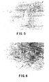
- human umbilical endothelial cells 10 5 or more cells per ml, were embedded in FG, the protein concentration of which was 4 mg/ml.
- the concentration of thrombin in the FG was adjusted to 0.6 NIH U/ml.
- the culture medium used in all of the experiments was M 199 (Sigma Chemical Co., St. Louis, MO) supplemented with 10% fetal bovine serum, 10 ⁇ g/ml streptomycin, 100 U/ml penicillin, 1 ng/ml FGF-1 and 10 U/ml heparin.
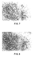
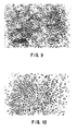
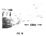
- Control cells As a control, an identical cell suspension was cultured on a surface coated with fibronectin at 10 ⁇ g/cm 2 . Control cells acquired a cobblestone shape and maintained this morphology for at least 5 days. Figures 9 and 10 show this situation at 24 and 48 hours, respectively.
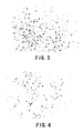
- PMEXNEO-3T3-2.2 cells are fibroblast cells that contain a modified genome with the potential to express genetically engineered proteins ( Forough et al., J. Biol. Chem. 268:2960-2968 (1993) ).
- 10 5 cells per well were cultured under three conditions: (1) embedded in FG; (2) on the surface of FG; and (3) in the absence of FG (controls).
- the experiments were carried out in duplicate in 24-well plates in DMEM media (Sigma Chemical Co., St. Louis, MO) supplemented with 10% FBS.
- the FG protein concentration was 4 mg/ml.
- the medium was supplemented with 1.5% FBS was used as negative controls.
- EC endothelial cell
- the modified FG was sterilely prepared by adding approximately 1 ng/cm 2 area of the inner and outer graft surfaces of human recombinant 125 I-HBGF-1, 20 ⁇ g/cm 2 porcine intestinal mucosal heparin, and 2.86 mg/cm 2 fibrinogen to 2.86 x 10 -2 U/cm 2 reconstituted, commercially available.
- human thrombin 1000 U/ml to induce polymerization.
- the 125 I-HBGF-1 was specifically prepared as follows. Fibrinogen was reconstituted by adding 500 mg of fibrinogen into 25 ml of PBS to produce a fibrinogen concentration of 20 mg/ml of PBS. Three ml of this solution which contained 60 mg fibrinogen were placed into 12 Eppendorf plastic tubes and maintained at -70°C. Each of these aliquots was used individually.
- the thrombin was reconstituted by diluting a commercially available preparation thereof (Armour Pharmaceutical Co., Kankakee, IL) at a concentration of 1000 U/ml by a factor of 1:10 in sterile solution to produce a concentration of 100 U/ml. This thrombin solution was again diluted 1:10 to produce a solution of 10 U/ml.
- the bovine heparin (Upjohn. Kalamazoo, MI) was reconstituted by diluting the preparation at a concentration of 1000 U/ml by a factor of 1:1000 using normal saline.
- One end of the expanded PTFE graft was placed over a plastic 3-way stopcock nozzle and was secured there with a 2-0 silk tie.
- the PTFE was then encircled with a 3 x 3 cm square of ParafilmTM which was then crimped there with a straight hemostat to establish a watertight seal.
- a second 2-0 silk tie was positioned over the parafilm adjacent to the stopcock to form another seal.
- a straight hemostat was then used to clamp the distal 2 mm of the PTFE/parafilm to seal this end.
- Equal volumes of fibrinogen and thrombin solution prepared as described above were mixed and allowed to react for approximately 30 seconds which is when polymerization occurs.
- the thrombin-polymerized fibrin is then opaque. (This time factor is approximate and varies from one thrombin lot to another. The appropriate length of time to polymerization can be determined by viewing the opacity of the mixture).
- the fibrin/thrombin mixture was aspirated into a one cc syringe. (NOTE: The volume of this graft was 0.42 ml.
- a larger syringe For a graft with a larger volume one needs to use a larger syringe.
- the syringe was attached to the stopcock and the mixture was injected by hand over a period of 5 seconds until the liquid was seen to "sweat" through the PTFE interstices and filled the space between the PTFE and the ParafilmTM.
- the 3-way stopcock was closed to the PTFE graft for 3 minutes and a scalpel blade was used to cut the ligature at the end of the PTFE over the stopcock.
- the PTFE graft/parafilm was removed from the stopcock and a hemostat was used to remove the PTFE from the parafilm envelope. To clear residual growth factor-supplemented FG from the graft lumen.
- a number 3 embolectomy catheter was passed through the graft five times until the graft lumen was completely clear.
- the growth factor-supplemented FG-treated PTFE graft was allowed to dry overnight for about 12 hours under a laminar flow hood. The treated graft was then ready for implantation.
- this HBGF-supplemented FG was pressure perfused into a 34 mm (24 mm + 5 mm at each end) x 4 mm (internal diameter) thin-walled, expanded PTFE graft thereby coating the graft's luminal surface and extending through the nodes to the graft's outer surfaces.
- the lumen of the graft was cleared as stated above.
- These grafts were then interposed into the infrarenal abdominal aortas of 24, 3-5 Kg New Zealand white rabbits.
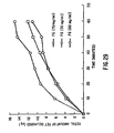
- the animals were sacrificed and specimens were explanted at 0 time (to correct for losses due to surgical manipulation) and after 5, 30, and 60 min, and 1, 7, 14, and 30 days. Residual radioactivity was determined by gamma counting. Remaining 125 I-HBGF-1, corrected for spontaneous decay, is expressed as a percentage of the zero time value.
- the second study evaluated the effects of the applied HBGF-1-supplemented FG suspension on: the rate of endothelialization of widely expanded 60 ⁇ m internodal distance expanded PTFE grafts implanted into canine aorta-iliac positions; the proliferative activity of these endothelial cells as a function of time; and the relative contributions of the HBGF-1 and the FG in stimulating the observed endothelial cell proliferation.
- Three groups of 50 x 4 mm non-reinforced expanded PTFE grafts were implanted in the aortailiac position of 12 dogs.
- Group 1 (n 6) contained 20 ⁇ g/cm 2 heparin.
- Plasma reduced platelets were prepared and pelleted. The supernatant plasma was removed. The pelleted platelets were washed, suspended in buffer containing 50 mM histidine and 0.15 M sodium chloride at pH 6.5, and treated with bovine thrombin. After treatment, the supernatant was collected by centrifugation and aliquots were frozen at -80°C. The extract was thawed and mixed with FG or other TSs.
- the platelet extract obtained in this manner was biologically active since it increased the incorporation of radioactive labeled thymidine into the DNA of proliferating NIH3T3 fibroblasts compared to the controls.
- mice Female C57BL/K s J-db/db mice were obtained, from Jackson Laboratories (Bar Harbor, ME) and were 8 to 12 weeks old at the start of the experiment. They were housed in separate cages after surgery in an animal care facility.
- mice are used as a model of impaired wound healing in diabetic humans because the metabolic abnormalities seen in these mice are similar to those found in human diabetics.
- the healing impairment characterized by markedly delayed cellular infiltration, granulation tissue formation. and time required for wound closure suggest that healing in this mouse model may be relevant to the healing impairment seen in human diabetes.
- the concentrated topical fibrinogen complex (TFC) used in this study was produced from fresh frozen pooled human plasma.
- the TFC product (American Red Cross-Baxter Hyland Division, Los Angeles, CA) was supplied in lyophilized form. After reconstitution with 3.3 ml of sterile water, the protein characteristics of the TFC solution used in this study were: total protein. 120 mg/ml; fibrinogen, 90 mg/ml; fibronectin, 13.5 mg/ml; Factor XIII, 17 U/ml; and plasminogen, 2.2 ⁇ g/ml.
- Topical bovine thrombin (5000-unit vial, Armour Pharmaceutical Co., Kankakee. IL) was reconstituted with 5 ml sterile water and was serially diluted in 80 mM calcium chloride solution (American Reagent Laboratories, Shirley, NY) to a concentration of 15 U/ml.
- Equal volumes of TFC and reconstituted thrombin were mixed to produce FG.
- 0.015 ml of TFC was mixed with 0.015 ml of thrombin.
- the FG that was produced had a protein concentration of approximately 60 mg/ml.
- a diluted FG with a protein concentration of approximately 1 mg/ml was also used.
- mice were anesthetized with a mixture consisting of 7 ml ketamine hydrochloride (100 mg/ml; Ketaset, Aveco Co., Inc., Fort Dodge, IA), 3 ml xylazine (20 mg/ml; Rompun, Mobey Corp., Shawnee, KA), and 20 ml physiological saline, at a dose of 0.1 ml per 100 g body wt, administered intramuscularly.
- the dorsal hair was clipped, and the skin was washed with povidone-iodine solution and wiped with 70% alcohol solution.
- Two full-thickness, round surgical wounds (6 mm diameter) were made on the lower back of the mouse, one on each side, equidistant from the midline.
- the medial edges of the two wounds were separated by a margin of at least 1.5 cm of unwounded skin.
- the dressing was a transparent semipermeable adhesive polyurethane dressing (OpsiteTM, Smith and Nephew, Massillon, OH). Tincture of Benzoin compound (Paddock Laboratories, Minneapolis, MN) was applied at the periphery of the wound area prior to application of the dressing. There was a margin of at least 0.5 cm of skin surrounding the wound edge over which no tincture of benzoin was applied to avoid the possible inflammatory effects of benzoin on the raw wound. No further treatments were applied to the wound for the duration of the experiment.
- mice were divided into 4 treatment groups, with each mouse serving as its own control:
- the animals were euthanized on Day 9 of the experiment.
- the wounds were excised down to the muscle layer, including a margin of 0.5 mm of unwounded skin, and were placed in buffered 10% formalin solution.
- the specimens were submitted to a histology laboratory for processing. Specimens were embedded in paraffin, and the midportion of the wound was cut in 5- ⁇ m sections. The slides were stained with hematoxylin and eosin, or with Masson's trichrome for histologic analysis.
- Each slide was given a histological score ranging from 1 to 15, with 1 corresponding to no healing and 15 corresponding to a scar with organized collagen fibers (Table 1).
- the scoring scale was based on scales used by previous investigators. The criteria used previously were modified and were further defined to more precisely reflect the extent of: reepithelialization, degree of cellular invasion, granulation tissue formation, collagen deposition, vascularity, and wound contraction.
- the histologic score was assigned separately by at least three analysts. The code describing the wound treatment was broken after the scoring was completed by all observers.
- the paired t test was used for comparison of paired means in the different treatment groups.
- the analyses were performed using the RS/1 Release 3.0 statistical software package (BBN Software Products Corporation).
- the sample mean differences were tested for analysis of variance using the Statistical Analysis Software (SAS) System.
- SAS Statistical Analysis Software
- mice (1) when applied over open wounds, FG at a concentration formulated for hemostasis (60 mg/ml) resulted in lower histological scores at Day 9 which indicated slower rates of wound healing compared to that of untreated wounds; (2) dilution of the FG protein concentration to 1 mg/ml resulted in a higher histological score at Day 9 which indicated a faster rate of wound healing: and (3) application of a semipermeable dressing (OpsiteTM) per se significantly retarded wound closure in this animal model by itself.
- FG a concentration formulated for hemostasis
- the total protein concentration of FG is an important variable when comparing the results of studies using FG. Beneficial effects of fibrin in promoting wound healing and tissue repair have been reported, but lower concentrations of fibrinogen have been used in the present studies than is commonly found in commercial preparations.
- FG at a concentration of 60 mg/ml delayed wound closure (Group I).
- HBGF-1B growth factor-supplemented FG The effect of HBGF-1B growth factor-supplemented FG on the rate of wound repair in diabetic mice was assessed.
- the methods used in this experiment were similar to those just described above.
- Two 6 mm full-thickness skin biopsies on the dorsal part of each of 6 test mice were filled with FG to which 5 ⁇ g of HBGF-1 ⁇ had been added.
- Identical biopsies in six mice were left untreated, and in six control mice were filled with unsupplemented FG. After 9 days, all of the mice were sacrificed and histological preparations of 5 micron thick slices from each of the wounds and surrounding skin were prepared and stained with hematoxylin and eosin.
- Human thrombin 1000 U vial was reconstituted with 3.3 ml sterile water, and was serially diluted in 40 mM calcium chloride solution (American Regent Laboratories, Shirley, NY) to a concentration of 15 U/ml. Human thrombin was used for preparing disks implanted which were onto calvarial defects.
- Topical bovine thrombin (5000 U vial, Armour Pharmaceutical Co., Kankakee, IL) was reconstituted with 5 ml sterile water, and was serially diluted in 40 mM calcium chloride solution to a concentration of 15 U/ml. Bovine thrombin was used for preparing implants for intramuscular bioassay.
- the fibrinogen should be present at a concentration of 1 to 120 mg/ml FG, more preferably from 3 to 60 mg/ml FG, most preferably from 10 to 30 mg/ml FG.
- DBM should be present at an approximate concentration of about 1 to 1000 mg/ml FG. more preferably from 50 to 500 mg/ml FG, most preferably from 300-500 mg/ml FG.
- the particle size of demineralized bone powder should be from 0.01 to 1000 microns, preferably from 20-500 microns and most preferably from 70-250 microns.
- the osteoinductive growth factor(s) or BMPs should be present at a concentration(s) of about 1 to 100 ⁇ g/ml wherein the concentration(s) is effective to accomplish its desired purpose.
- Growth factors which may be used as ostioinductive substances in this embodiment include, but are not limited to: osteogenin (BMP3); BMP-2; OP-1; HBGF-1; HBGF-2; BMP 2A, 2B and 7; FGF-1; FGF-4; and TGF- ⁇ .
- drugs such as antibiotics, can be used to supplement the TS for use in bone repair.
- Rat DBM was prepared as follows. The epiphyses of the long bones of rats were removed leaving only the diaphyses behind. The diaphyses were split, if necessary, and the bone marrow was then thoroughly flushed with deionized water (Milli-Q Water Purification SystemTM, Millipore Corporation, Bedford, MA). The diaphyses were then washed at room temperature. At 4°C, 1000 mls of deionized water was added to 100 g of bone. The mixture was stirred for 30 minutes and the water was decanted. This step was repeated for two hours.
- deionized water Milli-Q Water Purification SystemTM, Millipore Corporation, Bedford, MA
- the bone was then milled to make bone powder.
- the powder was seived and 74 to 420 micron size particles were collected.
- the pellets were then washed with 180 mls of deionized water by stirring to produce an even suspension.
- the suspension was then centrifuged for an additional 15 minutes.
- the supernatant was then decanted as before. The washing was repeated until the pH of the supernatant equaled the pH of the deionized water.
- pellets were then frozen at -180°C in a freezer. They were then lypholized using standard procedures.
- Disk-shaped implants 1 mm thick and 8 mm in diameter were produced using a 4-piece aluminum mold ( Figure 13 ). Twenty-five mg of rat DBM powder was added into the mold chamber. Thirty ⁇ l of TFC was then pipetted onto the DBM and mixed until the DBM had absorbed all of the solution. The concentrations of TFC which were used were 10, 20, 40, 80, or 120 mg/ml. Thirty ⁇ l of thrombin solution (15 U/ml in 40 mM calcium chloride solution) was then added to the DBM-TFC complex, was mixed, and was compressed into a disk-shape using a piston-shaped lid. It was determined that 25 mg of DBM powder had a volume of 20 ⁇ l.
- DBM-TFC complex Fifty mg was poured into an aluminum mold, to which 60 ⁇ l of TFC was then added to the DBM and mixed until fully absorbed. Sixty ⁇ l of thrombin was then added to the DBM-TFC complex, mixed and compressed into a disk-shape with a diameter of 1 cm and a thickness of 2 mm using a piston-shaped lid. The disk was then cut manually into the desired shape (triangle, square or donut).
- implants were placed in a sterile nylon bag having a mesh size of 70 microns and measuring 1 cm x 1 cm.
- mice Male Long-Evans rats were obtained from Charles River Laboratories (Wilmington, MA). For the intramuscular bioassay, 28 to 35 day old rats were used. Three month old rats were used for the craniotomy experiment.
- the animals were anesthetized with a mixture consisting of 10 ml ketamine hydrochloride (Vetalar, 100 mg/ml, Parke-Davis. Morris Plains, NJ), 5 ml xylazine (Rompun, 20 mg/ml, Mobay Corporation, Shawnee, KN), and 1 ml physiologic saline (0.9% NaCl), at a dose of 0.1 ml per 100 gm body weight, administered intramuscularly.
- the operative site of the animal was prepped with 70% alcohol solution, followed by povidone-iodine solution. The surgical procedure was then performed using aseptic technique.
- Intramuscular Bioassay A midline ventral incision was made and a space was created between the pectoralis muscles with blunt dissection. A nylon envelope containing the designated experimental material was inserted into the intramuscular space and secured with a 3-0 Dexon suture ( Figure 14 ). The same procedure was then repeated at the contralateral side. The skin was then closed with staples. The implants were harvested after four weeks, were x-rayed and were prepared for histology.
- Craniotomy Procedure A linear incision was made from the nasal bone to the mid-sagittal crest. Soft tissues were reflected gently and the periosteum was dissected from the craniotomy site (occipital, frontal, parietal bones). An 8-mm craniotomy was prepared with a trephine in a slow-speed rotary handpiece using copious saline irrigation as needed. The calvarial disk was dissected free while avoiding dural perforations and superior sagittal sinus intrusion. The 8-mm calvarial defect was either left untreated as control or filled with a 1 x 8-mm DBM or DBM-FG disk ( Figure 15 ). The skin was then closed with skin staples.
- each rat was identified by ear punches and returned to its cage where they were ambulatory within 2-3 hours.
- the rats were euthanized in a carbon dioxide chamber.
- a skin incision was made around the experimental recipient bed (i.e., pectoralis major or calvaria) and the soft tissues were reflected from the recipient beds.
- the craniotomies with 3-4 mm contiguous bone were recovered from the fronto-occipito-parietal complex.
- sharp and blunt dissection was used to recover the implanted nylon envelopes.
- the implants were radiographed using X-OMATLTM high contrast Kodak x-ray film (Eastman Kodak Company, Rochester, NY) in a Minishot Benchtop Cabinet x-ray system (TFI Corporation, West Haven, CT) at 30 kvp, 3 Ma, and 10 seconds. Gray-level densities of intramuscular and craniotomy site radiographs were analyzed using a Cambridge 920 Image Analysis SystemTM (Cambridge Instruments Limited, Cambridge, England).
- All DBM disks displayed radio-opaque images. Forty-five out of 48 implanted DBM-FG disks (93.75%) were radio-opaque. All DBM-FG disks, regardless of protein concentration (4-45 mg/ml) induced radio-opacity ( Figure 16 ). Radio-opacity measurements of some DBM disks ( Figure 16 ) were higher than DBM-FG disks but the other measurements were well within the range of measurements for DBM-FG disks. Thirty out of 32 FG disks which were not supplemented with DBM (93.75%) did not develop radio-opacity.
- DBM-FG disks in the form of squares, triangles or donuts were also markedly radio-opaque as compared to FG disks which were not supplemented with DBM.
- the original shapes of the implants were generally retained.
- the intramuscular bioassay was positive for DBM and DBM-FG implants, as evidenced by formation of ossicles with a central cavity filled with marrow and resorption of previously implanted DBM particles.
- X-rays showed DBM implants in a FG matrix to be generally more radio-opaque than DBM implants alone or untreated controls. There was no marked discernible difference between different concentrations of FG used to deliver DBM.
- the radiographs of untreated 8-mm diameter calvaria defects showed a negligible amount of radio-opacity.
- the second set of calvarial implants using DBM in 30 mg/ml FG matrix showed markedly increased radio-opacity within the craniotomy wounds of 3 or 4 month-old calvaria over 28 day calvaria ( Figure 17 ).
- Non-treated 8 mm craniotomy wounds showed only fibrous connective tissue developing across the craniotomy wound ( Figure 18 ). Histology of DBM implants showed DBM particles to be scattered all over the field. Some DBM particles migrated over and under the edges of host bone ( Figure 19 ). Most DBM particles were, however, within the confines of the craniotomy wound and were surrounded by loose connective tissue that was well vascularized. Active resorption of DBM by osteoclasts was noted. A lot of DBM particles were also noted to be populated by live cells. New osteoid and bone laid down by osteoblasts were quite evident.
- the histology of DBM implants in a FG matrix showed DBM particles localized within the craniotomy wound, surrounded by much denser and more cellular connective tissue ( Figures 20 and 21 ). Osteoid matrix and bony trabeculae formation were quite evident. More bone marrow was noted to have formed in craniotomy wounds implanted with DBM-FG disks than with DBM implants alone. There was also greater neovascularization with DBM-FG disks than with DBM implants alone or untreated controls. Osteoregeneration was evident at all concentrations of FG used to deliver DBM.
- FG The natural biocompatibility and biodegradability of FG are characteristics that make it an ideal delivery vehicle for DBM and BMPs. FG facilitated the shaping of DBM into the desired form to fill bony defects, maintained DBM within the defect. and may have been synergistic with DBM. Furthermore, soft tissue prolapse did not occur and bony contour was maintained. DBM-supplemented FG possessed an appropriate microarchitecture, biodegradation profile and release kinetics to support osteoblast recruitment and osteoregeneration.
- the DBM-FG matrix Since the shape of the DBM-FG matrix determined the morphology of the newly formed bone, when possible, the DBM-FG matrix should be made of a predetermined shape. However, the DBM-FG matrix in liquid form can be delivered or injected into an irregularly shaped defect where it will polymerize and encourage bone formation in the DBM-FG-filled area.
- TFC human topical fibrinogen concentrate
- Freeze-dried thrombin concentrate supplied by The American Red Cross/Baxter-Hyland, Inc., Glendale, CA, was reconstituted with 3.5 ml of a 40 mM solution of calcium chloride prepared in water for injection. The resulting solution contained approximately 250 U/ml.
- TET-FG was formulated by mixing the desired weight of TET with 1 ml of reconstituted TFC solution and with 1 ml of reconstituted thrombin solution in the presence of injection quality calcium chloride (purchased from American Reagent, Shirley, NY).
- the TET was in the free base form and was purchased from Sigma Chemical Company (St. Louis, MO).
- the TET-FG was formed by mixing TFC and thrombin through a DuofloTM dispenser (Hamaedics, CA) onto a Millipore membrane in a 12 mm diameter Millipore culture plate (Millipore Corporation, Bedford, MA). The mixture was allowed to set for one hour at 22°C. Six mm diameter disks containing the TET-FG and the Millipore membrane were cut from the latter using a 6 mm punch biopsy. The TET-FG-containing disks were used for the TET release studies.
- TET release from the TET-FG into phosphate buffered saline (PBS) or saliva was measured using 24-well cell culture plates (Coming Glass Works, Corning, NY) under two different sets of conditions. In one condition, the static mode, 2 ml of PBS or 0.75 ml of saliva was replaced daily in the 24-well cell culture plates. In the other condition, the continuous exchange mode, TET release from the TET-FG was measured with PBS having been exchanged at a rate of approximately 3 ml per day. The samples were stored at -20°C until analyzed. The saliva had been collected from 10 different people, had been pooled, and clarified by centrifugation at 5000g. It was then filtered through a 0.45 ⁇ m pore sized membrane and was stored at 4°C for daily use.
- PBS phosphate buffered saline
- saliva was measured using 24-well cell culture plates (Coming Glass Works, Corning, NY) under two different sets of conditions. In one condition, the static mode, 2 ml of
- the eluted TET was thawed and was analyzed spectrophotometrically at 320 nm and/or biologically by the inhibition of E . coli growth on agar plates.
- Ciprofloxacin HCl CIP
- Amoxicillin AMO
- Metronidazole MET
- FG containing CIP HCl, AMO or MET were prepared as before for TET.
- the AB-FG disks were placed in individual wells in a 24-well cell culture plate and were covered with 2 ml of PBS that was collected, replaced daily and stored at -20°C as before, until analyzed.
- the concentrations of CIP, AMO and MET in the eluates were measured spectrophotometrically at 275, 274 and 320 nm, respectively, and were compared to standard curves containing 0 to 50 ug/ml of the corresponding AB.
- the maintenance of the structural integrity of the FG and the TET-FG disks was estimated by visual observation and physical inspection by "poking" the disks with a fine spatula.
- the porous membrane which had been cut out while making the disks remained attached to the TET-FG and was used to help position the disks during the evaluation of their structural integrity. Pictures of top and lateral views of the disks were also taken and were used in the evaluation.
- the structural integrity of FG and TET-FG were measured under both sterile and non-sterile conditions.
- the PBS and saliva were stored frozen until analyzed.
- the same procedure was used except that the entire process was run under sterile conditions.
- the sterility of the system was tested by incubating 0.2 ml of sample and 2 ml of broth at 37°C and the turbidity of the broth was monitored for 48 hours. Lack of turbidity indicated sterility of the system.
- the stability of the CIP-, AMO-, and MET-FG were studied as above but under non-sterile conditions only.
- the antimicrobial activity of the AB released from the AB-FG was estimated by measuring the diameters of the zones of inhibition generated by the eluate from the 6mm diameter disks from the daily collected PBS or saliva surrounding the AB-FG.
- the eluates from unsupplemented FG served as controls.
- AB solutions of known concentration were used as standards.
- E . coli cultured on agar plates were used to measure the AB activity of the released TET, CIP and MET. To make the culture plates, 100 ⁇ l of the bacterial cell suspension, containing approximately 10 8 cells/ml, was mixed with 3 ml of top agar at 50°C and immediately poured onto the plate hard, bottom agar to make a uniform layer of cells. The plates were incubated at 37°C for 18 hours.
- TET concentrations which were obtained for different amounts of TET that had been incorporated into TET-FG are shown in Figure 22 .
- TET concentrations in the TET-FG of less than 50 mg/ml, the release of TET was completed in five days or less.
- TET concentrations in the TET-FG disks which contained TET concentrations of 100 and 200 mg/ml occurred for approximately two weeks, and more than three weeks, respectively.
- the structural integrity of the TET-FG disks was preserved for three to five weeks.
- the spectrophotometric data which were collected in the continuous exchange experiment are shown in Figure 23 . These data indicate that a continuous TET release from a TET-FG disk which originally contained a TET concentration of 100 mg/ml FG occurred over a two week period. The FG disk retained its structural integrity during this two week period, infra. The TET release data obtained in the continuous mode experiment also indicated that the rate of TET release opportunity depended on the concentration of TET which remained in the TET-FG disk.
- TET-FG containing 50 and 100 mg/ml of TET was measured in static experiments by determining the TET concentration in 0.75 ml of saliva that was replaced daily. These results ( Fig. 24 ) are similar to those obtained in PBS except that the concentration of TET was higher, most probably reflecting the smaller volume of saliva which was used to collect the released TET.
- the presence of TET in the FG matrix again unexpectedly prolonged the structural integrity of the TET-FG matrices for at least 15 days compared to that for the control FG disks which had begun to decay by 9 days and were almost completely decayed by 15 days ( Figure 25 ).
- CIP-, AMO- and MET-FG The antibiotic released from CIP-, AMO- and MET-FG is shown in Figure 27 .
- CIP was released at an apparent constant rate for approximately 4 weeks and then the rate decreased gradually for approximately one more week.
- the release of AMO and MET was complete within 3 days.
- the TET- and CIP-induced FG stabilization can be exploited for controlling the total release time not only for these ABs, but also for other drugs or "supplements" added to FG whose release rate and/or total release duration depends on the integrity of the FG matrix.
- TET-or CIP- induced FG stabilization can be exploited for controlling the total release time of TET, CIP and other drugs or supplements which have been added to the TET-FG or CIP-FG matrices.
- FG was supplemented with 50 mg/ml of TET free base and was shaped as 6 x 2.5 mm disks for this study.
- the protein concentration of FG was adjusted to 60 mg/ml.
- the disks were placed in 2 ml of PBS, pH 7.3 and were allowed to stand at 4, 23 and 37°C. To wash the disks, the PBS was replaced every 10 minutes, 6 times, with 2 ml of fresh PBS. Thereafter the PBS was replaced every hour for 4 hours.
- the TET concentrations in the collected samples were determined spectrophotometrically against a standard curve as before.
- FG supplemented with 1 mg/ml of TET HCl solution was prepared and was shaped as 6 x 2.5 mm disks for this study.
- the protein concentration of the FG was adjusted to 60, 30 and 15 mg/ml.
- Each disk was placed in 3 ml of distilled water. The water was replaced with the same volume of water every 10 minutes for a total of one hour.
- the TET concentration in the collected samples was determined spectrophotometrically against a standard curve as before.
- each one of 5 animals per group were injected intraperitoneally with 0.5 ml PBS (Group-I), FG (Group-II), TET-FG (Group-III) or CIP-FG (Group IV).
- FG and AB-FG was administered using a Hamaedics dispenser containing 0.25 ml of TFC at 120 mg/ml and 0.25 ml of human thrombin at 250 U/ml.
- the thrombin solution contained 50 mg of the respective AB.
- TET-FG and CIP-FG protected mice from death caused by S. aureaus 202A for at least 48 hours after the administration of the AB-supplemented FG.
- the fibrinogen was solubilized with sterile water or, for one group with water saturated with 5-FU at a concentration of 17 mg/ml.
- Thrombin solutions were made with sterile water, and then were diluted in 40 mM CaCl 2 to a concentration of 15 U/ml, or Thrombin was dissolved in 40 mM CaCl 2 saturated with 5-FU in a concentration of 17 mg/ml.
- Control FG clots did not contain 5-FU and were produced by mixing 200 ⁇ l of TFC solution (at 60 mg/ml) with 200 ⁇ l of Thrombin solution (at 15 U/ml) and allowing 20 minutes to polymerize. These clots were made in 12 by 75 mm test tubes and then were placed in 10 mls of 0.05 M Histidine, 0.15 M NaCl, pH 7.3 (Buffer).
- FG clots containing saturated levels of liquid 5-FU were produced by mixing 200 ⁇ l of TFC (60 mg/ml + 17 mg/ml 5-FU) with 200 ⁇ l Thrombin solution (15 U/ml + 17 mg/ml 5-FU) and allowing 20 minutes for the clots to fully polymerize.
- the addition of saturated levels of 5-FU in both the TFC and Thrombin solutions somewhat altered clot formation producing a clot that was translucent, as compared to the control FG clots which were quite opaque.
- the clots that were formed were physically the same as those made with FG alone except in color. Clots were formed in 12 by 75 mm test tubes and then placed in 10 ml of buffer.
- a second group of FG clots were made that contained an amount of solid anhydrous 5-FU equal to the amount included in clots formed with saturated solutions of 5-FU. These clots were formed by the addition of 7 mg of solid anhydrous 5-FU to 200 ⁇ l of TFC (60 mg/ml) and 200 ⁇ l of Thrombin (15 U/ml). Seven mg of 5-FU was placed in a 12 by 75 mm test tube. Two hundred ⁇ l of TFC was then added followed by 200 ⁇ l of Thrombin. The 3 components were then mixed by pipetting back and forth until a homogenous mixture was observed and further mixing was inhibited due to the clotting reaction. Clots were then placed in 10 ml of histidine buffer.
- the final group contained 50 mg of solid anhydrous 5-FU per clot. Due to the increased mass of 5-FU (50 mg instead of 7 mg) the previously used method did not work. Instead of producing a homogenous clot, a clot was formed with the majority of the 5-FU having settled to the bottom of the test tube. To avoid this problem the bottom of the test tube was first coated with 100 ⁇ l of TFC (60 mg/ml) and 100 ⁇ l of Thrombin (15 U/ml). This formed a clot which covered the concave bottom of the test tube. Next. 50 mg of solid anhydrous 5-FU was added to the surface of the 200 ⁇ l clot. Following this, 100 ⁇ l of TFC was added along with 100 ⁇ l of Thrombin.
- the two solutions were mixed using an automatic pipettor until the protein started to gel. When this occurred, the pipetting was ended and the clot was allowed to polymerize for 20 minutes.
- the final product was a clot that contained a dense core of approximately 50 mg of 5-FU. As with the other clots, these were then placed in 10 mls of buffer. The final total protein concentration of the FG in all groups was 30 mg/ml.
- Each group contained 10 replicates. Each duplicate was incubated at 37°C in 10 mls of buffer. Buffer was exchanged for 10 mls of fresh solution at 5, 10, 22, 33, 52, 75 and 114 hours. Aliquots of the eluate buffer were then examined in a spectrophotometer at a wavelength setting of 260 nm. Previous experiments had demonstrated that 5-FU absorbed strongly at this wavelength, while eluates from control FG clots did not.
- the duration of 5-FU delivery appeared to be a function of the mass of 5-FU loaded into the gel.
- the amount of 5-FU deliverable from the clots containing 5-FU in solution was limited by the solubility of the drug.
- the inclusion of amounts of solid anhydrous form equal to the amount present in the clots formed from liquid saturated with 5-FU resulted in nearly identical delivery kinetics, while the inclusion of greater amounts of 5-FU in the solid form than were possible using the liquid form, resulted in a tripling of the total duration of delivery, and typically a 10-fold increase in the duration of delivery of a given concentration of the drug.
- the result of this process is a sustainable delivery of the antiproliferative/cytotoxic drug 5-FU from fibrin clots for at least 10 times longer than is possible using the drug in the aqueous form.
- This technology i.e., the use of a solid form of the drug, preferably one with a low solubility and/or dissolution rate
- DMEM Dulbecco's Modified Eagle's Medium
- Antibiotic-Antimycotic solution was purchased from GIBCO (Grand Island, N. Y.).
- FGF-1 and -4 FGF-4 were a kind gift of Reginald Kidd, Plasma Derivatives Laboratory, American Red Cross, Rockville, MD, and Genetics Institute (Cambridge, MA), respectively.
- FGF-2 also known as basic FGF or bFGF
- FGF-2 also known as basic FGF or bFGF
- NIH/3T3 fibroblasts at passage 126 were purchased from the American Type Culture Collection, Rockville, MD. Cultures from passages 129-133 were used in the chemotaxis assays. Cultures were propagated in DMEM supplemented with 10% Calf serum and approximately 1% antibiotic antimycotic solution. Human dermal fibroblasts (HDFs) were purchased from Clonetics, Inc. (San Diego, CA) at passage 2. Cultures from passages 3-5 were used in the chemotaxis assays. Cultures were cultivated in DMEM supplemented with 20% FBS (Hyclone Laboratories, Inc., Logan. UT) and approximately 1% antibiotic antimycotic solution (Gibco, Grand Island. N.Y.).
- the procedure used to determine cellular chemotaxis was a combination of two known methodologies.
- a modification of Boyden's chamber was used as follows: Millicell-PCF (Millipore, Inc., Bedford, MA) (12.0 ⁇ m) 12.0 mm diameter inserts were placed in individual wells of 24 well plates to create the upper and lower chemotaxis chambers. Chemotaxis results were arrived at by performing checkerboard analysis for every combination of cells and growth factors. Concentrations ranging from .1, 1, 10, 100 ng/ml with/without added heparin (10 U/ml) were used for FGF-1, FGF-2 (no heparin) and FGF-4 with all the cell types mentioned in the materials section.
- Filters were mounted lower side facing up on slides in Crystal MountTM (Biomeda, Inc., Foster City, CA) solution and 10 random fields were counted per slide both visually at 400 x and at 200 x using an Image Analyzing System to automate the enumeration of the cells on the underside of the filters.
- Crystal MountTM Biomeda, Inc., Foster City, CA
- Chemotaxis chambers and cells were utilized as described above. Fifty ⁇ l of 8 mg/ml Topical Fibrinogen Complex (TFC, American Red Cross, Rockville, MD) was added to the bottom of 24 well plates. Forty ⁇ l of test growth factor +/- heparin at a final concentration of 10 U/ml (FGF-1, FGF-4 with heparin, FGF-2 alone) was added to the TFC and thoroughly mixed. Ten ⁇ l of bovine Thrombin (Armour Pharmaceutical Company, Kankakee, IL) was added and mixed throughly. The components were allowed to gel at room temperature for approximately 30 minutes. Total volume in the lower and upper chambers was made up to 0.5 ml each with DMEM + 0.1% BSA.
- TFC Topical Fibrinogen Complex
- the assay was performed at 37°C in a 5%CO 2 humidified chamber for approximately 24 hours. At the end of 24 hours, the filters were removed, fixed and stained and the number of cells on the underside of the filter were enumerated as described above.
- NIH 3T3 fibroblasts The ability of NIH 3T3 fibroblasts to migrate towards various well known chemotactic agents was determined to ensure that the cells used in this assay retained this capacity.
- Fibronectin was the most effective chemotactic agent tested for both NIH 3T3 and HDFs with maximal responses occuring at 20 ⁇ g/ml ( Figures 31 , Table 7). Thereafter, fibronectin at 20 ⁇ g/ml was used as the positive control for migration.
- the FGFs produced a profound chemotactic response in HDFs.
- a very good distinction was obtained between the negative control and the concentration of FGF which elicited a maximal migratory response: 18, 12 and 10 fold in response to FGF-1, -2 and -4, respectively.
- the stimulation of chemotaxis by growth factors was not as high for NIH 3T3 cells as it was for HDFs, possibly due to the high passage number of the available stock cultures of the NIH 3T3 cells as compared to the HDFs.
- FGF-1, FGF-2 and FGF-4 were found to be potent stimulators of fibroblast chemotaxis. Directed migration of fibroblasts by one or a combination of the above growth factors could result in fibroblast presence in the site of injury, thereby leading to fibroplasia and the laying down of collagen and an extracellular matrix. Thus, aside from it's well recognized angiogenic properties, FGF's may have a role in wound healing, acting either alone or in a combination with PDGF, IGF-I, TGF- ⁇ and/or other factors.
- Applicants' invention of incorporating FGFs into FG allows for the prolonged exposure of cells to the FGFs and can be applied to a wound.
- the resulting fibrin coating mimics the natural response to tissue injury, while delivering the growth factor directly to the wound site.
- FG which contained FGF-1 was used to line artificial vascular grafts (Example 8, herein). When these grafts were placed into the vessels of rabbits. the FGF-1 was released for a period of up to 28 days.
- the TS contains and delivers angiogenic substances, such as Fibroblast Growth Factor-1 (FGF-1), in an amount such that its concentration which is released from the supplemented TS is effective to induce angiogenesis.
- FGF-1 Fibroblast Growth Factor-1
- This embodiment is used in a controlled manner to revascularize body areas which have been deprived of an adequate blood supply such as cardiac, brain and muscle tissue, and the retina.
- This embodiment is used to restore or improve circulation to implanted organs or re-attached limbs.
- This embodiment can be used to generate a vascular network or "vascular bed" for: the generation of artificial organs or organoids, the delivery and/or localization of and/or nourishment of cells used in gene therapy, or as a target of gene therapy, for the nourishment and/or localization of cells for tissue augmentation.
- This embodiment also precludes the necessity of implantation of a device or substance which may induce a foreign body or other excessive inflammatory reaction which could compromise the blood vessel formation or the function of the underlying organ(s).
- the invention consists of a formulation of fibrinogen, (suitable for the formation of fibrin) with or without fibronectin and/or collagen, into which is placed an appropriate concentration of an angiogenic substance, such as FGF-1.
- the fibrinogen may also contain stabilizers to protect against the proteolytic activity of Thrombin.
- heparin sulfate (1-1000 U/ml) may be used as the stabilizer in the range of concentration of from 1 ng/ml to 1 mg/ml.
- the angiogenic substance is contained, in an appropriate concentration, in the thrombin, calcium, or water components.
- the concentration of the FGF-1 in the TS should be from 0.1 ng/ml to 1 mg/ml, more preferably from 1 ng/ml to 100 ⁇ g/ml, most preferably from 100 ng/ml to 10 ⁇ g/ml.
- the FGF-1, or other angiogenic substance will induce blood vessel formation within the body of the deposited FG.
- the FG will be naturally biodegraded leaving the intact blood vessel(s).
- the TS contains and delivers a cartilage promoting factor(s), such as cartilage-inducing factors-A and/or -B (CIF-A and CIF-B, respectively, which are also known as TGF-B 1 and TGF-B 2 , respectively) and/or another.
- factor(s) such as Osteoid-Inducing Factor (OIF) in an amount such that the concentration of the inducing factor(s) which is released from the supplemented TS is effective to induce cartilage formation.
- OIF Osteoid-Inducing Factor
- the concentration of the inducing factors should be 0.1 ng/ml to 1 mg/ml, more preferably from 1 ng/ml to 500 ng/ml, most preferably from 100 to 250 ng/ml.
- This embodiment may also contain drugs, such as antibiotics, and other growth factors, such as EGF, PDGF, and bFGF in the TS.
- the cartilage inducing substance is contained in an appropriate concentration in the fibrinogen or thrombin or calcium or water component(s) which are used to prepare the TS.
- the supplemented TS can either be pre-shaped to the desired final cartilage form prior to implantation or it can be implanted into the body of the recipient in the liquid form as the TS is mixed and polymerizes. The resulting form may then be sculpted as desired to produce the required shape of cartilage needed.
- the Cartilage Inducing TS (CI-TS) mixture can also be used to precoat a conventional implant, with the result being a conventional implant with a coating of living cartilage.
- the CI-TS is then implanted into the body of the recipient. This implantation can be heterotopic or orthotopic. After an appropriate interval, the CI-TS is be replaced by living cartilage with the form of the original CI-TS implant.
- Such implants can be used to replace damaged or lost cartilage, or to improve the tissue integration and/or function of an artificial implant.
- uses include the replacement or reconstruction of nasal or ear tissue, the generation of a functional joint surface on a bone implant grown in vivo , or the generation of a similar surface on an artificial implant.
- the repair of cartilage damaged by disease, such as rheumatoid arthritis, can also be accomplished using the CI-TS to produce a new and smooth cartilage surface to the arthus.
- Implants intended for space filling applications in Plastic/Reconstructive surgery can also be either formed from CI-TS, or coated with CI-TS to enhance tissue integration and reduce foreign body reactions.
- this invention is an advancement because it permits the generation of cartilaginous tissue which is required to fully mimic the body's natural make-up. This results in improved joint repair, artificial joints and other implants, both for orthopedic and other applications.
- this embodiment can be used: to produce improved orthopedic implants or improved plastic/reconstructive implants: for joint repair for traumatic, congenital or pathologically damaged or dysfunctional cartilage; to produce coatings of pacemaker implants and wires to increase their tissue integration and to reduce foreign body reactions. Similar coatings could also be applied to any implantable device for the similar purposes.
- This embodiment is a self-contained TS wound dressing, or bandage, which contains both the thrombin and fibrinogen components of the FG.
- the calcium is contained in either the thrombin and/or the fibrinogen component(s).
- Either or both of the thrombin or fibrinogen components can be, but does not have to be, supplemented with a growth factor(s), such as a FGF or bFGF, or a drug(s) such as, an analgesic, antibiotic or other drug(s), which can inhibit infection, promote wound healing and/or inhibit scar formation.
- the supplement(s) is at a concentration in the TS such that it will be effective for its intended purpose, e.g., an antibiotic will inhibit the growth of microbes, an analgesic will relieve pain.
- the thrombin and fibrinogen are separated from each other by an impermeable membrane, and the pair are covered with another such membrane.
- the thrombin and fibrinogen are contained in a quick evaporating gel (e.g., methylcellulose/alcohollwater).
- the bandage may be coated on the surface that is in contact with the gel in order to insure that the gel -pad remains in place during use. (See Figure 39 ).
- the membrane separating the two components is removed, allowing the two components to mix.
- the outer membrane is then removed and the bandage is applied to the wound site.
- the action of the thrombin and other components of the fibrinogen preparation cause the conversion of the fibrinogen to fibrin, just as they do with any application of FS. This results in a natural inhibition of blood and fluid loss from the wound, and the establishment of a natural barrier to infection.
- the thrombin component and the plastic film separating the Thrombin gel and the Fibrinogen gel may be omitted.
- the calcium that was previously in the Thrombin gel may or may not be included in the Fibrinogen gel as desired.
- the outer impervious plastic film is removed and the bandage applied, as previously described, directly to the wound site.
- the Thrombin and calcium naturally present at the wound site then-induce the conversion of fibrinogen to fibrin and inhibit blood and fluid loss from the wound as above.
- This embodiment has the advantage of being simpler, cheaper, and easier to produce. However, there may be circumstances in which a patient's wounds have insufficient thrombin. In those cases, the previous embodiment of the invention should be used.
- This embodiment is an advancement over the current technology as it permits the rapid application of TS to a wound without the time delay associated with solubilization and mixing of the components. It also requires no technical knowledge or skill to operate. These characteristics make it ideal for use in field applications, such as in trauma packs for soldiers, rescue workers, ambulance/paramedic teams, firemen, in first aid kits for the general public, and by emergency room personnel in hospitals. A small version may also be useful for use by the general public.
- This embodiment uses supplemented TS as a coating for the surfaces of orthopedic devices and other biomaterials which are to be implanted into an animal's body.
- these devices are urinary catheters, intravascular catheters, sutures, vascular prostheses, intraocular lenses, contact lenses, heart valves, shoulder/elbow/hip/knee replacement devices, total artificial hearts, etc.
- these biomaterials may become sites for bacterial adhesion and colonization, which eventually may lead to clinical infection that will endanger the life of the animal.
- the biomaterial is coated with a supplemented TS.
- the TS can be supplemented with: a growth factor(s); a drug(s), such as an antibiotic; BMP; and/or cultured cells, etc.
- antibiotics that may be incorporated into the TS include, but are not limited to: the penicillins; cephalosporins; tetracyclines; chloramphenicols; metronidazoles; and aminoglycosides.
- growth factors which may be incorporated into the TS include but are not limited to FGF, PDGF, TGF- ⁇ .
- BMPs which may be incorporated into the TS include, but are not limited to, BMP 1 through 8.
- DBM can also be added to the TS.
- cultured cells which may be incorporated into the TS include, but are not limited to, endothelial cells, osteoblasts, fibroblasts, etc.
- the supplement(s) may be contained in either the thrombin, fibrinogen, calcium or water component(s).
- concentration of the supplement in the TS is adequate such that it will be effective for its intended purpose, e.g., an antibiotic will inhibit the growth of microbes on the biomaterial, a growth factor will induce the growth of the desired cell type(s) in the TS and/or on the surface of the biomaterial.
- This invention is an improvement for existing biomaterial products, which include titanium and titanium alloy devices (such as fixation plates, shoulder/elbow/hip/knee replacement devices, osseointegrated dental implants, etc), solid silicone products (such as Silastic nasal implants, liquid and/or gel silicon products (such as breast implants and testicular implants), and natural or synthetic polymers used as conventional materials in healing a wound site, which may have various forms, such as monofilaments, fibrous assemblies (such as cotton, paper, nonwoven fabrics), films, sponges, bags, etc.
- titanium and titanium alloy devices such as fixation plates, shoulder/elbow/hip/knee replacement devices, osseointegrated dental implants, etc
- solid silicone products such as Silastic nasal implants, liquid and/or gel silicon products (such as breast implants and testicular implants)
- natural or synthetic polymers used as conventional materials in healing a wound site which may have various forms, such as monofilaments, fibrous assemblies (such as cotton, paper, nonwoven fabrics), films, sponges, bags, etc.
- FG is produced from 3 components: fibrinogen (for example as TFC); and thrombin, both of which may be in the lyophilized form; as well as calcium.
- fibrinogen for example as TFC
- thrombin both of which may be in the lyophilized form; as well as calcium.
- the lyophilized fibrinogen is reconstituted with sterile water, while the thrombin component is reconstituted with calcium chloride solution.
- a supplement may be added to any of the three components prior to mixing. Appropriate volumes of the fibrinogen and thrombin containing calcium are mixed to produce the FG.
- the FG is then applied to the biomaterial's surface as a coating thereof as, for example, by spraying, painting, etc. Alternatively, the implant is dipped in the FG while it is still liquid.
- a supplement may also be added to the FG before or after it has been coated on a biomaterial surface.
- a FG-coated implant is soaked in an antibiotic solution for a specified period of time so that the antibiotic will diffuse into the TS.
- Another example is coating a device with TS after which cultured cells are seeded onto the fibrin coating. Coating the surface of biomaterials, which will be implanted into an animal, with supplemented TS will serve several purposes. including: the inhibition of bacterial adhesion to the biomaterial; the inhibition of growth of bacteria adhered to the biomaterial; local immune stimulation and/or normalization; the promotion of would healing; and the promotion of engraftment of the biomaterial to the surrounding tissue.
Abstract
Description
- This invention is directed to supplemented TSs (TS), such as fibrin glue (FG), as well as to methods of their production and use. In one embodiment, this invention is directed to TSs which do not inhibit full thickness skin wound healing. The invention is directed to TSs which have been supplemented with a drug which is ciprofloxacin HCl, as well as to methods of their production and use.
- Wound healing, the repair of lesions, begins almost instantly after injury. It requires the successive coordinated function of a variety of cells and the close regulation of degradative and regenerative steps. The proliferation, differentiation and migration of cells are important biological processes which underlie wound healing, which also involves fibrin clot formation, resorption of the clot, tissue remodeling, such as fibrosis, endothelialization and epithelialization. Wound healing involves the formation of highly vascularized tissue that contains numerous capillaries, many active fibroblasts, and abundant collagen fibrils, but not the formation of specialized skin structures.
- The process of wound healing can be initiated by thromboplastin which flows out of injured cells. Thromboplastin contacts plasma factor VII to form factor X activator, which then, with factor V and in a complex with phospholipids and calcium, converts prothrombin into thrombin. Thrombin catalyzes the release of fibrinopeptides A and B from fibrinogen to produce fibrin monomers, which aggregate to form fibrin filaments. Thrombin also activates the transglutaminase, factor XIIIa, which catalyzes the formation of isopeptide bonds to covalently cross-link the fibrin filaments. Alpha2-antiplasmin is then bound by factor XIII onto the fibrin filaments to thereby protect the filaments from degradation by plasmin (see, for example, Doolittle et al., Ann. Rev. Biochem. 53:195-229 (1984)).
- When a tissue is injured, polypeptide growth factors, which exhibit an array of biological activities, are released into the wound where they play a crucial role in healing (see, e.g., Hormonal Proteins and Peptides (Li, C.H., ed.) ) and Brunt et al., Biotechnology 6:25-30 (1988)). These activities include recruiting cells, such as leukocytes and fibroblasts, into the injured area, and inducing cell proliferation and differentiation. Growth factors that may participate in wound healing include, but are not limited to: platelet-derived growth factors (PDGFs); insulin-binding growth factor-1 (IGF-1); insulin-binding growth factor-2 (IGF-2); epidermal growth factor (EGF); transforming growth factor-α (TGF-α); transforming growth factor-β (TGF-β); platelet factor 4 (PF-4); and heparin binding growth factors one and two (HBGF-1 and HBGF-2, respectively).
- PDGFs are stored in the alpha granules of circulating platelets and are released at wound sites during blood clotting (see, e.g., Lynch et al., J. Clin. Invest. 84:640-646 (1989)). PDGFs include: PDGF; platelet derived angiogenesis factor (PDAF); TGF-β; and PF-4, which is a chemoattractant for neutrophils (Knighton et al., in Growth Factors and Other Aspects of Wound Healing: Biological and Clinical Implications, Alan R. Liss, Inc., New York, New York, pp. 319-329 (1988)). PDGF is a mitogen, chemoattractant and a stimulator of protein synthesis in cells of mesenchymal origin, including fibroblasts and smooth muscle cells. PDGF is also a nonmitogenic chemoattractant for endothelial cells (see, for example, Adelmann-Grill et al., Eur. J. Cell Biol. 51:322-326 ( 1990)).
- IGF-1 acts in combination with PDGF to promote mitogenesis and protein synthesis in mesenchymal cells in culture. Application of either PDGF or IGF-1 alone to skin wounds does not enhance healing, but application of both factors together appears to promote connective tissue and epithelial tissue growth (Lynch et al., Proc. Natl. Acad. Sci. 76:1279-1283 (1987)).
- TGF-β is a chemoattractant for macrophages and monocytes. Depending upon the presence or absence of other growth factors, TGF-β may stimulate or inhibit the growth of many cell types. For example, when applied in vivo, TGF-β increases the tensile strength of healing dermal wounds. TGF-β also inhibits endothelial cell mitosis, and stimulates collagen and glycosaminoglycan synthesis by fibroblasts.
- Other growth factors, such as EGF, TGF-α, the HBGFs and osteogenin are also important in wound healing, EGF, which is found in gastric secretions and saliva, and TGF-α, which is made by both normal and transformed cells, are structurally related and may recognize the same receptors. These receptors mediate proliferation of epithelial cells. Both factors accelerate reepithelialization of skin wounds. Exogenous EGF promotes wound healing by stimulating the proliferation of keratinocytes and dermal fibroblasts (Nanney et al., J. Invest. Dermatol. 83:385-393 (1984) and Coffey et al., Nature 328:817-820 (1987)). Topical application of EGF accelerates the rate of healing of partial thickness wounds in humans (Schultz et al., Science 235:350-352 (1987)). Osteogenin, which has been purified from demineralized bone, appears to promote bone growth (see, e.g., Luyten et al., J. Biol. Chem. 264:13377 (1989)). In addition, platelet-derived wound healing formula, a platelet extract which is in the form of a salve or ointment for topical application, has been described (see, e.g., Knighton et al., Ann. Surg. 204:322-330 (1986)).
- The Heparin Binding Growth Factors (HBGFs), also known as Fibroblast Growth Factors (FGFs), which include acidic HBGF (aHBGF also known as HBFG-1 or FGF-1) and basic HBGF (bHBGF also known as HBGF-2), are potent mitogens for cells of mesodermal and neuroectodermal lineages, including endothelial cells (see, e.g., Burgess et al., Ann. Rev. Biochem. 58:575-606 (1989)). In addition, HBGF-1 is chemotactic for endothelial cells and astroglial cells. Both HBGF-1 and HBGF-2 bind to heparin, which protects them from proteolytic degradation. The array of biological activities exhibited by the HBGFs suggests that they play an important role in wound healing.
- Basic fibroblast growth factor (FGF-2) is a potent stimulator of angiogenesis and the migration and proliferation of fibroblasts (see, for example, Gospodarowicz et al., Mol. Cell. Endocinol. 46:187-204 (1986) and Gospodarowicz et al., Endo. Rev. 8:95-114 (1985)). Acidic fibroblast growth factor (FGF-1) has been shown to be a potent angiogenic factor for endothelial cells (Burgess et al., supra, 1989). However, it has not been established if any FGF growth factor is chemotactic for fibroblasts.
- Growth factors are, therefore, potentially useful for specifically promoting wound healing and tissue repair. However, their use to promote wound healing has yielded inconsistent results (see, e.g., Carter et al., in Growth Factors and Other Aspects of Wound Healing: Biological and Clinical Implications, Alan R. Liss, Inc., New York, New York, pp. 303-317 (1988)). For example, PDGF, IGF-1, EGF, TGF-α, TGF-β and FGF (also known as HBGF) applied separately to standardized skin wounds in swine had little effect on the regeneration of connective tissue or epithelium in the wounds (Lynch et al., J. Clin. Invest. 84:640-646 (1989)). Of the factors tested, TGF-β stimulated the greatest response alone. However, a combination of factors, such as PDGF-bb homodimer and IGF-1 or TGF-α produced a dramatic increase in connective tissue regeneration and epithelialization. (Id.) Tsuboi et al. have reported that the daily application of bFGF to an open wound stimulated wound healing in healing-impaired mice but not in normal mice (J. Exp. Med. 172:245-251 (1990)). On the other hand, the application to human skin wounds of crude preparations of porcine or bovine platelet lysate, which presumably contained growth factors, increased the rate at which the wounds closed, the number of cells in the healing area, the growth of blood vessels, the total rate of collagen deposition and the strength of the scar tissue (Carter et al., supra).
- The reasons for such inconsistent results are not known, but might be the result of difficulty in applying growth factors to a wound in a manner in which they can exhibit their normal array of biological activities. For example, it is now believed that some growth factor receptors must be occupied for at least 12 hours to produce a maximal biologic effect (Presta et al., Cell Regul. 2:719-726 (1991) and Rusnati et al., J. Cell Physiol. 154:152-161 (1993)). Because of such inconsistent results, the role played by exogenously applied growth factors in stimulating wound healing is not clear. Further, a means by which growth factors might be applied to wounds to produce prolonged contact between the wound and the growth factor(s) is not presently known.
- Surgical adhesives and TSs which contain plasma proteins are used for sealing internal and external wounds, such as in bones and skin, to reduce blood loss and maintain hemostasis. Such TSs contain blood clotting factors and other blood proteins. FG, also called fibrin sealant, is a gel similar to a natural clot which is prepared from plasma. The precise components of each FG are a function of the particular plasma fraction which is used as a starting material. Fractionation of plasma components can be effected by standard protein purification methods, such as ethanol, polyethylene glycol, and ammonium sulfate precipitation, ion exchange, and gel filtration chromatography. Typically FG contains a mixture of proteins including traces of albumin, fibronectin and plasminogen. In Canada, Europe and possibly elsewhere, commercially available FG typically also contains aprotinin as a stabilizer.
- FGs generally are prepared from: (1) a fibrinogen concentrate, which contains fibronectin, Factor XIII, and von Willebrand factor; (2) dried human or bovine thrombin; and (3) calcium ions. Commercially prepared FGs generally contain bovine components. The fibrinogen concentrate can be prepared from plasma by cryoprecipitation followed by fractionation, to yield a composition that forms a sealant or clot upon mixture with thrombin and an activator of thrombin such as calcium ions. The fibrinogen and thrombin concentrates are prepared in lyophilized form and are mixed with a solution of calcium chloride immediately prior to use. Upon mixing, the components are applied to a tissue where they coagulate on the tissue surface and form a clot that includes cross-linked fibrin. Factor XIII, which is present in the fibrinogen concentrate, catalyzes the cross-linking.
-
Australian Patent 75097/87 U.S. Patent Nos. 4,427,650 and4,427,651 , describes the preparation of an enriched plasma derivative in the form of a powder or sprayable preparation for enhanced wound closure and healing that contains fibrinogen, thrombin and/or prothrombin, and a fibrinolysis inhibitor, and may also contain other ingredients, such as a platelet extract. Rose et al.,U.S. Patent Nos. 4,627,879 and4,928,603 , disclose methods for preparing cryoprecipitated suspensions that contain fibrinogen and Factor XIII and their use to prepare a FG.JP 1-99565 U.S. Patent No. 4,714.457 ) and Morse et al. (U.S. Patent No. 5,030,215 ) disclose methods to produce autologous FG. In addition, improved FG delivery systems have been disclosed elsewhere (Miller et al.,U.S. Patent No. 4,932,942 and Morse et al.,PCT Application WO 91/09641 - IMMUNO AG (Vienna, Austria) and BEHRINGWERKE AG (Germany) (Gibble et al., Transfusion 30:741-747 (1990)) presently have FGs on the market in Europe and elsewhere (see, e.g.,
U.S. Patent Nos. 4,377,572 and4,298,598 , which are owned by IMMUNO AG). TSs are not commercially available in the U.S. However, the American National Red Cross and BAXTER/HYLAND (Los Angeles, CA) have recently co-developed a FG (ARC/BH FG) which is now in clinical studies. - The TSs which are used clinically outside of the U.S. pose certain clinical risks and have not been approved by the Food and Drug Administration for use in the USA. For example, the TSs available in Europe contain proteins of non-human origin such as aprotinin and bovine thrombin. Since these proteins are of non-human origin, people may develop allergic reactions to them. In Europe heat inactivation is used to inactivate viruses which may be present in the components of the FG. However, this heat inactivation method may produce denatured proteins in the FG which may also be allergenic. In addition, there is concern that this inactivation method will not inactivate prions which cause bovine spongiform enchephalopathy, "mad cow disease." which may be present in the TS due to the use of bovine proteins therein. Since this disease is believed to have already crossed from sheep, in which it is called "scrapies," to cows, it is not an insignificant concern that it could infect humans.
- The ARC/BH FG has advantages over the TSs available in Europe because it does not contain bovine proteins. For example, the ARC/BH TS contains human thrombin instead of bovine thrombin and does not contain aprotinin. Since the ARC/BH FG does not contain bovine proteins it should be less allergenic in humans than those TSs available in Europe. In addition, the ARC/BH FG is virally inactivated by a solvent detergent method which produces fewer denatured proteins and thus is less allergenic than those available in Europe. Therefore, the ARC/BH FG possesses advantages over the TSs which are now commercially available in other countries.
- FG is primarily formulated for clinical topical application and is used to control bleeding, maintain hemostasis and promote wound healing. The clinical uses of FG have recently been reviewed (Gibble et al., Transfusion 30:741-747 (1990); Lerner et al., J. Surg. Res. 48:165-181 (1990)). By sealing tissues FG prevents air or fluid leaks, induces hemostasis, and may contribute to wound healing indirectly by reducing or preventing events which may interfere with wound healing such as bleeding, hematomas, infections, etc. Although FG maintains hemostasis and reduces blood loss, it has not yet been shown to possess true wound healing properties. Because FG is suitable for both internal and external injuries, such as bone and skin injuries, and is useful to maintain hemostasis, it is desirable to enhance its wound healing properties.
- FG with a fibrinogen concentration of approximately 39 g/l and a thrombin concentration of 200-600 U/ml has produced clots with significantly increased stress, energy absorption and elasticity values (Byrne et al., Br. J. Surg. 78:841-843 (1991)). Perforated Teflon cylinders filled with fibrin clot (5 mg/ml) and implanted subcutaneously stimulated the formation of granulation tissue, including an increased precipitation of collagen, when compared to empty cylinders (Hedelin et al., Eur. Surg. Res. 15:312 (1983)).
- The sequence of bone induction was first described by Urist et al. using demineralized cortical bone matrix (Clin. Orthop. Rel. Res. 71:271 (1970) and Proc. Natl. Acad. Sci. USA 70:3511 (1973)). Implanted subcutaneously in allogeneic recipients, demineralized cortical bone matrix releases factors which act as local mitogens to stimulate the proliferation of mesenchymal cells (Rath et al., Nature (Lond.) 278:855 (1979)). New bone formation occurs between 12 and 18 days postimplantation. Ossicle development replete with hematopoietic marrow lineage occurred by day 21 (Reddi, A., In Ertracellular Matrix Biochemistry (Piez et al., ed.) Elsevier, New York, NY, pp. 375-412 (1984)).
- Demineralized bone matrix (DBM) is a source of osteoinductive proteins known as bone morphogenetic proteins (BMP), and growth factors which modulate the proliferation of progenitor bone cells (see, e.g., Hauschka et al., J. Biol. Chem. 261:12665-12674 (1986) and Canalis et al., J. Clin. Invest. 81:277-281 (1988)). Eight BMPs have now been identified and are abbreviated BMP-1 through BMP-8. BMP-3 and BMP-7 are also known as osteogenin and osteogenic protein-1 (OP-1), respectively.
- Unfortunately, DBM materials have little clinical use unless combined with particulate marrow autografts. There is a limit to the quantity of DBM that can be surgically placed into a recipient's bone to produce a therapeutic effect. In addition, resorption has been reported to be at least 49% (Toriumi et al., Arch. Otolaryngo. Head Neck Surg. 116:676-680 (1990)).
- DBM powder and osteogenin may be washed away by tissue fluids before their osteoinductive potential is expressed. In addition, seepage of tissue fluids into DBM-packed bone cavities or soft-tissue collapse into the wound bed are two factors that may significantly affect the osteoinductive properties of DBM and osteogenin. Soft-tissue collapse into the wound bed may likewise inhibit the proper migration of osteocompetent stem cells into the wound bed.
- Human DBM in powder form is currently used by American dentists to pack jaw bone cavities created during oral surgery. However, DBM in powder form is difficult to use.
- Purified BMPs have osteoinductive effects in animals when delivered by a variety of means including FG (Hattori, T., Nippon. Seikeigeka. Gakkai. Zasshi. 64:824-834 (1990); Kawamura et al., Clin. Orthop. Rel. Res. 235:302-310 (1988); Schlag et al., Clin. Orthop. Rel. Res. 227:269-285 (1988) and Schwarz et al., Clin. Orthop. Rel. Res. 238:282-287 (1989)) and whole blood clots (Wang et al., J. Cell. Biochem. 15F:Q20 Abstract (1990)). However, Schwarz et al. (supra.) demonstrated neither a clear positive or negative effect of FG on ectopic osteoinduction or BMP-dependent osteoregeneration. Kawamura et al. (supra.) found a synergistic effect when partially purified BMP in FG was tested in an ectopic non-bony site. Therefore, these results are inconsistent and confusing.
- TS also can serve as a "scaffold" which cells can use to move into a wounded area to generate new tissues. However, commercially available preparations of FG and other TSs are too dense to allow cell migration into and through them. This limits their effectiveness in some in vivo uses.
- In one type of bone wound, called bone nonunion defects, there is a minimal gap above which no new bone formation occurs naturally. Clinically, the treatment for these situations is bone grafting. However, the source of bone autografts is usually limited and the use of allogeneic bones involves a high risk of viral contamination. Because of this situation, the use of demineralized, virally inactivated bone powder is an attractive solution.
- Artificial vascular prostheses are frequently made out of polytetrafluoroethylene (PTFE) and are used to replace diseased blood vessels in humans and other animals. To maximize patency rates and minimize the thrombogenicity of vascular prostheses various techniques have been used including seeding of nonautologous endothelial cells onto the prothesis. Various substrates which adhere both to the vascular graft and endothelial cells have been investigated as an intermediate substrate to increase endothelial cell seeding. These substrates include preclotted blood (Herring et al., Surgery 84:498-504 (1978)), FG (Rosenman et al., J. Vasc. Surg. 2:778-784 (1985); Schrenk et al., Thorac. Cardiovasc. Surg. 35:6-10 (1986); Köveker et al., Thorac. Cardiovasc. Surgeon 34: 49-51 (1986) and Zilla et al., Surgery 105:515-522 (1989)), fibronectin (see, e.g., Kesler et al., J. Vasc. Surg. 3:58-64 (1986); Macarak et al., J. Cell Physiol. 116:76-86 (1983) and Ramalanjeona et al., J. Vasc. Surg. 3:264-272 (1986)), or collagen (Williams et al., J. Surg. Res. 38:618-629 (1985)). However, one general problem with these techniques is that nonautologous cells were used for the seeding (see, e.g., Schrenk et al., supra) thus raising the possibility of tissue rejection. In addition, a confluent endothelium is usually never established and requires months to do so if it is. As a result of this delay, there is a high occlusion rate of vascular prostheses (see, e.g., Zilla et al., supra).
- Angiogenesis is the induction of new blood vessels. Certain growth factors such as HBGF-1 and HBGF-2 are angiogenic. However, their in vivo administration attached to: collagen sponges (Thompson et al., Science 241:1349-1352 (1988)); beads (Hayek et al., Biochem. Biophys. Res. Commun. 147:876-880 (1987)); solid PTFE fibers coated with collagen arranged in a sponge-like structure (Thompson et al., Proc. Natl. Acad. Sci. USA 86:7928-7932 (1989)); or by infusion (Puumala et al.. Brain Res. 534:283-286 (1990)) resulted in the generation of random. disorganized blood vessels. These growth factors have not been used successfully to direct the growth of a new blood vessel(s) at a given site in vivo. In addition. fibrin gels (0.5-10 mg/ml) implanted subcutaneously in plexiglass chambers induce angiogenesis within 4 days of implantation, compared to empty chambers, or chambers filled with sterile culture medium (Dvorak et al., Lab. Invest. 57:673 (1987)).
- An efficacious, site-directed, drug delivery system is greatly needed in several areas of medicine. For example, localized drug delivery is needed in the treatment of local infections, such as in periodontitis, where the systemic administration of antimicrobial agents is ineffective. The problem after systemic administration usually lies in the low concentration of the antimicrobial agent which can be achieved at the target site. To raise the local concentration a systemic dose increase may be effective but also may produce toxicity, microbial resistance and drug incompatibility. To circumvent some of these problems, several alternative methods have been devised but none are ideal. For example, collagen and/or fibrinogen dispersed in an aqueous medium as an amorphous flowable mass, and a proteinaceous matrix composition which is capable of stable placement, have also been shown to locally deliver drugs (Luck et al.,
U.S. Reissue Patent 33,375 ; Luck et al.,U.S. Patent 4,978,332 ). - A variety of antibiotics (AB) have been reported to be released from FG, but only at relatively low concentrations and for relatively short periods of time ranging from a few hours to a few days (Kram et al., J. Surg. Res. 50:175-178 (1991)). Most of the ABs have been in freely water soluble forms and have been added into the TS while it was being prepared. However, the incorporation of TET HCl and other freely water soluble forms of ABs into FG has interfered with fibrin polymerization during the formation of the AB-supplemented FG (Schlag et al., Biomaterials 4:29-32 (1983)). This interference limited the amount and concentration of the TET HCl that could be achieved in the AB-FG mixture and appeared to be AB concentration dependent. The relatively short release time of the AB from the FG may reflect the relatively short life of the AB-supplemented TS or the form and/or quantity of the AB in the AB-TS.
- For some clinical applications controlled, localized drug release is desirable. As discussed above, some drugs, especially ABs, have been incorporated into and been released from TSs such as FG. However, there is little or no control over the duration of the drug release which apparently is at least partially a reflection of the relatively short life of the drug-supplemented FG. Therefore, a means to stabilize FG and other TSs to allow for extended, localized drug release is desirable and needed, as are new techniques for the incorporation and extended release of other supplements from TS.
- In a first aspect the invention provides a supplemented tissue sealant composition that promotes localized delivery of a drug, comprising: (i) a tissue sealant comprising fibrin glue, and (ii) the drug in solid form, wherein said drug is ciprofloxacin HCl; wherein said tissue sealant composition does not contain a growth factor, and wherein said drug is capable of being released long term.
- In one embodiment, the sealant does not inhibit full thickness skin wound healing.
- In another embodiment, the total protein concentration of the sealant is less than 30 mg/ml.
- In another embodiment, the total protein concentration is less than 30 mg/ml.
- In another embodiment, the total protein concentration is greater than 30 mg/ml.
- In various further aspects, the invention provides: a biomaterial treated with a supplemented tissue sealant composition of the first aspect of the invention; an ex vivo process for the preparation of a treated biomaterial, the process comprising applying a supplemented tissue sealant composition of the first aspect of the invention to a biomaterial; and the use of a supplemented tissue sealant composition according to the first aspect of the invention for the preparation of an agent for the treatment and/or prevention of infection, the treatment of neoplasia, the acceleration of wound healing, or the treatment and/or prevention of cardiovascular disease.
- The present invention also relates to a process for the localized delivery of at least one drug to a tissue, comprising applying to the tissue a TS which contains ciprofloxacin HCl.
- In the embodiments of this invention, the TS may be FG.
- In the various embodiments of the invention FG may be made from the mixing of topical fibrinogen complex (TFC), human thrombin and calcium chloride. Varying the concentration of the TFC has the most significant effect upon the density of the final FG matrix. Varying the concentration of the thrombin has an insignificant effect upon the total protein concentration of the final FG, but has a profound effect upon the time required for the polymerization of the fibrinogen component of the TFC into fibrin. While this effect is well known, it is not generally appreciated that it may be used to maximize the effectiveness of the FG, when it is used alone or supplemented. Because of this effect one can alter the time between the mixing of the FG components and the setting of the FG. Thus, one can allow the FG to How more freely into deep crevices in a wound, permitting it to fill the wound completely before the FG sets. Alternatively, one can allow the FG to set quickly enough to prevent it from exiting the wound site, especially if the wound is leaking fluid under pressure (i.e., blood, lymph, intercellular fluid, etc). This property is also important to keep the FG from clogging delivery devices with long passages, i.e., catheters, endoscopes, etc., which is important to allow the application of the FG or supplemented FG to sites in the body that are only accessible by surgery. This effect is also important in keeping the insoluble supplements in suspension and preventing them from settling in the applicator or in the tissue site.
- As used herein, TFC is a lyophilized mixture of human plasma proteins which have been purified and virally inactivated. When reconstituted TFC contains:
- Total Protein: 100-130 mg/ml
- Fibrinogen (as clottable protein) 80% of total protein (minimum)
- Albumin(human): 5-25 mg/ml
- Plasminogen: 5 mg/ml
- Factor XIII: 10-40 Units/ml
- Polysorbate-80: 0.3% (maximum)
- pH: 7.1-7.5.
- The reconstituted TFC may also contain trace amounts of fibronectin.
- As used herein, human thrombin is a lyophilized mixture of human plasma proteins, which have been purified and vitally inactivated. When reconstituted it contains:
- Thrombin Potency: 300 ± 50 International Units/ml
- Albumin(Human): 5 mg/ml
- Glycine: 0.3 M ± .05 M
- pH: 6.5-7.1.
- Calcium chloride is added in sufficient concentration to activate the Thrombin. As long as there is sufficient calcium, its concentration is not important.
- A cosmetic use for the compositions of this invention is to treat wrinkles and scars instead of using silicone or other compounds to do so. In this embodiment, for example, the TS may contain fat cells. The TSs of this invention may be used to aid the integration of a graft. whether artificial or natural. into an animal's body as for example when the graft is composed of natural tissue. The TSs of this invention can be used to combat some of the major problems associated with certain conditions such as periodontitis, namely persistent infection, bone resorption, loss of ligaments and premature re-epithelialization of the dental pocket.
- Drug(s) used in the compositions of the invention may be added to the TS to accelerate wound healing, and combat infection, neoplasia, and/or other disease processes. These drugs may include, but are not limited to: antibiotics, such as tetracycline and ciprofloxacin; antiproliferative/cytotoxic drugs, such as 5-fluorouracil (5-FU), taxol and/or taxotere; antivirals, such as gangcyclovir, zidovudine, amantidine, vidarabine, ribaravin, trifluridine, acyclovir, dideoxyuridine and antibodies to viral components or gene products; cytokines, such as α- or β- or γ-Interferon, α- or β-tumor necrosis factor, and interleukins; colony stimulating factors; erythropoietin; antifungals, such as diflucan, ketaconizole and nystatin; antiparasitic agents, such as pentamidine; anti-inflammatory agents, such as α-1-anti-trypsin and α-1-antichymotrypsin; steroids; anesthetics; analgesics; and hormones. Other compounds which may be added to the TS include, but are not limited to: vitamins and other nutritional supplements; hormones; glycoproteins; fibronectin; peptides and proteins; carbohydrates (both simple and/or complex); proteoglycans; anciangiogenins; antigens; oligonucleotides (sense and/or antisense DNA and/or RNA); antibodies (for example, to infectious agents, tumors, drugs or hormones); and gene therapy reagents. Genetically altered cells and/or other cells may also be included in the TSs of this invention. In addition, anything which does not destroy the TS can be added to the TSs of this invention.
- In the compositions of the invention, ciprofloxacin HCl is included in solid form. The compositions of the invention do not contain a growth factor
- The studies reported herein unexpectedly demonstrate that the inclusion of compounds such as the free base TET or ciprofloxacin HCl, in FG or the treatment of FG therewith confers extended longevity to the supplemented FG. This phenomenon can be exploited to increase the duration of a drug's release from the TS. Alternatively, this phenomenon can be exploited to modulate the release of another drug(s) other than the compound used to stabilize the FG, which is (are) also incorporated into the TET-FG, and/or to cause the FG to persist for a greater period in vivo or in vitro.
- In general, poorly water soluble forms of a drug, such as the free base of TET, increase the delivery of the drug from the TS more than freely water soluble forms thereof. Therefore, the drug may be bound to an insoluble carrier, such as fibrinogen or activated charcoal, within the TS to prolong the delivery of the drug from the supplemented TS.
- In another embodiment, the supplemented TS can be used in organoids.
- In another embodiment, this invention provides a composition that promotes the localized delivery of a poorly water soluble form of an antibiotic(s) and other drug(s), comprising a TS and an effective concentration of at least one poorly water soluble form of an antibiotic.
- The present invention has several advantages over the previously used TS compositions and methods. The first advantage is that the drug-supplemented TSs of the present invention have many of the characteristics of an ideal biodegradable carrier, namely: they can be formulated to contain only human proteins thus eliminating or minimizing immunogenicity problems and foreign-body reactions; their administration is versatile; and their removal from the host's tissues is not required because they' are degraded by the host's own natural fibrinolytic system.
- A second advantage is that the present invention provides a good way to effectively deliver drugs for a prolonged period of time to an internal or external wound. It is now believed that some growth factor receptors must be occupied for at least 12 hours to produce a maximal biological effect.
- A third advantage of the present invention is that animal cells can migrate into and through, and grow in the TSs of the present invention. This aids engraftment of the cells to neighboring tissues and prostheses. Based on the composition of the TSs which are available in Europe, it is expected that this is not possible with these formulations. Instead, animal cells must migrate around or digest commercially available TS. Since the importation into the U.S. of commercially available TSs from Europe is illegal (their use in the USA has not been approved by the U.S. FDA) Applicants cannot readily verify this.
- A fourth advantage is that because of its initial liquid nature, the TS of the present invention can cover surfaces more thoroughly and completely than many previously available delivery systems. This is especially important for the use of the present invention in coating biomaterials and in the endothelialization of vascular prostheses because the supplemented FG will coat the interior, exterior and pores of the vascular prosthesis. As a result of this, plus the ability of endothelial cells to migrate into and through the TS, engraftment of autologous endothelial cells will occur along the whole length of the vascular prosthesis, thereby decreasing its thrombogenicity and antigenicity. With previously used TSs, engraftment started at the ends of the vascular prosthesis and proceeded, if at all, into the interior of the same, thus allowing a longer period for thrombogenicity and antigenicity to develop. Previously used TSs for vascular prostheses also primarily were seeded with nonautologous cells which could be rejected by the body and could be easily washed off by the shearing force of blood passing through the prosthesis.
- A fifth advantage is that the supplemented TS of this invention can be molded and thus can be custom made into almost any desired shape.
- A sixth advantage is that the AB-supplemented FG of this invention, has unexpectedly increased the longevity and stability of the FG compared to that of the unsupplemented FG. This increased stability continues even after appreciable quantities of the AB are no longer remaining in the FG. For example, soaking a newly formed FG clot in a solution of CIP HCl. produces a FG clot which is stable and preserved even after substantially all the CIP has left the FG clot. While not wishing to be bound by any theory as to how this effect is produced, it is believed that CIP, inhibits plasminogen which is in the TFC and breaks down the FG. Once the plasminogen is inhibited, its continued inhibition does not appear to depend on appreciable quantities of CIP remaining in the FG. As a result of this stabilizing effect, one can expect an increased storage shelf life of the TS, and possibly an increased persistance in vivo.
- The seventh advantage of the present invention is a direct result of the prolonged longevity and stability of the TS. As a result of this unexpected increase in stability of the TS, AB-supplemented FG can be used to produce localized, long term delivery of a drug(s). This delivery will continue even after the stabilizing drug CIP, has substantially left the TS. Inclusion of a solid form, preferably a poorly water soluble form of a drug such as free base, into a TS that has been stabilized by CIP, then allows the stabilized TS to deliver that drug locally for an extended period of time. To the best of Applicants' knowledge, no one else has used a compound to stabilize a TS in order to produce prolonged, localized drug delivery.
- An eighth advantage of Applicants' invention is that it allows site-directed angiogenesis to occur in vivo. While others have demonstrated localized non-specific angiogenesis, supra, to the best of Applicants' knowledge, no one else has shown site-directed angiogenesis.
-
-
Fig. 1 shows Western blots of gels on which samples containing HBGF-1β had been incubated with 250 U/ml thrombin in the presence of increasing concentrations of heparin. Solutions containing HBGF-1β (10 µg/ml), thrombin (250 µg/ml), and increasing concentrations of heparin (0, 0.5, 5, 10, 20, and 50 units/ml) were incubated at 37°C for 72 hours. Aliquots were periodically removed from each of the incubating mixtures and were loaded onto 8% SDS polyacrylamide gels that were prepared and run as described by Laemmli (Nature 227:680 (1970)). The gel was then electroblotted onto nitrocellulose and the band corresponding to HBGF-1β was identified using an affinity-purified polyclonal rabbit antiserum to HBGF-1β.
The concentrations of heparin in the incubating mixtures were: panel A) 0 units/ml; panel B) 0.5 U/ml; Panel C) 5 U/ml; panel D) 10 U/ml; panel E) 20 U/ml; and panel F) 50 U/ml. In the gels pictured in each of panels A-F, each lane contains the following:lane 1 contains SDS-PAGE low molecular weight standards;lane 2 contains biotinylated standards;lane 3 contains 10 µg/ml HBGF-1β;lane 4 contains 250 U/ml thrombin; and lanes 5-13 contain samples removed from the incubating mixtures attimes -
Fig. 2 shows the incorporation of 3H-thymidine as a function of relative HBGF-1β concentration. Samples of the HBGF-1β were incubated, as described inFigure 1 and Example 2, in the presence of 250 U/ml thrombin and 5 U/ml heparin for 0, 24 or 72 hours. Dilutions of these samples were then added to NIH 3T3 cells, which were plated as described in Example 3. CPM is plotted v. HBGF-1 concentration. -
Fig. 3 . Typical pattern of human umbilical vein endothelial cells after 7 days' growth on FG supplemented with 100 ng/ml of active, wild-type FGF-1. Note the large number of cells and their elongated shape. Compare with the paucity of cells grown on unsupplemented FG (Figure 5 ). -
Fig. 4 . Typical pattern of human umbilical vein endothelial cells after 7 days' growth on FG supplemented with 10 ng/ml of active, wild-type FGF-1. Note the large number of cells and their elongated shape. Compare with the paucity of cells grown on unsupplemented FG (Figure 5 ). -
Fig. 5 . Typical pattern of human umbilical vein endothelial cells after 7 days' growth on unsupplemented FG. Note the small number of cells, compared to the number of cells inFigures 3 and 4 , which indicates a slower proliferation rate. -
Fig. 6 . Typical pattern of human umbilical vein endothelial cells after 7 days' growth on FG supplemented with 100 ng/ml of inactive, mutant FGF-1. Note the small number of cells, compared to the number of cells inFigures 3 and 4 , which indicates a slower proliferation rate. -
Fig. 7 . Typical pattern of human umbilicalendothelial cells 24 hours after having been embedded in FG at a concentration of 105 cells per ml of FG. The protein and thrombin concentrations of the FG were 4 mg/ml and 0.6 NIH units/ml, respectively. Note, their elongated, multipodial morphology and that they formed a cellular network where they came in contact with each other. Compare with the cobblestone shape of similar cells grown in fibronectin (Fig. 9 .) -
Fig. 8 . Typical pattern of human umbilical endothelial cells 48 hours after having been embedded in FG at a concentration of 105 cells per ml of FG. The culture conditions were as described inFig. 7 . Note the further accentuated, elongated and multipodial morphology and increasing development of cellular networks. Compare with the cobblestone shaped cells grown in fibronectin (Fig. 10 ) and note the lack of a cellular network in the latter. -
Fig. 9 . Typical pattern of human umbilicalendothelial cells 24 hours after having been cultured on a surface coated with fibronectin. Note the cobblestone shape of the cells and the lack of cellular networks. Compare toFigure 7 . -
Fig. 10 . Typical pattern of human umbilical endothelial cells 48 hours after having been cultured in a commonly used film of fibronectin. Note the cobblestone shape of the cells and the lack of cellular networks. Compare toFigure 8 . -
Fig. 11 . Micrographs of cross sections of PTFE vascular grafts that were explanted from dogs after 7 days (panels A, C, E) or 28 days (panels B, D, F). Prior to implantation, the grafts were either untreated (A and B), coated with FG alone (C and D), or coated with FG supplemented with heparin and HBGF-1 (E and F).
Untreated controls (A & B) showed minimal mesenchymal tissue ingrowth, with both their interstices filled with, and their luminal surfaces coated with fibrin coagulum. The FG-treated grafts showed mesenchymal tissue ingrowth in only the outer half of the grafts' interstices, with the rest being filled with fibrin coagulum. Very few interstitial capillaries were present. In contrast, the grafts treated with FG containing FGF-1 showed more abundant interstitial ingrowth and by 28 days showed numerous capillaries, myofibroblasts and macrophages, with inner capsules consisting of several layers of myofibroblasts beneath confluent endothelial cell layers. Results of similar grafts after 128 days of implantation were similar, with greater numbers of capillaries in the FG + FGF-1 group (data not shown). -
Fig. 12 . Scanning electron micrographs of the inner lining of the vascular grafts described inFigure 11 after 28 days of implantation. The grafts were either untreated (G), coated with FG alone (H), or coated with FG supplemented with heparin and HBGF-1 (I). Untreated control grafts (G) showed sparse areas of endothelial cell coverage amidst areas of thrombus containing red blood cells, platelets, and areas of exposed PTFE graft material (visible in the center and top of the picture). Grafts coated with FG alone (H) showed islands of endothelial cells amidst areas of fibrin coagulum. In contrast, grafts treated with FG + FGF-1 showed confluent endothelial cells oriented along the direction of blood flow. -
Fig. 13 . Preparation of disc-shapedimplants 1 mm thick and 8 mm in diameter prepared using an aluminum mold. -
Fig. 14 . Diagram illustrating intramuscular bioassay for the induction of bone formation in rats by DBM alone, by FG implants or by DBM-FG. -
Fig. 15 . Diagram illustrating the induction of bone formation in calvarial implants by DBM-FG. -
Fig. 16 . Radio-opacity data at 28 days postoperative from intramuscular implants of DBM-FG, DBM or FG. -
Fig. 17 . Radio-opacity data from DBM-FG (30 mg/ml) calvarial implants at 28 days, 3 months and 4 months postoperative. -
Fig. 18. Figure 18A is a photograph of a craniotomy site at 28 days post surgery in a treated animal. -
Figure 18B is a photograph of the calvarial wound from an untreated control at 28 days postoperative. Note that only fibrous connective tissue has developed across the craniotomy wound. -
Fig. 19 . Photograph from the craniotomy wounds of animals which were treated with DBM particles only. -
Fig. 20 . Photograph of new bone formed in the craniotomy site in response to DBM-FG (15 mg/ml). -
Fig. 21 . Photograph of new bone formed in the craniotomy site in response to DBM-FG (15 mg/ml). Note that typically more bone marrow formed in craniotomy wounds that had been implanted with DBM-FG disks than with DBM implants alone. -
Fig. 22 . The release of TET from 3 x 6 mm diameter disks of FG at 37°C. The concentration of the released TET was measured spectrophotometrically in 2 ml of PBS supernatant that had been replaced daily. Two of these "static" in vitro experiments were carried out with identical results. The results of one of them is shown here. -
Fig. 23 . The release of TET from 3 x 6 mm diameter disks of FG at 37°C. The disks contained 100 mg/ml of TET and were placed in closed vessels filled with 2ml of PBS. The TET concentration was measured spectrophotometrically in the PBS effluent which had been continuously exchanged at a rate of 3 ml/day. The volume of the PBS supernatant had been kept constant at approximately 2 ml. The data are the average of two experiments. -
Fig. 24 . The release of TET into saliva from 3 x 6 mm diameter disks containing 50 or 100 TET mg/ml FG at 37°C. The TET concentration was measured spectrophotometrically in 0.75 ml of saliva supernatant that had been replaced daily. The saliva used in these experiments had been pooled from ten donors, centrifuged, filtered and kept at 4°C. -
Fig. 25 . The stability of TET-supplemented FG was increased compared to that of control FG. Photographs of 3 x 6 mm diameter FG matrixes without TET and with 50 and 100 mg/ml TET over a period of 15 days. The disks had been kept in 0.75 ml of saliva which had been changed daily. The saliva had been pooled from 10 donors. It had then been centrifuged, filtered and stored at 4°C before use in this experiment. Note that at nine days, the FG matrix which did not contain TET had decayed more than the matrices which contained either 50 or 100 mg/ml of TET Also note that at 15 days, the FG matrix which did not contain TET had almost totally decayed, whereas the FG matrices which contained 50 or 100 mg/ml of TET were almost unchanged. Therefore, the inclusion of 50 or 100 mg/ml of TET dramatically prolonged the longevity of FG matrices in saliva in vitro. -
Fig. 26 . Antibacterial activity of TET released from TET-supplemented FG. Two ml PBS surrounding the 3 x 6 mm TET-supplemented FG disks was replaced daily. For testing the antimicrobial activity of the released TET, 6 mm paper disks impregnated with the collected eluates were incubated for 18 hours at 37°C on agar plates containing E. coli. Then the diameter of the zone of inhibition was measured. -
Fig. 27 . The release of ciprofloxacin, amoxicillin and metronidazole from FG matrices. Individual 3 x 6 mm diameter disks containing 100 mg/ml of the respective antibiotics were immersed in 2 ml of phosphate-buffered saline at 37°C. The supernatant was replaced daily and the antibiotic concentration was measured spectrophotometrically at 275, 274 and 320 nm, respectively. -
Fig. 28 . The release of TET from TET-supplemented FG disks was proportional to the temperature of the PBS bathing the TET-FG disks. -
Fig. 29 . The effect of FG protein concentration on the release of TET from TET-FG. Note that higher FG protein concentrations resulted in a slower TET release rate from the TET-FG. -
Fig. 30 . The release of 5-FU from 5-FU-supplemented FG was prolonged by the use of solid forms of 5-FU. -
Fig. 31 . Dose-response relationship of the chemotactic response of NIH 3T3 fibroblasts to Fibronectin. A step gradient of increasing concentrations of Fibronectin was added to the lower wells of the modified Boyden's chambers. The data are expressed as means +/- S.E. of migrated cells per high power field and demonstrate that, as a function of dose, fibronectin induced the chemotaxis of NIH 3T3 cells toward it. -
Fig. 32 . Dose-response relationship of the chemotactic response of NIH 3T3 fibroblasts to FGF-1. A step gradient of increasing concentrations of FGF-1 was added to the lower wells of the modified Boyden's chambers in the presence of heparin. The data are expressed as the means +/- S.E. of migrated cells per high power field and demonstrate that, as a function of dose, FGF-1 induced the chemotaxis of fibroblasts toward it. -
Fig. 33 . Dose-response relationship of the chemotactic response of NIH 3T3 fibroblasts to FGF-2. A step gradient of increasing concentrations of FGF-2 was added to the lower wells of the modified Boyden's chambers. The data are expressed as the means +/- S.E. of migrated cells per high power field and demonstrate that, as a function of dose, FGF-2 induced the chemotaxis of fibroblasts toward it. -
Fig. 34 . Dose-response relationship of the chemotactic response of NIH 3T3 fibroblasts to FGF-4. A step gradient of increasing concentrations of FGF-4 was added to the lower wells of the modified Boyden's chambers in the presence of heparin. The data are expressed as the means +/- S.E. of migrated cells per high power field and demonstrate that, as a function of dose. FGF-4 induced the chemotaxis of fibroblasts toward it. -
Fig. 35 . Dose-response relationship of the chemotactic response of human dermal fibroblasts (HDFs) to FGF-1. A step gradient of increasing concentrations of FGF-1 was added to the lower wells of the modified Boyden's chambers in the presence of heparin. The data are expressed as the means +/- S.E. of migrated cells per high power field and demonstrate that, as a function of dose, FGF-1 induced the chemotaxis of HDFs toward it. -
Fig. 36 . Dose-response relationship of the chemotactic response of HDFs to FGF-2. A step gradient of increasing concentrations of FGF-2 was added to the lower wells of the modified Boyden's chambers. The data are expressed as the means +/- S.E. of migrated cells per high power field and demonstrate that, as a function of dose, FGF-2 induced the chemotaxis of HDFs toward it. -
Fig. 37 . Dose-response relationship of the chemotactic response of HDFs to FGF-4. A step gradient of increasing concentrations of FGF-4 was added to the lower wells of the modified Boyden's chambers. The data are expressed as the means +/- S.E. of migrated cells per high power field and demonstrate that, as a function of dose, FGF-4 induced the chemotaxis of HDFs toward it. -
Fig. 38 . Dose-response relationship of the chemotactic response of HDFs to FGF-4 in solution and in FG. FGF-4 was incorporated into FG and placed in the bottom well of chemotaxis chambers. The amount of FGF in the FG was sufficient to result in the indicated concentrations when evenly distributed throughout the FG and medium in the lower chamber. Negative controls included medium alone and FG without FGF. Medium containing FGF-4 at a concentration of 10 ng/ml in the lower chamber was utilized as a positive control. The data are expressed as the means +/- S.E. of migrated cells per high power field and demonstrate that, as a function of dose, FGF-4 released from FG induced the chemotaxis of HDFs toward the FG. -
Fig. 39 . Diagram of a self-contained TS Wound Dressing. - Unless defined otherwise, all technical and scientific terms used herein have the same meaning as is commonly understood by one of skill in the art to which this invention belongs. All patents and publications mentioned herein are incorporated by reference.
- As used herein, a wound includes damage to any tissue in a living organism. The tissue may be an internal tissue, such as the stomach lining or a bone, or an external tissue, such as the skin. As such a wound may include, but is not limited to, a gastrointestinal tract ulcer, a broken bone, a neoplasia, and cut or abraided skin. A wound may be in a soft tissue, such as the spleen, or in a hard tissue, such as bone. The wound may have been caused by any agent, including traumatic injury, infection or surgical intervention.
- As used herein, TS is a substance or composition that, upon application to a wound, seals the wound, thereby reducing blood loss and maintaining hemostasis. As used herein, FG is a composition, prepared from recombinant or plasma proteins, that upon application to a wound forms a clot, thereby sealing the wound, reducing blood loss and maintaining hemostasis. FG. supra, is a form of TS.
- As used herein, supplemented TS includes any TS that, without substantial modification, can serve as a carrier vehicle for the delivery of a growth factor, drug or other compound, or mixtures thereof, and that, by virtue of its adhesive or adsorptive properties, can maintain contact with the site for a time sufficient for the supplemented TS to produce its desired effect. for example to promote wound healing.
- As used herein, a potentiating compound is a compound that mediates or otherwise increases the biological activity of a growth factor in the TS. Heparin is an example of a compound that potentiates the biological activity of HBGF-1.
- As used herein, an inhibiting compound is a compound that inhibits, interferes with, or otherwise destroys a deleterious activity of a component of the TS that would interfere with or inhibit the biological activity of a growth factor or factors in the TS. Inhibiting compounds may exert their effect by protecting the growth factor from degradation. An inhibiting compound does not, however, inhibit any activities that are essential for the desired properties, such as, for example, wound healing of the growth factor-supplemented TS. An example of an inhibiting compound is heparin.
- As used herein, a growth factor includes any soluble factor that regulates or mediates cell proliferation, cell differentiation, tissue regeneration, cell attraction, wound repair and/or any developmental or proliferative process. The growth factor may be produced by any appropriate means including extraction from natural sources, production through synthetic chemistry, production through the use of recombinant DNA techniques and any other techniques, including virally inactivated, growth factor(s)-rich platelet releasate, which are known to those of skill in the art. The term growth factor is meant to include any precursors, mutants, derivatives, or other forms thereof which possess similar biological activity(ies), or a subset thereof, of those of the growth factor from which it is derived or otherwise related.
- As used herein. HBGF-1, which is also known to those of skill in the art by alternative names, such as endothelial cell growth factor (ECGF) and FGF-1, refers to any biologically active form of HBGF-1, including HBGF-1β, which is the precursor of HBGF-1α and other truncated forms, such as FGF.
U.S. Patent No. 4,868,113 to Jaye et al., herein incorporated by reference, sets forth the amino acid sequences of each form of HBGF. HBGF-1 thus includes any biologically active peptide, including precursors, truncated or other modified forms, or mutants thereof that exhibit the biological activities, or a subset thereof, of HBGF-1. - Other growth factors may also be known to those of skill in the art by alternative nomenclature. Accordingly, reference herein to a particular growth factor by one name also includes any other names by which the factor is known to those of skill in the art and also includes any biologically active derivatives or precursors, truncated mutant, or otherwise modified forms thereof.
- As used herein, biological activity refers to one or all of the activities that are associated with a particular growth factor in vivo and/or in vitro. Generally, a growth factor exhibits several activities, including mitogenic activity (the ability to induce or sustain cellular proliferation) and also nonmitogenic activities, including the ability to induce or sustain differentiation and/or development. In addition, growth factors are able to recruit or attract particular cells from which the proliferative and developmental processes proceed. For example, under appropriate conditions HBGF-1 can recruit endothelial cells and direct the formation of vessels therefrom. By virtue of this activity, growth factor-supplemented TS may thereby provide a means to enhance blood flow and nutrients to specific sites.
- As used herein, extended longevity means at least a two fold increase in the visually observable, useful in vitro lifespan of a TS.
- As used herein, demineralized bone matrix (DBM) means the organic matrix of bone that remains after bone is decalcified with hydrochloric or another acid.
- As used herein, bone morphogenetic. proteins (BMPs) means a group of related proteins originally identified by their presence in bone-inductive extracts of DBM. At least 8 related members have been identified and are call BMP-1 through BMP-8. The BMPs are also known by other names. BMP-2 is also known as BMP-2A. BMP-4 is also known as BMP-2B, BMP-3 is also known as osteogenin. BMP-6 is also known as Vgr-1. BMP-7 is also known as OP-1. Bone morphogenetic proteins is meant to include, but is not limited to BMP-1 through BMP-8.
- As used herein, augmentation means using a supplemented or unsupplemented TS to change the internal or external surface contour of a component of an animal's body.
- As used herein, a damaged bone is a bone which is broken, fractured, missing a portion thereof, or otherwise not healthy, normal bone.
- As used herein, a deficient bone is a bone which has an inadequate shape or volume to perform its function.
- As used herein, bone or DBM which is to be used to supplement a TS can be in the form of powder, suspension, strips or blocks or other forms as necessary to perform its desired function.
- As used herein, organoid means a structure that may be composed of natural, artificial, or a combination of natural and artificial elements, that wholly or in part, replaces the function of a natural organ. An example would be an artificial pancreas consisting of a network of capillaries surrounded by cells transfected with an expression vector containing the gene for insulin. Such an organoid would function to release insulin into the bloodstream of a patient with
Type 1 Diabetes. - As a first step when practicing any of the embodiments of the invention disclosed herein, the supplement and TS must be selected. The supplement and TS may be prepared by methods known to those of skill in the art, may be purchased from a supplier thereof, or may be prepared according to the methods of this application. In a preferred embodiment, drug-or DBM-supplemented FG is prepared.
- In any of the embodiments of the present invention the supplement may be added to the fibrinogen, the thrombin, the calcium and/or the water component(s) before they are mixed to form the TS. Alternatively, the supplement(s) can be added to the components as they are being mixed to form the TS.
- In embodiments of the present invention, the calcium and/or thrombin may be supplied endogenously from body fluids as, for example, those in a wound.
- In certain embodiments of this invention such as, but not limited to, vascular prostheses, and in bone and cartilage augmentation, TS which allows cells to migrate into and/or through it may preferably be used.
- Any TS, such as commercially available FG, may be used in some embodiments of this invention. For example, FGs which are well known to those of skill in the art (see, e.g.,
U.S. Patent Nos.: 4,627,879 ;4.377.572 ; and4.298.598 . all herein incorporated by reference) may be purchased from a supplier or manufacturer thereof, such as IMMUNO AG (Vienna, Austria) and BEHRINGWERKE AG (Germany). For these uses, such as localized drug delivery, the particular composition of the selected TS is not critical as long as it functions as desired. Commercially available FGs may be supplemented with antibiotics and/or other drugs for use in the embodiments of this invention including, but not limited to: in vitro cellular proliferation and/or differentiation; drug delivery; etc. - For the experiments exemplified herein, FG was prepared from cryoprecipitate from fresh frozen plasma. The components of the FG that were used included: fibrinogen concentrate; thrombin; and calcium ions.
- In a preferred embodiment of this invention, the total protein concentration in the prepared FG is from about 0.01 to 500 mg/ml of FG. In a more preferred embodiment, the total protein concentration in the prepared FG is from about 1 to 120 mg/ml FG. In the most preferred embodiment, the total protein concentration in the prepared FG is from about 4 to 30 mg/ml FG.
- In a preferred embodiment of this invention, the fibrinogen concentration used to prepare the FG is from about 0.009 to 450 mg/ml of solution. In a more preferred embodiment, the fibrinogen concentration in this preparatory solution is from about 0.9 to 110 mg/ml. In the most preferred embodiment, the fibrinogen concentration in this preparatory solution is from about 3 to 30 mg/ml.
- In a preferred embodiment, the thrombin concentration used to prepare the FG is 0.01-350 U/ml. In a more preferred embodiment, the thrombin concentration is 1-175 U/ml. In the most preferred embodiment, the thrombin concentration is 2-4 U/ml.
- It is important that the calcium ion concentration be sufficient to allow for activation of the thrombin. In a preferred embodiment, the USP calcium chloride concentration is 0-100 mM. In a more preferred embodiment, the USP calcium chloride concentration is 1-40 mM. In the most preferred embodiment, the USP calcium chloride concentration is 2-4 mM. In some embodiments of this invention, the calcium may be supplied by the tissue or body fluids as, for example, in the wound dressing embodiment.
- In preparing the TS, sterile water for injection should be used.
- Although the concentration(s) of drugs and other compounds will vary depending on the desired objective, the concentrations must be great enough to allow them to be effective to accomplish their stated purpose. In a preferred embodiment of this invention the CIP concentration is from 0.01 to 300 mg/ml FG. In a more preferred embodiment of this invention the CIP concentration is 0.01-200 mg/gl. In the most preferred embodiment of this invention the CIP concentration is 1-150 mg/ml. The amount of the supplements to be added can be empirically determined by one of skill in the art by testing various concentrations and selecting that which is effective for the intended purpose and the site of application.
- The growth-factor(s), or mixture thereof, may be prepared by any method known to those of skill in the art or may be purchased commercially. Any growth factor may be selected including, but not limited to, for example, growth factors that stimulate the proliferation and/or attraction of certain cell types, such as endothelial cells, fibroblasts, epithelial cells, smooth muscle cells, hepatocytes, and keratinocytes, and/or growth factors which inhibit the growth of the same cell types and smooth muscle cells. Such selection may be dependent upon the particular tissue site for which the growth factor-supplemented TS will be applied and/or the type of effect desired. For example, an EGF-supplemented TS may be preferred for application to wounds in the eye and for treating gastric ulcers while an osteogenin-supplemented TS may preferred for application to bone fractures and bone breaks in order to promote healing thereof.
- In one example HBGF-1β was prepared and added to FG. HBGF-1β, or HBGF-1α, or any other active form of HBGF-1, can be purified from natural sources, from genetically engineered cells that express HBGF-1 or a derivative thereof, or by any method known to those of skill in the art.
- HBGF-1β has been prepared using recombinant DNA methodology (Jaye et al.,
U.S. Patent No. 4,868,113 ; Jaye et al., J. Biol. Chem. 262:16612-16617 (1987)). Briefly, DNA encoding HBGF-1β was cloned into a prokaryotic expression vector, a pUC9 derivative, and expressed intracellularly in E. coli. The expressed peptide was then released from the cells by pressure, using a cell disrupter operated on high compression-decompression cycles. After disruption, cell,debris was removed by filtration and HBGF-1β was purified from the supernatant using standard methods of protein purification including affinity chromatography on heparin Sepharose™ followed by ion-exchange chromatography on CM-Sepharose™. - In addition to HBGF-1, described above, other growth factors that may be added to the FG include, but are not limited to, HBGF-2, IGF-1, EGF, TGF-β, TGF-α, any platelet-derived growth factor or extract, BMPs, and mixtures of any growth factors. For example, platelet-derived extracts, which serve as rich sources of growth factors, may be added to the TS in addition to or in place of other growth factors, such as HBGF-1.
- In a another example, a platelet-derived extract, prepared by any method known to those of skill in the art, is added to a TS. Such an extract has been prepared from plasma derived platelets for use with FG.
- PDWHF may be prepared and added to FG (Knighton et al., Ann. Surg. 204:322-330 (1986)). Briefly, to prepare PDWHG, blood is drawn into anticoagulant solution and platelet-rich plasma is prepared by refrigerated centrifugation. The platelets are isolated and stimulated with thrombin, which releases the contents of the alpha granule contents. The platelets are removed and an effective concentration of the remaining extract is added to a TS.
- Since they are essentially plasma fractions, the TS contemplated for use with growth factors contain numerous components, some of which may interfere with the biological activity of the selected growth factor(s). For example, thrombin, which is an essential component of FG, can act as a proteolytic enzyme and specifically cleave HBGF-1β. Therefore, it may be necessary to include additional compounds, such as protease or other inhibitors. that protect the selected growth factor(s) from the action of other components in the TS which interfere with or destroy the biological activity of the growth factor(s).
- Selection of the particular inhibiting compound(s) may be empirically determined by using methods, discussed below, that assess the biological activity of the growth factor(s) in the TS. Methods to assess biological activity are known to those of skill in the art.
- In addition, in order for certain growth factors to exhibit their biological activities, it may be necessary to include compounds that potentiate or mediate the desired activity. For example, heparin potentiates the biological activity of HBGF-I in vivo (see, e.g., Burgess et al., Annu. Rev. Biochem. 58:575-606 (1989)).
- The supplemented TS of the present invention contain at least ciprofloxacin HCl in solid form, as described elsewhere herein. They may also contain other drugs, chemicals, and proteins. These may include, but are not limited to: antibiotics such as TET, ciprofloxacin, amoxicillin, or metronidazole, anticoagulants, such as activated protein C, heparin, prostracyclin (PGI2), prostaglandins, leukotrienes, antithrombin III, ADPase, and plasminogen activator; steroids, such as dexamethasone. inhibitors of prostacyclin, prostaglandins, leukotrienes and/or kinins to inhibit inflammation; cardiovascular drugs, such as calcium channel blockers; chemoattractants; local anesthetics such as bupivacaine; and antiproliferative/antitumor drugs such as 5-fluorouracil (5-FU), taxol and/or taxotere. These supplemental compounds may also include polyclonal, monoclonal or chimeric antibodies, or functional derivatives or fragments thereof. They may be antibodies which, for example, inhibit smooth muscle proliferation, such as antibodies to PDGF, and/or TGF-β, or the proliferation of other undesirable cell types within and about the area treated with the TS. These antibodies can also be useful in situations where anti-cancer, antiplatelet or anti-inflammatory activity is needed. In general, any antibody whose efficacy would be improved by site-directed delivery may benefit from being used with this TS delivery system.
- In order to ascertain whether a particular growth factor-supplemented TS promotes wound healing and to select optimal concentrations of the growth factor(s) to do the same, the composition may be tested by any means known to those of skill in the art (see, e.g., Tsuboi et al., J. Exp. Med. 172:245-251 (1990); Ksander et al., J. Am. Acad. Dermatol. 22:781-791 (1990); and Greenhalgh et al., Am. J. Path. 136:1235 (1990)). Any method including both in vivo and in vitro assays, by which the activity of the selected growth factor(s) in the TS composition can be assessed may be used. For example, the activity of HBGF-1β has been assessed using two independent in vitro assays. In the first, the proliferation of endothelial cells that had been suspended in a shallow fluid layer covering a plastic surface which had been impregnated with growth factor-supplemented FG was measured. In the second, the incorporation of 3H-thymidine in cultured fibroblasts in the presence of HBGF-1 was measured.
- In an in vivo assay, FG that had been supplemented with HBGF-1β has been tested for its ability to promote healing in vivo using mice as a model system. In this method identical punch biopsies were made in the dorsal region of the mice, which were then separated into test, treated control and untreated control groups. The wounds in the mice in the test group were treated with the growth factor-supplemented TS. The wounds in the mice in the treated control group were treated with unsupplemented TS. The wounds in the untreated group were not treated with TS. After a time sufficient for detectable wound healing to proceed, generally a week to ten days, the mice were sacrificed and the wound tissue was microscopically examined to histologically assess the extent of wound repair in each group.
- The ability of the growth factor-supplemented TS to induce cell proliferation and to recruit cells may also be assessed by in vitro methods known to those of skill in the art. For example, the in vitro assays described above for measuring the biological activity of growth factors and described in detail in the Examples, may be used to test the activity of the growth factor in the TS composition. In addition, the effects of adding inhibiting and/or potentiating compounds can also be assessed.
- Generally, the necessity for adding inhibiting and/or potentiating compounds can be empirically determined. For example, in the experiments described below, the HBGF-1β in HBGF-1-supplemented FG was specifically cleaved in a stochastic manner, suggesting that a component of the FG preparation, most likely thrombin, was responsible. Heparin, which is known to bind to HBGF-1 and protect it from certain proteolytic activities, was added to the HBGF-1-supplemented FG. The addition of relatively low concentrations of heparin protected HBGF-1β from cleavage that would destroy its biological activity in the FG. Therefore, TS compositions that include HBGF-1 may include heparin or some other substance that inhibits the cleavage of HBGF-1 by thrombin or other proteolytic components of the FG.
- Similarly, the ability of a selected inhibitor to protect a growth factor from degradation by TS components may be assessed by any method known to those of skill in the art. For example, heparin has been tested for its ability to inhibit cleavage of HBGF-1 by thrombin, which is an essential component of FG. To do so, mixtures of various concentrations of heparin and HBGF-1-supplemented FG have been prepared, and incubated for various times. The biological activity of HBGF-1 in the mixture has been tested and the integrity of the HBGF-1 has been ascertained using western blots of SDS gels. Relatively low concentrations, about a 1:1 molar ratio of heparin: HBGF-1, are sufficient to protect HBGF-1 from degradation in FG.
- It can also be empirically determined whether a particular compound can be used to potentiate, mediate or enhance the biological activity of a growth factor(s) in TS.
- Prior to clinical use, the growth factor and TS, or the growth factor-supplemented TS is pasteurized or otherwise treated to inactivate any pathogenic contaminants therein, such as viruses. Methods for inactivating contaminants are well-known to those of skill in the art and include, but are not limited to, solvent-detergent treatment and heat treatment (see, e.g., Tabor et al., Thrombosis Res. 22:233-238 (1981) and Piszkiewicz et al., Transfusion 28:198-199 (1988)).
- The supplemented TS is applied directly to the wound, other tissue or other desired location. Typically for external wounds it can be applied directly by any means, including spraying on top of the wound. It can also be applied internally, such as during a surgical procedure. When it is applied internally, such as to bones, the clot gradually dissolves over time.
- The following examples are included for illustrative purposes only and are not intended to limit the scope of the invention.
- Those examples marked with an asterisk * at least in part relate to unsupplemented TS or growth factor-supplemented TS, which are outside the scope of the present invention. To this extent, the examples are comparative examples, useful for understanding the invention.
- An 800 ml culture of recombinant E. coli containing a plasmid that included DNA encoding HBGF-1β was.prepared. After induction and culturing for 24 hours at 37°C, the cells were centrifuged and the supernatant was discarded. The cell pellet was resuspended in 25 mls of 20 mM phosphate buffer, containing 0.15 M NaCl, pH 7.3. The suspended cells were disrupted with a cell disrupter and the cell debris was separated from the resulting solution by centrifugation at 5000 g for 20 min.
- The pellet was discarded and the supernatant containing the solubilized HBGF-1β and other bacterial proteins was loaded onto a 2.6 cm diameter by 10 cm high column of Heparin-Sepharose™ (Pharmacia Fine Chemicals. Upsala, Sweden). The column was washed with 5 column volumes of 0.15 M NaCl in 20 mM phosphate buffer, pH 7.3, and then was eluted with a 0.15 M NaCl in 20 mM phosphate buffer to 2.0 M NaCl gradient.
- The eluate was monitored by UV absorption at 280 nm. Three peaks of UV absorbing material eluted and were analyzed by SDS polyacrylamide gel electrophoresis. Peak number three electrophoresed as a single band ,at about 17,400 daltons and contained substantially pure HBGF-1β.
- In order to further insure that the HBGF-1β was free of contaminating bacterial proteins, peak number three, which contained the growth factor activity, was dialyzed overnight against 20 mM histidine, 0.15 M NaCl, pH 7.5. Two mg of protein was loaded onto a 1 ml CM-Sepharose™ (Pharmacia, Upsala, Sweden) ion exchange column. The column was washed with 10 bed volumes (0.5 ml/min) of 20 mM histidine, 0.15 M NaCl, pH 7.5 and eluted with a gradient of 0.15 M NaCl to 1.0 M NaCl in 20 mM histidine, pH 7.5. The eluate was monitored by UV absorption at 280 nm and HBGF-1β was identified by SDS polyacrylamide gel electrophoresis.
- This purified HBGF-1 was used to supplement FG in subsequent examples.
- It was necessary to add an ingredient to the FG that would inhibit or prevent the digestion of HBGF-1β by thrombin (Lobb, Biochem. 27:2572-2578 (1988)), which is a component of FG. Heparin, which adsorbs to HBGF-1. was selected and tested to determine whether it could protect HBGF-1 from digestion by thrombin and any other proteolytic components of the FG. The stability of HBGF-1 in the presence of increasing concentrations of heparin was assessed.
- Solutions containing HBGF-1β (10 µg/ml), thrombin (250 U/ml), and increasing concentrations of heparin (0, 0.5, 5, 10, 20, and 50 U/ml) were incubated at 37°C. Aliquots were periodically removed from the incubating solutions and were frozen and stored at -70°C for further testing.
- After the incubation was complete, the samples were thawed and separated on 15% SDS polyacrylamide gels under reducing conditions according to the method of Laemmli (Nature 227:680 (1970)). The gel was then electroblotted onto nitrocellulose and the band corresponding to HBGF-1 was identified using an affinity-purified polyclonal rabbit antiserum to HBGF-1. The Western blots are shown in
Fig. 1 on which the HBGF-1β band at 17,400 mw can be seen. The results indicated that in the presence of concentrations of heparin as low as 5 U/ml, HBGF-1β was protected from digestion by thrombin. In addition, as described in Example 3, its biological activity was not altered. - The biological activity of HBGF-1 in the incubation mixture that contained 5 U/ml of heparin, and was described in Example 2, was measured using an 3H-thymidine incorporation assay with NIH 3T3 cells.
- NIH 3T3 cells were introduced into 96 well plates and were incubated at 37°C under starvation conditions in Dulbecco's Modified Medium (DMEM; GIBCO. Grand Island, New York) with 0.5% fetal bovine serum (BCS; GIBCO. Grand Island, New York) until the cells reached 30 to 50% confluence. Two days later, varying dilutions of HBGF-1 from the samples prepared in Example 2 were added to each well without changing the medium. Diluent (incubation buffer) was added in place of growth factor for the negative controls and DMEM with 10% BCS, which contains growth factors needed for growth, was added in place of the HBGF-1 sample for the positive controls.
- After incubation at 37°C for 18 hours, 0.25 µCi of 3H-thymidine, specific activity 6.7 µCi/mol, was added to each well and the incubation was continued at 37°C for an additional 4 hours. The plates were rinsed with phosphate-buffered saline (PBS) and fixed with 0.5 ml cold 10% trichloracetic acid (TCA) for 15 min at 4°C. The TCA was removed, the plates were rinsed with PBS and the acid-precipitable material was solubilized with 0.5 ml/well of 0.1 N sodium hydroxide for 1 hour at room temperature. The samples were transferred to scintillation vials and 10 ml of scintillation fluid (New England Nuclear, Aquasure™) was added per vial.
- The results, which are shown in
Figure 2 , demonstrated that HBGF-1, which had been incubated in the presence of thrombin and heparin, retained its biological activity. The observed concentration dependence of thymidine incorporation was independent of incubation time and was typical of that expected for the dependence of the proliferation of cells as a function of growth factor concentration. Growth factors typically exhibit an optimal concentration at which cell proliferation is maximal. - The biological activity of HBGF-1 in the presence of thrombin and heparin was also measured by observing endothelial cell proliferation. The surfaces of petri dishes were impregnated with the HBGF-1 supplemented FG. A shallow layer of endothelial cells was added and the number of cells was measured. Over time the number of cells increased. In addition. the cells appeared to be organizing into vessels.
- Therefore, HBGF-1 retains its biological activities in FG that includes heparin, which protects HBGF-1 from the degradative activity of thrombin and may also potentiate the biological activity of the HBGF-1 in the growth factor-supplemented FG.
- A FG clot was formed in a 5 ml plastic test tube by mixing 0.3 ml of the fibrinogen complex containing 10 U/ml heparin and thrombin and 40 mM CaCl2. Four test tubes were set up as follows:
- (A) 0.5 U/ml thrombin and 10 µg/ml HBGF-1;
- (B) 0.5 U/ml thrombin and 50 µg/ml HBGF-1;
- (C) 5 U/ml thrombin and 10 µg/ml HBGF-1; and
- (D) 5 U/ml thrombin and 50 µg/ml HBGF-1.
- The results of the experiment demonstrated that HBGF-1 diffusion out of the clot is a function of time and its concentration in the clot, and that the concentration of thrombin in the clot does not affect the rate at which HBGF-1 is released from the clot.
- To study the in vitro effects of acidic fibroblast growth factor (FGF-1)-supplemented FG on human endothelial cells. suspensions of these cells were added to 10 cm diameter petri dishes that contained evenly spread layers of 2.5 ml of FG containing approximately 9 mg of fibrinogen per ml and 0.25 NIH units of thrombin per ml. The FG was supplemented in the following ways:
- (A) No added growth factor;
- (B) Supplemented with 100 ng/ml of active, wild-type FGF-1;
- (C) Supplemented with 100 ng/ml of inactive, mutant FGF-1; or
- (D) Supplemented with 10 ng/ml of active, wild-type FGF-1.
- The cells became elongated and proliferated efficiently when in contact with FG supplemented with biologically active FGF-1 (
Figures 3 and 4 ). In contact with unsupplemented FG (Figure 5 ) or with FG supplemented with biologically inactive mutant FGF-1 (Figure 6 ), the cells become elongated but proliferated relatively slowly. - To study their growth, human umbilical endothelial cells, 105 or more cells per ml, were embedded in FG, the protein concentration of which was 4 mg/ml. The concentration of thrombin in the FG was adjusted to 0.6 NIH U/ml. The culture medium used in all of the experiments was M 199 (Sigma Chemical Co., St. Louis, MO) supplemented with 10% fetal bovine serum, 10 µg/ml streptomycin, 100 U/ml penicillin, 1 ng/ml FGF-1 and 10 U/ml heparin.
- Within 24 hours in FG the cells became elongated, multipodial and formed a cellular network when they came in contact with each other (
Fig. 7 ). This growth continued for at least 5 days.Figure 8 shows this situation at 48 hours. - As a control, an identical cell suspension was cultured on a surface coated with fibronectin at 10 µg/cm2. Control cells acquired a cobblestone shape and maintained this morphology for at least 5 days.
Figures 9 and 10 show this situation at 24 and 48 hours, respectively. - PMEXNEO-3T3-2.2 cells are fibroblast cells that contain a modified genome with the potential to express genetically engineered proteins (Forough et al., J. Biol. Chem. 268:2960-2968 (1993)). To determine the behavior of these cells in FG, 105 cells per well were cultured under three conditions: (1) embedded in FG; (2) on the surface of FG; and (3) in the absence of FG (controls). The experiments were carried out in duplicate in 24-well plates in DMEM media (Sigma Chemical Co., St. Louis, MO) supplemented with 10% FBS. The FG protein concentration was 4 mg/ml. In identical experiments the medium was supplemented with 1.5% FBS was used as negative controls.
- In the presence of media supplemented with 10% FBS, the cells in all 3 groups grew and became confluent. In the negative control experiments in which the media was supplemented with 1.5% FBS, the cells grew and survived for at least five days in the presence of FG, but not without it. However, their growth was faster in FG supplemented with 10% FBS than in that supplemented with 1.5% FBS. In the absence of FG, in the media supplemented with 1.5% FBS. the cells died within 48 hours. The criteria for survival was the ability of the tested cells to proliferate upon transfer to fresh media supplemented with 10% FBS.
- Two studies demonstrated that pretreatment of blood-contacting biomaterials with endothelial cell (EC) mitogens enhanced endothelialization. The first study examined the in vivo washout characteristics of HBGF-1-supplemented FG suspension applied to expanded PTFE grafts implanted into rabbit aortas. In the second study similar grafts were implanted into the aortaileac position in dogs. HBGF-1, an angiogenic factor, was used in studies. Other growth factors such as a FGF, FGF-4 and/or OP-1 can also be used as a supplement(s) for the vascular grafts.
- In general, the modified FG was sterilely prepared by adding approximately 1 ng/cm2 area of the inner and outer graft surfaces of human recombinant 125I-HBGF-1, 20 µg/cm2 porcine intestinal mucosal heparin, and 2.86 mg/cm2 fibrinogen to 2.86 x 10⁻² U/cm2 reconstituted, commercially available. human thrombin (1000 U/ml) to induce polymerization.
- The 125I-HBGF-1 was specifically prepared as follows. Fibrinogen was reconstituted by adding 500 mg of fibrinogen into 25 ml of PBS to produce a fibrinogen concentration of 20 mg/ml of PBS. Three ml of this solution which contained 60 mg fibrinogen were placed into 12 Eppendorf plastic tubes and maintained at -70°C. Each of these aliquots was used individually.
- The thrombin was reconstituted by diluting a commercially available preparation thereof (Armour Pharmaceutical Co., Kankakee, IL) at a concentration of 1000 U/ml by a factor of 1:10 in sterile solution to produce a concentration of 100 U/ml. This thrombin solution was again diluted 1:10 to produce a solution of 10 U/ml.
- The bovine heparin (Upjohn. Kalamazoo, MI) was reconstituted by diluting the preparation at a concentration of 1000 U/ml by a factor of 1:1000 using normal saline.
- One and 48/100 (1.48) ml of the reconstituted fibrinogen, 63 µL of the reconstituted heparin, plus 15.66 µL of 125I-HBGF-1 were mixed in a glass scintillation tube. This mixture was then aspirated into a 3 ml plastic syringe. Five ml of the reconstituted thrombin was placed into a glass scintillation tube.
- One end of the expanded PTFE graft was placed over a plastic 3-way stopcock nozzle and was secured there with a 2-0 silk tie. The PTFE was then encircled with a 3 x 3 cm square of Parafilm™ which was then crimped there with a straight hemostat to establish a watertight seal. A second 2-0 silk tie was positioned over the parafilm adjacent to the stopcock to form another seal. A straight hemostat was then used to clamp the distal 2 mm of the PTFE/parafilm to seal this end.
- Equal volumes of fibrinogen and thrombin solution prepared as described above were mixed and allowed to react for approximately 30 seconds which is when polymerization occurs. The thrombin-polymerized fibrin is then opaque. (This time factor is approximate and varies from one thrombin lot to another. The appropriate length of time to polymerization can be determined by viewing the opacity of the mixture). The fibrin/thrombin mixture was aspirated into a one cc syringe. (NOTE: The volume of this graft was 0.42 ml. For a graft with a larger volume one needs to use a larger syringe.) The syringe was attached to the stopcock and the mixture was injected by hand over a period of 5 seconds until the liquid was seen to "sweat" through the PTFE interstices and filled the space between the PTFE and the Parafilm™. The 3-way stopcock was closed to the PTFE graft for 3 minutes and a scalpel blade was used to cut the ligature at the end of the PTFE over the stopcock. The PTFE graft/parafilm was removed from the stopcock and a hemostat was used to remove the PTFE from the parafilm envelope. To clear residual growth factor-supplemented FG from the graft lumen. a
number 3 embolectomy catheter was passed through the graft five times until the graft lumen was completely clear. The growth factor-supplemented FG-treated PTFE graft was allowed to dry overnight for about 12 hours under a laminar flow hood. The treated graft was then ready for implantation. - Alternatively, this HBGF-supplemented FG was pressure perfused into a 34 mm (24 mm + 5 mm at each end) x 4 mm (internal diameter) thin-walled, expanded PTFE graft thereby coating the graft's luminal surface and extending through the nodes to the graft's outer surfaces. The lumen of the graft was cleared as stated above. These grafts were then interposed into the infrarenal abdominal aortas of 24, 3-5 Kg New Zealand white rabbits. In the first study, the animals were sacrificed and specimens were explanted at 0 time (to correct for losses due to surgical manipulation) and after 5, 30, and 60 min, and 1, 7, 14, and 30 days. Residual radioactivity was determined by gamma counting. Remaining 125I-HBGF-1, corrected for spontaneous decay, is expressed as a percentage of the zero time value.
- The washout of 125I-HBGF-1 followed classic kinetics with a rapid initial loss with the reestablishment of circulation (%/min = -24.1 between 5 and 60 minutes) followed by a slow loss after 1 hr (%/min = -0.03) with 13.4% ± 6.9% remaining after 1 week and 3.8% ± 1.1% remaining after 30 days.
- The second study evaluated the effects of the applied HBGF-1-supplemented FG suspension on: the rate of endothelialization of widely expanded 60 µm internodal distance expanded PTFE grafts implanted into canine aorta-iliac positions; the proliferative activity of these endothelial cells as a function of time; and the relative contributions of the HBGF-1 and the FG in stimulating the observed endothelial cell proliferation. Three groups of 50 x 4 mm non-reinforced expanded PTFE grafts were implanted in the aortailiac position of 12 dogs. Group 1 (n = 6) contained 20 µg/cm2 heparin. 2.86 mg/cm2 fibrinogen and 2.86 x 10⁻² U/cm2 of human thrombin plus 1 ng/cm2 of HBGF-1. Group 2 (n = 3) contained the same FG without HBGF-1. Group 3 (n = 3) consisted of identical but untreated control grafts. Tritiated thymidine (3H-TdR; 0.5 µCi/kg) was injected in 10 hours before explantation. Grafts were explanted at 7 and 28 days for light and electron microscopy, Factor VIII immunohistochemistry, and en face autoradiography for endothelial cell proliferation in random high power fields. Each graft was viewed by three observers who did not know from which treatment group the graft came. Differences in endothelial cell proliferation were statistically analyzed by two-way ANOVA and independent t-tests.
- At 7 days 33% of both the FG and HBGF-1-supplemented FG grafts demonstrated non-contiguous foci of endothelial cells (
Fig. 11 ). The surface of the control grafts remained a fibrin coagulum. At 28 days, every HBGF-1-supplemented FG showed extensive capillary ingrowth and confluent endothelialized blood contacting surfaces, which were not seen in any specimen of the other two groups (Figs. 11 and12). Figure 12 demonstrates that untreated grafts at 28 days had few visible endothelial cells on their surface (Panel G). Grafts treated with FG alone had about 33% of their surface covered with endothelial cells indicating that FG treatment alone encouraged some reendothelialization (Panel H). However, grafts treated with FG supplemented with HBGF-1 (Panel I) appeared to be completely (>95%) covered with endothelial cells which display the characteristic cobblestone morphology of endothelial cells. Thus, the combination of growth factors delivered by FG was able to encourage essentially the complete covering of the vascular graft with a non-thrombogenic endothelial cell lining. En face autoradiography revealed a statistically significant increase (p < .05) in 3H-TdR incorporation into the DNA of endothelial cells in the HBGF-1-supplemented FG grafts at 28 days vs. all other groups both as a function of time and of graft treatment. - These data demonstrate that pressure perfusion of an HBGF-1-supplemented FG suspension into 60 µ internodal distance expanded PTFE grafts promotes endothelialization via capillary ingrowth and increased endothelial cell proliferation.
- These studies demonstrate enhanced spontaneous re-endothelialization of small diameter vascular grafts, and also a method for stimulating a more rapid confluence of transplanted endothelial cells.
- Plasma reduced platelets were prepared and pelleted. The supernatant plasma was removed. The pelleted platelets were washed, suspended in buffer containing 50 mM histidine and 0.15 M sodium chloride at pH 6.5, and treated with bovine thrombin. After treatment, the supernatant was collected by centrifugation and aliquots were frozen at -80°C. The extract was thawed and mixed with FG or other TSs.
- The platelet extract obtained in this manner was biologically active since it increased the incorporation of radioactive labeled thymidine into the DNA of proliferating NIH3T3 fibroblasts compared to the controls.
- To evaluate the effect of platelet extract on wound healing, experiments identical to those carried out below in Example 10 with HBGF-1beta were carried out with platelet extract in diabetic mice. From the results of these experiments is clear that, given the low concentration of growth factors in the platelet extract, a dose larger than 100 µg of platelet extract protein per wound needs to be used to promote wound healing.
- Female C57BL/KsJ-db/db mice were obtained, from Jackson Laboratories (Bar Harbor, ME) and were 8 to 12 weeks old at the start of the experiment. They were housed in separate cages after surgery in an animal care facility.
- These mice are used as a model of impaired wound healing in diabetic humans because the metabolic abnormalities seen in these mice are similar to those found in human diabetics. In addition, the healing impairment characterized by markedly delayed cellular infiltration, granulation tissue formation. and time required for wound closure suggest that healing in this mouse model may be relevant to the healing impairment seen in human diabetes.
- The concentrated topical fibrinogen complex (TFC) used in this study was produced from fresh frozen pooled human plasma. The TFC product (American Red Cross-Baxter Hyland Division, Los Angeles, CA) was supplied in lyophilized form. After reconstitution with 3.3 ml of sterile water, the protein characteristics of the TFC solution used in this study were: total protein. 120 mg/ml; fibrinogen, 90 mg/ml; fibronectin, 13.5 mg/ml; Factor XIII, 17 U/ml; and plasminogen, 2.2 µg/ml.
- Topical bovine thrombin (5000-unit vial, Armour Pharmaceutical Co., Kankakee. IL) was reconstituted with 5 ml sterile water and was serially diluted in 80 mM calcium chloride solution (American Reagent Laboratories, Shirley, NY) to a concentration of 15 U/ml.
- Equal volumes of TFC and reconstituted thrombin were mixed to produce FG. In order to fill a round 6-mm-diameter full thickness wound, 0.015 ml of TFC was mixed with 0.015 ml of thrombin. The FG that was produced had a protein concentration of approximately 60 mg/ml.
- A diluted FG with a protein concentration of approximately 1 mg/ml was also used.
- The mice were anesthetized with a mixture consisting of 7 ml ketamine hydrochloride (100 mg/ml; Ketaset, Aveco Co., Inc., Fort Dodge, IA), 3 ml xylazine (20 mg/ml; Rompun, Mobey Corp., Shawnee, KA), and 20 ml physiological saline, at a dose of 0.1 ml per 100 g body wt, administered intramuscularly. The dorsal hair was clipped, and the skin was washed with povidone-iodine solution and wiped with 70% alcohol solution. Two full-thickness, round surgical wounds (6 mm diameter) were made on the lower back of the mouse, one on each side, equidistant from the midline. The medial edges of the two wounds were separated by a margin of at least 1.5 cm of unwounded skin.
- Immediately after the wounding had been performed, FG and/or a dressing was placed over the designated wound. The dressing was a transparent semipermeable adhesive polyurethane dressing (Opsite™, Smith and Nephew, Massillon, OH). Tincture of Benzoin compound (Paddock Laboratories, Minneapolis, MN) was applied at the periphery of the wound area prior to application of the dressing. There was a margin of at least 0.5 cm of skin surrounding the wound edge over which no tincture of benzoin was applied to avoid the possible inflammatory effects of benzoin on the raw wound. No further treatments were applied to the wound for the duration of the experiment.
- The mice were divided into 4 treatment groups, with each mouse serving as its own control:
- Group I: The wound on one side of the animal was treated with FG (60 mg/ml) while the contralateral wound received no treatment. Both wounds were covered with Opsite™.
- Group II: Diluted FG (1.0 mg/ml) was topically applied to the wound on one side while the contralateral wound received no treatment. Both wounds were covered with Opsite™.
- Group III: FG (60 mg/ml) was topically applied over both wounds. The wound on one side was left uncovered while the contralateral wound was covered with Opsite™.
- Group IV: No topical treatment was applied over the wounds. The wound on one side of the animal was left uncovered while the wound on the contralateral side was covered with Opsite™.
- The animals were euthanized on
Day 9 of the experiment. The wounds were excised down to the muscle layer, including a margin of 0.5 mm of unwounded skin, and were placed in buffered 10% formalin solution. The specimens were submitted to a histology laboratory for processing. Specimens were embedded in paraffin, and the midportion of the wound was cut in 5-µm sections. The slides were stained with hematoxylin and eosin, or with Masson's trichrome for histologic analysis. - Each slide was given a histological score ranging from 1 to 15, with 1 corresponding to no healing and 15 corresponding to a scar with organized collagen fibers (Table 1). The scoring scale was based on scales used by previous investigators. The criteria used previously were modified and were further defined to more precisely reflect the extent of: reepithelialization, degree of cellular invasion, granulation tissue formation, collagen deposition, vascularity, and wound contraction. The histologic score was assigned separately by at least three analysts. The code describing the wound treatment was broken after the scoring was completed by all observers.
Table 1 Criteria for Scoring of Histologic Sections Score Criteria 1-3 Epithelialization None to very minimal Cellular content None to very minimal (mainly inflammatory cells) Granulation tissue None to sparse amount at wound edges Collagen deposition None Vascularity None 4-6 Epithelialization Minimal (less than half of wound diameter) to moderate (more than half of wound diameter) Cellular content Predominantly inflammatory cells, few fibroblasts Granulation tissue None to thin layer at wound center, thicker at wound edges Collagen deposition Few collagen fibers Vascularity Few capillaries 7-9 Epithelialization Completely epithelialized; thin layer Cellular content More fibroblasts, still with inflammatory cells Granulation tissue 7, sparse at wound center, mainly adipose tissue underneath epithelium 8, thin layer at wound center;few collagen fibers 9, thicker layer; more collagen10-12 Epithelialization Thicker epithelial layer Cellular content Predominantly fibroblasts Granulation tissue Uniformly thick Collagen deposition Moderate to extensive collagen deposited, but less mature when compared to collagen of unwounded skin margin Vascularity Moderate to extensive neovascularization 13-15 Epithelialization Thick epithelium Cellular content Fewer number of fibroblasts in dermis Granulation tissue Uniformly thick Dense, organized, oriented collagen fibers Few well-defined capillary systems - The values of the histological scores of the analysts were averaged and were expressed as the mean ± standard error of the mean.
- The paired t test was used for comparison of paired means in the different treatment groups. The analyses were performed using the RS/1 Release 3.0 statistical software package (BBN Software Products Corporation).
- The sample mean differences were tested for analysis of variance using the Statistical Analysis Software (SAS) System.
- In Group I both wounds on each mouse were covered with Opsite™. Under these conditions, the topical application of FG with a protein concentration of 60 mg/ml to only one side of the animal resulted in statistically lower mean histological scores (3.06) for the FG side compared to the untreated wounds (5.26) (P<0.005) (Table 2).
Table 2 The Effect of FG (60 mg/ml) on Wound Closure (Group I) Treatment Histologic score N FG + Opsite™ 3.06 ± 0.7 15 Opsite™ alone 5.26 ± 2.21 15 - In this group, both paired wounds which were covered with Opsite™, topical application of dilute FG (protein concentration of 1 mg/ml) resulted in a mean histological score (4.0) that was not statistically different from that for untreated wounds (4.36) (P=0.17) (Table 3).
Table 3 The Effect of Dilute FG (1 mg/ml) on Wound Closure (Group II) Treatment Histologic score N Dilute FG + Opsite™ 4.00 ± 0.77 11 Opsite™ alone 4.36 ± 0.67 11 - In this group of paired wounds both treated with FG with a protein concentration of 60 mg/ml, the application of Opsite™ to one side resulted in a mean histological score (4.2) which was not statistically different from that for wounds which were left uncovered (4.93) (P=0.11) (Table 4).
Table 4 The Effect of Opsite™ on Paired Wounds Treated with FG (Group III) Treatment Histologic score N Opsice™ + FG 4.20 ± 1.93 15 FG but no Opsite™ 4.93 ± 1.09 15 - In this group of paired wounds which did not receive topical treatment of FG, application of Opsite™ to one side resulted in a significantly lower mean histological score (4.92), as compared to that for wounds which were left uncovered (6.31) (P < 0.0005) (Table 5).
- ANOVA of the treatment effects on sample mean differences was significant at < 0.0001.
Table 5 The Effect of Opsite™ on the Closure of Paired Untreated Open Wounds (Group IV) Treatment Histologic score N Opsite™ (no FG) 4.92 ± 1.26 13 No Opsite™ (no FG) 6.31 ± 1.25 13 - The results of this study indicated that in mice (1) when applied over open wounds, FG at a concentration formulated for hemostasis (60 mg/ml) resulted in lower histological scores at
Day 9 which indicated slower rates of wound healing compared to that of untreated wounds; (2) dilution of the FG protein concentration to 1 mg/ml resulted in a higher histological score atDay 9 which indicated a faster rate of wound healing: and (3) application of a semipermeable dressing (Opsite™) per se significantly retarded wound closure in this animal model by itself. - The total protein concentration of FG is an important variable when comparing the results of studies using FG. Beneficial effects of fibrin in promoting wound healing and tissue repair have been reported, but lower concentrations of fibrinogen have been used in the present studies than is commonly found in commercial preparations.
- FG at a concentration of 60 mg/ml delayed wound closure (Group I). The total protein concentration of FG which is commercially available in Europe, after mixture of the fibrinogen and thrombin components, is 37.5 to 57.5 mg/ml. These data indicate that FG as presently formulated for hemostatic and adhesive indications retards healing when applied to open skin wounds. This effect may be due to (1) mechanical obstruction to the migration or proliferation of cellular elements that actively participate in the wound healing process, (2) mechanical inhibition of wound contraction or (3) a chemical inhibitory effect of one or more FG components on wound healing. Mechanical obstruction and inhibition of wound closure may be the more likely explanation, since at
Day 9 there is persistence of a solid fibrinogen-based clot on the wound surface. - In order to help determine if this was the cause, the total protein concentration of FG was diluted to 1 mg/ml. Topical application of this dilute FG resulted in a histological score that was not significantly different from that for untreated wounds (Group II), suggesting that lower total protein concentrations do not significantly inhibit the wound healing process.
- It is also worth noting that the mean histological score for covered wounds treated with the same concentration of FG (60 mg/ml) but belonging to different treatment groups (Groups I and III) had significantly different values (3.06 for Group I vs. 4.2 for Group III). These data demonstrated that animal to animal variation makes it difficult to derive definitive conclusions from different animals subjected to the same treatment variables because some animals may heal faster or slower than the others despite receiving the same treatment. This is reflected in the range of standard errors for the mean scores. For this reason each animal served as its own control, e.g. wounds in the same animal were compared to each other. By having the control wounds in the same animal as the test wounds, the effects of interanimal variability was minimized. These data also show that an adhesive dressing such as Opsite™ significantly delayed wound closure. It should be noted, however, that in partial thickness skin wounds in pigs the protein concentration of the FG does not appear to be related to the rate of wound healing.
- The effect of HBGF-1B growth factor-supplemented FG on the rate of wound repair in diabetic mice was assessed. The methods used in this experiment were similar to those just described above. Two 6 mm full-thickness skin biopsies on the dorsal part of each of 6 test mice were filled with FG to which 5 µg of HBGF-1β had been added. Identical biopsies in six mice were left untreated, and in six control mice were filled with unsupplemented FG. After 9 days, all of the mice were sacrificed and histological preparations of 5 micron thick slices from each of the wounds and surrounding skin were prepared and stained with hematoxylin and eosin.
- The extent of wound repair in each sample, which was not identified as to the treatment group from which it came, was "blindly" evaluated by each of three trained analysts, who assessed collagen deposition, reepithelialization, thickness of the granulation tissue and the density of inflammatory cells, fibroblasts and capillaries. Each sample was scored from 1 to 15, ranging from no to complete repair. The samples from the wounds treated with unsupplemented FG were consistently given the lowest scores and those from the untreated wounds or wounds treated with the growth factor-supplemented FG were given the highest scores.
- Concentrated human TFC (Baxter Hyland Division, San Pedro, CA) and human thrombin (Baxter Hyland Division, Glendale, CA) was produced for the American Red Cross from screened fresh frozen pooled human plasma. Both components underwent viral inactivation using the solvent detergent method (New York Blood Center) during their production and were supplied in lyophilized form. After reconstitution with 3.3 ml of sterile water, the protein characteristics of the TFC solution were: total protein = 120 mg/ml; fibrinogen = 90 mg/ml; fibronectin = 13.5 mg/ml; Factor XIII = 17 U/ml; and plasminogen = 2.2 ug/ml.
- Human thrombin (1000 U vial) was reconstituted with 3.3 ml sterile water, and was serially diluted in 40 mM calcium chloride solution (American Regent Laboratories, Shirley, NY) to a concentration of 15 U/ml. Human thrombin was used for preparing disks implanted which were onto calvarial defects.
- Topical bovine thrombin (5000 U vial, Armour Pharmaceutical Co., Kankakee, IL) was reconstituted with 5 ml sterile water, and was serially diluted in 40 mM calcium chloride solution to a concentration of 15 U/ml. Bovine thrombin was used for preparing implants for intramuscular bioassay.
- In practicing this embodiment of this invention the fibrinogen should be present at a concentration of 1 to 120 mg/ml FG, more preferably from 3 to 60 mg/ml FG, most preferably from 10 to 30 mg/ml FG. DBM should be present at an approximate concentration of about 1 to 1000 mg/ml FG. more preferably from 50 to 500 mg/ml FG, most preferably from 300-500 mg/ml FG. The particle size of demineralized bone powder should be from 0.01 to 1000 microns, preferably from 20-500 microns and most preferably from 70-250 microns. The osteoinductive growth factor(s) or BMPs should be present at a concentration(s) of about 1 to 100 µg/ml wherein the concentration(s) is effective to accomplish its desired purpose. Growth factors which may be used as ostioinductive substances in this embodiment include, but are not limited to: osteogenin (BMP3); BMP-2; OP-1; HBGF-1; HBGF-2;
BMP 2A, 2B and 7; FGF-1; FGF-4; and TGF-β. In addition, drugs, such as antibiotics, can be used to supplement the TS for use in bone repair. - Rat DBM was prepared as follows. The epiphyses of the long bones of rats were removed leaving only the diaphyses behind. The diaphyses were split, if necessary, and the bone marrow was then thoroughly flushed with deionized water (Milli-Q Water Purification System™, Millipore Corporation, Bedford, MA). The diaphyses were then washed at room temperature. At 4°C, 1000 mls of deionized water was added to 100 g of bone. The mixture was stirred for 30 minutes and the water was decanted. This step was repeated for two hours.
- At 4°C, one litre of cold absolute ethanol (Quantum Chemical Corporation, U.S.I. Division, Tuscola, IL) was added for every 100 g of bone. After stirring for 15 minutes, the ethanol was decanted. This was repeated four times for a total of one hour's duration.
- Under a fume hood, 500 ml of diethyl ether (Mallinckrodt Speciality Chemicals, Paris, KY) was added to the bone to cover it. This was stirred gently for 15 minutes and the ether was then decanted. An additional 500 mls of ether was added to the bone and the mixture was stirred for 15 minutes. The ether was again decanted. The bone was left under the fume hood for the evaporation of the ether to occur. Defatted bone can be stored indefinitely in an ultralow freezer (-135°C).
- The bone was then milled to make bone powder. The powder was seived and 74 to 420 micron size particles were collected.
- Ten gram aliquots of the bone powder were placed in 250 ml centrifuge bottles. Eighty mls of 0.5 N HCl was added to each bottle slowly in order to avoid frothing. The contents of each bottle were then stirred gently. After 15 minutes, an additional 100 mls more of 0.5 N HCl was added to each bottle over the course of 10 minutes. The bottles were then stirred gently for an additional 35 minutes. The total time that the powder was in the HCl did not exceed one hour.
- Each mixture was then spun in a centrifuge at 3000 rpm at 4°C for 15 minutes. The pH of the supernatant was then checked. If the pH was greater than 2, the supernatant was poured down a chemical sink without disturbing the pellet(s). If the pH of the supernatant was less than 2, the supernatant was poured off into a hazardous waste container. If the pellet(s) were loose, the centrifuge time was increased to 30 minutes. These steps were repeated until the pH of the supernatant was equal to 0.5 N HCl.
- The pellets were then washed with 180 mls of deionized water by stirring to produce an even suspension. The suspension was then centrifuged for an additional 15 minutes. The supernatant was then decanted as before. The washing was repeated until the pH of the supernatant equaled the pH of the deionized water.
- The pellets were then frozen at -180°C in a freezer. They were then lypholized using standard procedures.
- Disk-shaped
implants 1 mm thick and 8 mm in diameter were produced using a 4-piece aluminum mold (Figure 13 ). Twenty-five mg of rat DBM powder was added into the mold chamber. Thirty µl of TFC was then pipetted onto the DBM and mixed until the DBM had absorbed all of the solution. The concentrations of TFC which were used were 10, 20, 40, 80, or 120 mg/ml. Thirty µl of thrombin solution (15 U/ml in 40 mM calcium chloride solution) was then added to the DBM-TFC complex, was mixed, and was compressed into a disk-shape using a piston-shaped lid. It was determined that 25 mg of DBM powder had a volume of 20 µl. After DBM had been added to the FG, the final protein concentrations were as follows:Table 6 TFC (mg/ml) Thrombin (µ/ml) DBM (mg) FG, Total protein conc. (mg/ml) 120 15 25 45 80 15 25 30 40 15 25 15 20 15 25 8 10 15 25 4 - Disk implants composed of DBM alone or FG alone (4, 8, 15 and 45 mg/ml total protein concentrations) were likewise made using the same mold.
- Fifty mg of DBM was poured into an aluminum mold, to which 60 µl of TFC was then added to the DBM and mixed until fully absorbed. Sixty µl of thrombin was then added to the DBM-TFC complex, mixed and compressed into a disk-shape with a diameter of 1 cm and a thickness of 2 mm using a piston-shaped lid. The disk was then cut manually into the desired shape (triangle, square or donut).
- For the intramuscular bioassay experiment, implants were placed in a sterile nylon bag having a mesh size of 70 microns and measuring 1 cm x 1 cm.
- Male Long-Evans rats were obtained from Charles River Laboratories (Wilmington, MA). For the intramuscular bioassay, 28 to 35 day old rats were used. Three month old rats were used for the craniotomy experiment.
- The animals were anesthetized with a mixture consisting of 10 ml ketamine hydrochloride (Vetalar, 100 mg/ml, Parke-Davis. Morris Plains, NJ), 5 ml xylazine (Rompun, 20 mg/ml, Mobay Corporation, Shawnee, KN), and 1 ml physiologic saline (0.9% NaCl), at a dose of 0.1 ml per 100 gm body weight, administered intramuscularly. The operative site of the animal was prepped with 70% alcohol solution, followed by povidone-iodine solution. The surgical procedure was then performed using aseptic technique.
- Intramuscular Bioassay. A midline ventral incision was made and a space was created between the pectoralis muscles with blunt dissection. A nylon envelope containing the designated experimental material was inserted into the intramuscular space and secured with a 3-0 Dexon suture (
Figure 14 ). The same procedure was then repeated at the contralateral side. The skin was then closed with staples. The implants were harvested after four weeks, were x-rayed and were prepared for histology. - Disk-shaped implants were placed randomly and consisted of the following: DBM alone (n = 12); FG alone at different concentrations (4 mg/ml, n = 14; 8 mg/ml, n = 3; 15 mg/ml, n = 3; and 45 mg/ml, n = 12), and DBM-FG complex (4 mg/ml, n = 12; 8 mg/ml, n = 12; 15 mg/ml. n = 12; and 45 mg/ml, n = 12). There were four each of the square-, triangle- and donut-shaped implants.
- Craniotomy Procedure. A linear incision was made from the nasal bone to the mid-sagittal crest. Soft tissues were reflected gently and the periosteum was dissected from the craniotomy site (occipital, frontal, parietal bones). An 8-mm craniotomy was prepared with a trephine in a slow-speed rotary handpiece using copious saline irrigation as needed. The calvarial disk was dissected free while avoiding dural perforations and superior sagittal sinus intrusion. The 8-mm calvarial defect was either left untreated as control or filled with a 1 x 8-mm DBM or DBM-FG disk (
Figure 15 ). The skin was then closed with skin staples. - Following surgery, each rat was identified by ear punches and returned to its cage where they were ambulatory within 2-3 hours.
- The first set of calvarial implants consisted of DBM alone (25 mg, n = 3) or DBM in a FG matrix (15 mg/ml, n = 2; 30 mg/ml, n = 3; and 45 mg/ml, n = 3), and were retrieved after 28 days. The second set of calvarial implants consisted of 25 mg DBM in a 30 mg/ml FG matrix and were retrieved at different postoperative times (28 days, n = 10; 3 months, n = 9; and 4 months, n = 5).
- At the indicated times, the rats were euthanized in a carbon dioxide chamber. A skin incision was made around the experimental recipient bed (i.e., pectoralis major or calvaria) and the soft tissues were reflected from the recipient beds. In orthotopic sites, the craniotomies with 3-4 mm contiguous bone were recovered from the fronto-occipito-parietal complex. In heterotopic sites, sharp and blunt dissection was used to recover the implanted nylon envelopes.
- The implants were radiographed using X-OMATL™ high contrast Kodak x-ray film (Eastman Kodak Company, Rochester, NY) in a Minishot Benchtop Cabinet x-ray system (TFI Corporation, West Haven, CT) at 30 kvp, 3 Ma, and 10 seconds. Gray-level densities of intramuscular and craniotomy site radiographs were analyzed using a Cambridge 920 Image Analysis System™ (Cambridge Instruments Limited, Cambridge, England).
- All retrieved specimens (soft and hard tissues) were immediately placed into appropriately labeled vials containing preservative solution and were submitted to a histology laboratory for processing. Histologic specimens were 4.5 micrometer-thick sections through the coronal diameter. For each recipient site, one section was prepared with hematoxylin and eosin stain (for photomicrography and examination of cell and stromal detail) and the other section was prepared with a von Kossa stain.
- All DBM disks displayed radio-opaque images. Forty-five out of 48 implanted DBM-FG disks (93.75%) were radio-opaque. All DBM-FG disks, regardless of protein concentration (4-45 mg/ml) induced radio-opacity (
Figure 16 ). Radio-opacity measurements of some DBM disks (Figure 16 ) were higher than DBM-FG disks but the other measurements were well within the range of measurements for DBM-FG disks. Thirty out of 32 FG disks which were not supplemented with DBM (93.75%) did not develop radio-opacity. - DBM-FG disks in the form of squares, triangles or donuts were also markedly radio-opaque as compared to FG disks which were not supplemented with DBM. The original shapes of the implants were generally retained.
- The intramuscular bioassay was positive for DBM and DBM-FG implants, as evidenced by formation of ossicles with a central cavity filled with marrow and resorption of previously implanted DBM particles.
- X-rays showed DBM implants in a FG matrix to be generally more radio-opaque than DBM implants alone or untreated controls. There was no marked discernible difference between different concentrations of FG used to deliver DBM. The radiographs of untreated 8-mm diameter calvaria defects showed a negligible amount of radio-opacity.
- The second set of calvarial implants using DBM in 30 mg/ml FG matrix showed markedly increased radio-opacity within the craniotomy wounds of 3 or 4 month-old calvaria over 28 day calvaria (
Figure 17 ). - Non-treated 8 mm craniotomy wounds showed only fibrous connective tissue developing across the craniotomy wound (
Figure 18 ). Histology of DBM implants showed DBM particles to be scattered all over the field. Some DBM particles migrated over and under the edges of host bone (Figure 19 ). Most DBM particles were, however, within the confines of the craniotomy wound and were surrounded by loose connective tissue that was well vascularized. Active resorption of DBM by osteoclasts was noted. A lot of DBM particles were also noted to be populated by live cells. New osteoid and bone laid down by osteoblasts were quite evident. - The histology of DBM implants in a FG matrix showed DBM particles localized within the craniotomy wound, surrounded by much denser and more cellular connective tissue (
Figures 20 and 21 ). Osteoid matrix and bony trabeculae formation were quite evident. More bone marrow was noted to have formed in craniotomy wounds implanted with DBM-FG disks than with DBM implants alone. There was also greater neovascularization with DBM-FG disks than with DBM implants alone or untreated controls. Osteoregeneration was evident at all concentrations of FG used to deliver DBM. - The natural biocompatibility and biodegradability of FG are characteristics that make it an ideal delivery vehicle for DBM and BMPs. FG facilitated the shaping of DBM into the desired form to fill bony defects, maintained DBM within the defect. and may have been synergistic with DBM. Furthermore, soft tissue prolapse did not occur and bony contour was maintained. DBM-supplemented FG possessed an appropriate microarchitecture, biodegradation profile and release kinetics to support osteoblast recruitment and osteoregeneration.
- Overall, the data indicated that DBM delivered in FG at any of the tested FG protein concentrations induced as much bone formation as the DBM did alone. Moreover, when DBM was configured with FG to a particular preoperative form, the induced bone closely retained the original shape postoperatively.
- Since the shape of the DBM-FG matrix determined the morphology of the newly formed bone, when possible, the DBM-FG matrix should be made of a predetermined shape. However, the DBM-FG matrix in liquid form can be delivered or injected into an irregularly shaped defect where it will polymerize and encourage bone formation in the DBM-FG-filled area.
- Three-and-one-half ml of water for injection was injected into a vial of lyophilized human topical fibrinogen concentrate (TFC), supplied by The American Red Cross. The protein concentration of the resulting solution was approximately 120 mg/ml.
- Freeze-dried thrombin concentrate, supplied by The American Red Cross/Baxter-Hyland, Inc., Glendale, CA, was reconstituted with 3.5 ml of a 40 mM solution of calcium chloride prepared in water for injection. The resulting solution contained approximately 250 U/ml.
- TET-FG was formulated by mixing the desired weight of TET with 1 ml of reconstituted TFC solution and with 1 ml of reconstituted thrombin solution in the presence of injection quality calcium chloride (purchased from American Reagent, Shirley, NY). The TET was in the free base form and was purchased from Sigma Chemical Company (St. Louis, MO). The TET-FG was formed by mixing TFC and thrombin through a Duoflo™ dispenser (Hamaedics, CA) onto a Millipore membrane in a 12 mm diameter Millipore culture plate (Millipore Corporation, Bedford, MA). The mixture was allowed to set for one hour at 22°C. Six mm diameter disks containing the TET-FG and the Millipore membrane were cut from the latter using a 6 mm punch biopsy. The TET-FG-containing disks were used for the TET release studies.
- The release of TET from the TET-FG into phosphate buffered saline (PBS) or saliva was measured using 24-well cell culture plates (Coming Glass Works, Corning, NY) under two different sets of conditions. In one condition, the static mode, 2 ml of PBS or 0.75 ml of saliva was replaced daily in the 24-well cell culture plates. In the other condition, the continuous exchange mode, TET release from the TET-FG was measured with PBS having been exchanged at a rate of approximately 3 ml per day. The samples were stored at -20°C until analyzed. The saliva had been collected from 10 different people, had been pooled, and clarified by centrifugation at 5000g. It was then filtered through a 0.45 µm pore sized membrane and was stored at 4°C for daily use.
- In order to measure the concentration and biological activity of the TET which had been released from the TET-FG disks, the eluted TET was thawed and was analyzed spectrophotometrically at 320 nm and/or biologically by the inhibition of E. coli growth on agar plates. To calibrate these assays, standard curves covering TET concentrations of from 0 to 50 and 0 to 500 µg/ml, respectively, were used.
- FG containing CIP HCl, AMO or MET were prepared as before for TET. To monitor the release of these AB from the corresponding AB-FG into the immediate environment, the AB-FG disks were placed in individual wells in a 24-well cell culture plate and were covered with 2 ml of PBS that was collected, replaced daily and stored at -20°C as before, until analyzed. The concentrations of CIP, AMO and MET in the eluates were measured spectrophotometrically at 275, 274 and 320 nm, respectively, and were compared to standard curves containing 0 to 50 ug/ml of the corresponding AB.
- The maintenance of the structural integrity of the FG and the TET-FG disks was estimated by visual observation and physical inspection by "poking" the disks with a fine spatula. The porous membrane which had been cut out while making the disks remained attached to the TET-FG and was used to help position the disks during the evaluation of their structural integrity. Pictures of top and lateral views of the disks were also taken and were used in the evaluation.
- The structural integrity of FG and TET-FG were measured under both sterile and non-sterile conditions. For the non-sterile experiments, the PBS and saliva were stored frozen until analyzed. For the sterile experiments, the same procedure was used except that the entire process was run under sterile conditions. The sterility of the system was tested by incubating 0.2 ml of sample and 2 ml of broth at 37°C and the turbidity of the broth was monitored for 48 hours. Lack of turbidity indicated sterility of the system. The stability of the CIP-, AMO-, and MET-FG were studied as above but under non-sterile conditions only.
- The antimicrobial activity of the AB released from the AB-FG was estimated by measuring the diameters of the zones of inhibition generated by the eluate from the 6mm diameter disks from the daily collected PBS or saliva surrounding the AB-FG. The eluates from unsupplemented FG served as controls. AB solutions of known concentration were used as standards. E. coli cultured on agar plates were used to measure the AB activity of the released TET, CIP and MET. To make the culture plates, 100 µl of the bacterial cell suspension, containing approximately 108 cells/ml, was mixed with 3 ml of top agar at 50°C and immediately poured onto the plate hard, bottom agar to make a uniform layer of cells. The plates were incubated at 37°C for 18 hours.
- The release of TET from TET-FG disks into the surrounding PBS in the "static" experiments was measured spectrophotometrically by determining the TET concentration achieved in the 2 ml of PBS which was replaced daily. The TET concentrations which were obtained for different amounts of TET that had been incorporated into TET-FG are shown in
Figure 22 . At TET concentrations in the TET-FG of less than 50 mg/ml, the release of TET was completed in five days or less. However, the release of TET from TET-FG disks which contained TET concentrations of 100 and 200 mg/ml occurred for approximately two weeks, and more than three weeks, respectively. The structural integrity of the TET-FG disks was preserved for three to five weeks. These results demonstrated that the TET release was independent of the FG degradation and that the rate of TET release depended on the amount of TET which remained in the TET-FG disks. - The spectrophotometric data which were collected in the continuous exchange experiment are shown in
Figure 23 . These data indicate that a continuous TET release from a TET-FG disk which originally contained a TET concentration of 100 mg/ml FG occurred over a two week period. The FG disk retained its structural integrity during this two week period, infra. The TET release data obtained in the continuous mode experiment also indicated that the rate of TET release opportunity depended on the concentration of TET which remained in the TET-FG disk. - While not wishing to be bound by theory, it is believed that the initial high TET concentrations observed in these experiments were probably a consequence of the diffusion of TET from at or near the disk's surface. That is, as the TET "trapped" at these locations was exhausted, the rate of solubilization and/or diffusion decreased in a fashion that was most probably determined by the TET concentration gradient and by the shape or configuration of the FG.
- Temperature and FG protein concentration also played a role in determining the TET diffusion rate from the TET-FG disks (see Examples 13 and 14), but these two parameters were kept constant in these experiments.
- The release of TET into saliva from TET-FG containing 50 and 100 mg/ml of TET was measured in static experiments by determining the TET concentration in 0.75 ml of saliva that was replaced daily. These results (
Fig. 24 ) are similar to those obtained in PBS except that the concentration of TET was higher, most probably reflecting the smaller volume of saliva which was used to collect the released TET. In addition, the presence of TET in the FG matrix again unexpectedly prolonged the structural integrity of the TET-FG matrices for at least 15 days compared to that for the control FG disks which had begun to decay by 9 days and were almost completely decayed by 15 days (Figure 25 ). - The antimicrobial effects on E. coli growth of several TET concentrations in PBS are shown in
Figure 26 . The lowest TET concentration detectable by this method was approximately 5 µg/ml. These results clearly indicate that the released TET has antimicrobial activity. These TET data corroborate those obtained by spectrophotometry and indicate that the amount of TET incorporated into the FG determines the TET concentration in the solution surrounding the TET-FG. These data also demonstrate that the amount of TET in the FG can be tailored to maintain the desired TET concentration in the medium surrounding the TET-FG at or above the minimum desired TET concentration. - The longevity of control FG and AB-FG disks was evaluated by visual assessment of the disks. The porous membrane, cut during the making of the disks, remained attached to the FG and nelped to position the disks during their integrity evaluation. Top views of disks containing no TET (controls), and 50 or 100 mg of TET per ml of FG are shown in
Figure 25 atdays - The antibiotic released from CIP-, AMO- and MET-FG is shown in
Figure 27 . CIP was released at an apparent constant rate for approximately 4 weeks and then the rate decreased gradually for approximately one more week. The release of AMO and MET was complete within 3 days. - The antimicrobial activity of released CIP and MET (data not shown) parallels the profiles determined spectrophotometrically for identical AB-FG disks.
- The results for CIP-FG were similar to those for TET-FG. The results for AMO- and MET-FG were similar to those obtained for the FG control. No significant change in the FG longevity was observed between sterile and non-sterile experiments.
- The results demonstrated that poorly water soluble forms of CIP and TET provide a combination of factors that increase significantly the maximum AB load, release period and longevity of the FG matrix into which they are mixed. Alternatively, the FG disks can be stabilized by immersing them in solutions of AB such as TET or CIP.
- The results also clearly showed that the AB delivered by AB-FG preserved its antimicrobial activity as demonstrated by the inhibition of E. coli growth. These results demonstrated that TET and CIP supplementation of FG and other TS can overcome the degradation of FG as a limiting factor in drug delivery therefrom. That is these ABs stabilized the FG so that their release periods and the released AB concentrations can be controlled using AB concentrations in the FG. Using these procedures TET and CIP can be loaded into FG and their release can be controlled for a period of days or weeks at effective antimicrobial concentrations.
- The TET- and CIP-induced FG stabilization can be exploited for controlling the total release time not only for these ABs, but also for other drugs or "supplements" added to FG whose release rate and/or total release duration depends on the integrity of the FG matrix.
- These results have clinical applications in periodontal and other conditions where FG can serve as a localized drug delivery system. The TET-or CIP- induced FG stabilization can be exploited for controlling the total release time of TET, CIP and other drugs or supplements which have been added to the TET-FG or CIP-FG matrices.
- FG was supplemented with 50 mg/ml of TET free base and was shaped as 6 x 2.5 mm disks for this study. The protein concentration of FG was adjusted to 60 mg/ml. The disks were placed in 2 ml of PBS, pH 7.3 and were allowed to stand at 4, 23 and 37°C. To wash the disks, the PBS was replaced every 10 minutes, 6 times, with 2 ml of fresh PBS. Thereafter the PBS was replaced every hour for 4 hours. The TET concentrations in the collected samples were determined spectrophotometrically against a standard curve as before.
- The results demonstrated that the rate of TET release was proportional to the temperature (
Figure 28 ). - FG supplemented with 1 mg/ml of TET HCl solution was prepared and was shaped as 6 x 2.5 mm disks for this study. The protein concentration of the FG was adjusted to 60, 30 and 15 mg/ml. Each disk was placed in 3 ml of distilled water. The water was replaced with the same volume of water every 10 minutes for a total of one hour. The TET concentration in the collected samples was determined spectrophotometrically against a standard curve as before.
- The data (
Figure 29 ) show that the TET release rate was highest from the FG with the lowest total protein concentration and vice versa. That is, the TET release rate was inversely proportional to the FG protein concentration. - To test the antimicrobial activity of TET and CIP released from TET-and CIP-FG, the capacity of these AB-supplemented FGs to protect mice from induced peritonitis was evaluated. Experimentally, at
day 1, each one of 5 animals per group were injected intraperitoneally with 0.5 ml PBS (Group-I), FG (Group-II), TET-FG (Group-III) or CIP-FG (Group IV). FG and AB-FG was administered using a Hamaedics dispenser containing 0.25 ml of TFC at 120 mg/ml and 0.25 ml of human thrombin at 250 U/ml. In the case of TET-and CIP-FG, the thrombin solution contained 50 mg of the respective AB. Atday 2, all the animals were injected intraperitoneally with 2x10⁸ (Experiment 1) or 4x10⁸ (Experiment 2) colony forming units (cfu) of S. aureus 202A. Results: (Experiment 1,Experiment 2. Animals surviving at 48 hours after infection): Group I, 0 and 1 survivors; Group II, 3 and 1; Group III, 3 and 5; and Group IV, 5 and 4 survivors. Most survivors lived through the duration of the experiment (2 weeks) but some died or were intentionally killed because they were sick. - These data demonstrated that TET-FG and CIP-FG protected mice from death caused by S. aureaus 202A for at least 48 hours after the administration of the AB-supplemented FG.
- The fibrinogen was solubilized with sterile water or, for one group with water saturated with 5-FU at a concentration of 17 mg/ml. Thrombin solutions were made with sterile water, and then were diluted in 40 mM CaCl2 to a concentration of 15 U/ml, or Thrombin was dissolved in 40 mM CaCl2 saturated with 5-FU in a concentration of 17 mg/ml.
- Control FG clots did not contain 5-FU and were produced by mixing 200 µl of TFC solution (at 60 mg/ml) with 200 µl of Thrombin solution (at 15 U/ml) and allowing 20 minutes to polymerize. These clots were made in 12 by 75 mm test tubes and then were placed in 10 mls of 0.05 M Histidine, 0.15 M NaCl, pH 7.3 (Buffer).
- FG clots containing saturated levels of liquid 5-FU were produced by mixing 200 µl of TFC (60 mg/ml + 17 mg/ml 5-FU) with 200 µl Thrombin solution (15 U/ml + 17 mg/ml 5-FU) and allowing 20 minutes for the clots to fully polymerize. The addition of saturated levels of 5-FU in both the TFC and Thrombin solutions somewhat altered clot formation producing a clot that was translucent, as compared to the control FG clots which were quite opaque. The clots that were formed were physically the same as those made with FG alone except in color. Clots were formed in 12 by 75 mm test tubes and then placed in 10 ml of buffer.
- A second group of FG clots were made that contained an amount of solid anhydrous 5-FU equal to the amount included in clots formed with saturated solutions of 5-FU. These clots were formed by the addition of 7 mg of solid anhydrous 5-FU to 200 µl of TFC (60 mg/ml) and 200 µl of Thrombin (15 U/ml). Seven mg of 5-FU was placed in a 12 by 75 mm test tube. Two hundred µl of TFC was then added followed by 200 µl of Thrombin. The 3 components were then mixed by pipetting back and forth until a homogenous mixture was observed and further mixing was inhibited due to the clotting reaction. Clots were then placed in 10 ml of histidine buffer.
- The final group contained 50 mg of solid anhydrous 5-FU per clot. Due to the increased mass of 5-FU (50 mg instead of 7 mg) the previously used method did not work. Instead of producing a homogenous clot, a clot was formed with the majority of the 5-FU having settled to the bottom of the test tube. To avoid this problem the bottom of the test tube was first coated with 100 µl of TFC (60 mg/ml) and 100 µl of Thrombin (15 U/ml). This formed a clot which covered the concave bottom of the test tube. Next. 50 mg of solid anhydrous 5-FU was added to the surface of the 200 µl clot. Following this, 100 µl of TFC was added along with 100 µl of Thrombin. The two solutions were mixed using an automatic pipettor until the protein started to gel. When this occurred, the pipetting was ended and the clot was allowed to polymerize for 20 minutes. The final product was a clot that contained a dense core of approximately 50 mg of 5-FU. As with the other clots, these were then placed in 10 mls of buffer. The final total protein concentration of the FG in all groups was 30 mg/ml.
- Each group contained 10 replicates. Each duplicate was incubated at 37°C in 10 mls of buffer. Buffer was exchanged for 10 mls of fresh solution at 5, 10, 22, 33, 52, 75 and 114 hours. Aliquots of the eluate buffer were then examined in a spectrophotometer at a wavelength setting of 260 nm. Previous experiments had demonstrated that 5-FU absorbed strongly at this wavelength, while eluates from control FG clots did not.
- The results are shown in
Figure 30 . Control clots containing no 5-FU gave no significant readings. Clots made with 7 mg of 5-FU either in the form of saturated solutions of 5-FU or an equivalent amount of solid anhydrous 5-FU completed their delivery of 5-FU between 5 to 10 hours, while the clots containing 50 mg of solid anhydrous 5-FU continued to deliver 5-FU for at least 75 hours. Peak levels in all cases occurred at the 5 hour time point. - While not wishing to be bound by theory, it is believed that the duration of 5-FU delivery appeared to be a function of the mass of 5-FU loaded into the gel. As a result, the amount of 5-FU deliverable from the clots containing 5-FU in solution was limited by the solubility of the drug. Thus the inclusion of amounts of solid anhydrous form equal to the amount present in the clots formed from liquid saturated with 5-FU resulted in nearly identical delivery kinetics, while the inclusion of greater amounts of 5-FU in the solid form than were possible using the liquid form, resulted in a tripling of the total duration of delivery, and typically a 10-fold increase in the duration of delivery of a given concentration of the drug. It would be expected that the inclusion of still greater amounts of the solid anhydrous 5-FU would also result in even greater delivery times. In other experiments, we have found that the amount of 5-FU included in the clots can be increased at least 5-fold and probably higher, and that the 5-FU-FG mixture can also be formulated into an injectable form (data not shown). It would further be expected that the use of an analog or other form of 5-FU that was less soluble in the surrounding aqueous medium than the anhydrous form, and/or had a slower dissolution rate, would result in a further increase in delivery times.
- The result of this process is a sustainable delivery of the antiproliferative/cytotoxic drug 5-FU from fibrin clots for at least 10 times longer than is possible using the drug in the aqueous form. This technology (i.e., the use of a solid form of the drug, preferably one with a low solubility and/or dissolution rate) should be generally applicable regardless of the matrix in which the drug particles are suspended, or the drug itself.
- Dulbecco's Modified Eagle's Medium (DMEM) was purchased from Sigma Chemical Co., St. Louis, MO. Antibiotic-Antimycotic solution was purchased from GIBCO (Grand Island, N. Y.). Recombinant fibroblast growth factor-1 (FGF-1) and -4 (FGF-4) were a kind gift of Reginald Kidd, Plasma Derivatives Laboratory, American Red Cross, Rockville, MD, and Genetics Institute (Cambridge, MA), respectively. Recombinant Fibroblast growth factor-2 (FGF-2, also known as basic FGF or bFGF) was purchased from Upstate Biotechnology, Inc. (Lake Placid, NY) All plasticware required for sterile propagation of cultures as well as the chemotaxis assays were purchased from Fisher Scientific (Newark, DE). Millicell-PCF (12.0 µm) inserts were purchased from Millipore, Inc. (Bedford, MA). Heparin was obtained from the UpJohn Company (Kalamazoo, MI).
- NIH/3T3 fibroblasts at passage 126 were purchased from the American Type Culture Collection, Rockville, MD. Cultures from passages 129-133 were used in the chemotaxis assays. Cultures were propagated in DMEM supplemented with 10% Calf serum and approximately 1% antibiotic antimycotic solution. Human dermal fibroblasts (HDFs) were purchased from Clonetics, Inc. (San Diego, CA) at
passage 2. Cultures from passages 3-5 were used in the chemotaxis assays. Cultures were cultivated in DMEM supplemented with 20% FBS (Hyclone Laboratories, Inc., Logan. UT) and approximately 1% antibiotic antimycotic solution (Gibco, Grand Island. N.Y.). - The procedure used to determine cellular chemotaxis was a combination of two known methodologies. A modification of Boyden's chamber was used as follows: Millicell-PCF (Millipore, Inc., Bedford, MA) (12.0 µm) 12.0 mm diameter inserts were placed in individual wells of 24 well plates to create the upper and lower chemotaxis chambers. Chemotaxis results were arrived at by performing checkerboard analysis for every combination of cells and growth factors. Concentrations ranging from .1, 1, 10, 100 ng/ml with/without added heparin (10 U/ml) were used for FGF-1, FGF-2 (no heparin) and FGF-4 with all the cell types mentioned in the materials section. Briefly, cultures were trypsinized and placed in DMEM + 0.1% Bovine Serum Albumin (BSA) (Sigma Chemical Co., St. Louis. MO) for approximately one hour at 37°C in a 5% CO2 humidified chamber. Two to 2.5 x 10⁵ cells in 50 µl were added per insert to the upper chamber of the setup of the 24 well plates. Treatments were added as mentioned above. The assay was kept at 37°C in a 5% CO2 humidified chamber for 4 hours. All combinations tested were performed in triplicate. At the end of 4 hours, the plates were removed from the incubator and the filters were stained following the protocol for staining included with the Millicell-PCF inserts. Briefly, the fluid surrounding the inside and outside of the Millicell-PCF inserts was removed. Three percent glutaraldehyde (Sigma Chemical Co., St. Louis. MO) was added to the outside and inside of the inserts for approximately 20 minutes. Following removal of the 3.0% glutaraldehyde, 0.5% Triton X-100 (E.M. Science. Cherry Hill. N.J.) was added for 5-7 minutes. On removal of the 0.5% Triton X-100, Fisher's Hematoxylin Solution Gill's Formulation (Fisher Scientific, Newark, DE) No.1 was added for about 10 minutes. This solution was washed off in running distilled water for about 5 minutes. Using a cotton swab the upper side of the filter was swabbed to remove cells which had not migrated. Filters were mounted lower side facing up on slides in Crystal Mount™ (Biomeda, Inc., Foster City, CA) solution and 10 random fields were counted per slide both visually at 400 x and at 200 x using an Image Analyzing System to automate the enumeration of the cells on the underside of the filters.
- As required, checkerboard analysis was carried out to determine random migration, and positive and negative chemotaxis. Growth factors were added to the upper and/or lower chambers to observe whether cells migrated towards the GF alone (chemotaxis), whether migration was random irrespective of whether the growth factor was added to the upper or lower well (chemokinesis) or whether cell migration was against the chemotactic gradient (negative chemotaxis).
- Chemotaxis chambers and cells were utilized as described above. Fifty µl of 8 mg/ml Topical Fibrinogen Complex (TFC, American Red Cross, Rockville, MD) was added to the bottom of 24 well plates. Forty µl of test growth factor +/- heparin at a final concentration of 10 U/ml (FGF-1, FGF-4 with heparin, FGF-2 alone) was added to the TFC and thoroughly mixed. Ten µl of bovine Thrombin (Armour Pharmaceutical Company, Kankakee, IL) was added and mixed throughly. The components were allowed to gel at room temperature for approximately 30 minutes. Total volume in the lower and upper chambers was made up to 0.5 ml each with DMEM + 0.1% BSA. The concentration of the FGF's added to the TFC was adjusted to produce the desired overall concentration as determined by:

- The ability of NIH 3T3 fibroblasts to migrate towards various well known chemotactic agents was determined to ensure that the cells used in this assay retained this capacity. Fibronectin was the most effective chemotactic agent tested for both NIH 3T3 and HDFs with maximal responses occuring at 20 µg/ml (
Figures 31 , Table 7). Thereafter, fibronectin at 20 µg/ml was used as the positive control for migration. - Maximum stimulation of migration of NIH 3T3 fibroblasts by FGF-1 was observed at 10 ng/ml in the presence of 10 U/ml of heparin (
Figure 32 ). Checkerboard analysis revealed that FGF-1 was chemotactic for NIH 3T3 cells (Table 8). - Maximum stimulation of migration of NIH 3T3 fibroblasts by FGF-2 was observed at 1 ng/ml of FGF-2 (
Figure 33 ). Checkerboard analysis showed that FGF-2 was chemotactic for NIH 3T3 cells (data not shown). - Maximum stimulation of migration of NIH 3T3 fibroblasts by FGF-4 was observed at 10 ng/ml (
Figure 34 ). Checkerboard analysis revealed that FGF-4 was chemotactic for NIH 3T3 cells. - Maximum stimulation of migration of HDFs by FGF-1 was observed from 1 to 10 ng/ml (
Figure 35 ). Checkerboard analysis showed that FGF-1 was chemotactic for HDFs (Table 9). - Maximum stimulation of migration of HDFs by FGF-2 was observed at 10 ng/ml (
Figure 36 ). Checkerboard analysis revealed that FGF-2 was chemotactic for HDFs (data not shown). - Maximum stimulation of migration of HDFs by FGF-4 was observed at 10 ng/ml (
Figure 37 ). Checkerboard analysis showed that FGF-4 was chemotactic for HDFs (data not shown). - Maximal migratory response to FGF released from FG was elicited at an incorporated and total concentration of FGF-4 in FG of 1 ng/ml (
Figure 38 ). Similar results were also found when FGF-1 and FGF-2 were incorporated into the FG (data not shown) except that the concentration of FGF-2 that elicited the peak chemotactic response was 0.01 mg/ml.Table 7 Concentration of Fibronectin In Lower Compartment Concentration of Fibronectin In Upper Compartment 0 µg/ml 10 µg/ml 20 µg/ml 50 µg/ml 0 µg/ml 48.53 +/-4.695 62.3 +/-3.269 69.6 +/-12.25 62.0 +/-2.616 10 µg/ml 68.03 +/-10.793 47.53 +/-5.605 64.86 +/-7.961 74.66 +/-3.946 20 µg/ml 90.53 +/-5.203 88.73 +/-4.152 56.9 +/-3.289 76.23 +/-1.8190 50 µg/ml 72.43 +/-8.276 91.3 +/-1.003 63.26 +/-3.835 57.46 +/-2.287 Table 8 Concentration of FGF-1 In Lower Compartment Concentration of FGF-1 In Upper Compartment 0 ng/ml 1 ng/ml 5 ng/ml 10 ng/ml 0 ng/ml 32.1 +/-6.328 53.93 +/-4.152 27.27 +/-3.873 25.96 +/-4.151 1 ng/ml 59.46 +/-6.89 36.9 +/-5.728 22.1 +/-9.232 35.86 +/-2.074 5 ng/ml 64.867 +/-1.75 41.44 +/-1.866 24.84 +/-4.337 41.6 +/-6.717 10 ng/ml 70.83 +/-2.752 39.73 +/-2.428 39.73 +/-2.428 41.83 +/-6.879 Table 9 Concentration of FGF-1 in Lower Compartment Concentration of FGF-1 In Upper Compartment 0 ng/ml 0.1 ng/ml 1 ng/ml 10 ng/ml 100 ng/ml 0 ng/ml 1.96 +/- .602 48. +/- 1.965 1.3 +/- 0.351 4.6 +/- 2.424 2.4 +/- 2.59 0.1 ng/ml 66.46 +/- 3.304 3.5 +/- 1.550 22.0 +/- 6.621 9.7 +/- 7.758 11.6 +/- 8.609 1 ng/ml 90.0 +/- 5.776 27.7 +/- 8.10 52.6 +/- 2.775 9.7 +/- 3.553 1.83 +/- 2.2 10 ng/ml 92.4 +/- 29.307 55.1 +/- 7.151 44.2 +/- 11.844 16.2 +/- 4.781 21.2 +/- 6.42 100 ng/ml 65.4 +/- 22.055 53.7 +/- 7.3118 54.9 +/- 18.599 49.166 +/- 9.152 4.66 +/- 3.3 - The FGFs produced a profound chemotactic response in HDFs. For every chemotactic assay performed with HDFs, a very good distinction was obtained between the negative control and the concentration of FGF which elicited a maximal migratory response: 18, 12 and 10 fold in response to FGF-1, -2 and -4, respectively.
- The stimulation of chemotaxis by growth factors was not as high for NIH 3T3 cells as it was for HDFs, possibly due to the high passage number of the available stock cultures of the NIH 3T3 cells as compared to the HDFs.
- FGF-1, FGF-2 and FGF-4 were found to be potent stimulators of fibroblast chemotaxis. Directed migration of fibroblasts by one or a combination of the above growth factors could result in fibroblast presence in the site of injury, thereby leading to fibroplasia and the laying down of collagen and an extracellular matrix. Thus, aside from it's well recognized angiogenic properties, FGF's may have a role in wound healing, acting either alone or in a combination with PDGF, IGF-I, TGF-β and/or other factors.
- Previous studies into the use of FGF's to speed wound healing have not yielded significant results (Carter et al., 1988). This may be due to a requirement for the prolonged exposure of cells to the factors in vivo for a maximal response (Presta et al., Cell Regul. 2:719-726 (1991) and Rusnati et al., J. Cell. Physiol. 154:152-161 (1993)). Unfortunately, it is difficult to deliver growth factors to wounds for such long time periods under conditions that would not interfere with the healing process.
- Applicants' invention of incorporating FGFs into FG allows for the prolonged exposure of cells to the FGFs and can be applied to a wound. The resulting fibrin coating mimics the natural response to tissue injury, while delivering the growth factor directly to the wound site. In a previous study by Applicant's. FG which contained FGF-1 was used to line artificial vascular grafts (Example 8, herein). When these grafts were placed into the vessels of rabbits. the FGF-1 was released for a period of up to 28 days. In further studies involving canine grafts, the effect of the incorporation of FGF-1 into the graft walls was the total endothelialization of the artificial grafts within the same period (Greisler et al., Surgery 112:244-255 (1992)). Thus, this form of application can elicit a profound biological effect in vivo. In this study, Applicants' have demonstrated that fibroblasts can be attracted towards FGF's released from FG. This property will be useful in treating wounds with GF-supplemented TS.
- This embodiment permits the directed generation of new blood vessels in a controlled manner within the body. In this embodiment, the TS contains and delivers angiogenic substances, such as Fibroblast Growth Factor-1 (FGF-1), in an amount such that its concentration which is released from the supplemented TS is effective to induce angiogenesis.
- This embodiment is used in a controlled manner to revascularize body areas which have been deprived of an adequate blood supply such as cardiac, brain and muscle tissue, and the retina. This embodiment is used to restore or improve circulation to implanted organs or re-attached limbs. This embodiment can be used to generate a vascular network or "vascular bed" for: the generation of artificial organs or organoids, the delivery and/or localization of and/or nourishment of cells used in gene therapy, or as a target of gene therapy, for the nourishment and/or localization of cells for tissue augmentation. This embodiment also precludes the necessity of implantation of a device or substance which may induce a foreign body or other excessive inflammatory reaction which could compromise the blood vessel formation or the function of the underlying organ(s).
- The invention consists of a formulation of fibrinogen, (suitable for the formation of fibrin) with or without fibronectin and/or collagen, into which is placed an appropriate concentration of an angiogenic substance, such as FGF-1. The fibrinogen may also contain stabilizers to protect against the proteolytic activity of Thrombin. In the case of FGF-1, heparin sulfate (1-1000 U/ml) may be used as the stabilizer in the range of concentration of from 1 ng/ml to 1 mg/ml. Alternatively the angiogenic substance is contained, in an appropriate concentration, in the thrombin, calcium, or water components. This formation is then mixed with thrombin and rapidly applied within the body in a line connecting the desired sites, or to a single site. The fibrinogen-thrombin mix then polymerizes to form FG. The FGF-1, or other angiogenic substance, remains trapped in the FG matrix, either as a free form or bound to the stabilizer or another component of the mixture. In one embodiment, the concentration of the FGF-1 in the TS should be from 0.1 ng/ml to 1 mg/ml, more preferably from 1 ng/ml to 100 µg/ml, most preferably from 100 ng/ml to 10 µg/ml. The FGF-1, or other angiogenic substance, will induce blood vessel formation within the body of the deposited FG. The FG will be naturally biodegraded leaving the intact blood vessel(s).
- This embodiment permits the controlled generation of new cartilage as well as the guided regeneration of damaged cartilage within the body. In this embodiment the TS contains and delivers a cartilage promoting factor(s), such as cartilage-inducing factors-A and/or -B (CIF-A and CIF-B, respectively, which are also known as TGF-B1 and TGF-B2, respectively) and/or another. factor(s) such as Osteoid-Inducing Factor (OIF) in an amount such that the concentration of the inducing factor(s) which is released from the supplemented TS is effective to induce cartilage formation. In one embodiment the concentration of the inducing factors should be 0.1 ng/ml to 1 mg/ml, more preferably from 1 ng/ml to 500 ng/ml, most preferably from 100 to 250 ng/ml. This embodiment may also contain drugs, such as antibiotics, and other growth factors, such as EGF, PDGF, and bFGF in the TS. The cartilage inducing substance is contained in an appropriate concentration in the fibrinogen or thrombin or calcium or water component(s) which are used to prepare the TS.
- The supplemented TS can either be pre-shaped to the desired final cartilage form prior to implantation or it can be implanted into the body of the recipient in the liquid form as the TS is mixed and polymerizes. The resulting form may then be sculpted as desired to produce the required shape of cartilage needed. The Cartilage Inducing TS (CI-TS) mixture can also be used to precoat a conventional implant, with the result being a conventional implant with a coating of living cartilage.
- Using any of the techniques described above, the CI-TS is then implanted into the body of the recipient. This implantation can be heterotopic or orthotopic. After an appropriate interval, the CI-TS is be replaced by living cartilage with the form of the original CI-TS implant.
- Such implants can be used to replace damaged or lost cartilage, or to improve the tissue integration and/or function of an artificial implant. Examples of such uses include the replacement or reconstruction of nasal or ear tissue, the generation of a functional joint surface on a bone implant grown in vivo, or the generation of a similar surface on an artificial implant. The repair of cartilage damaged by disease, such as rheumatoid arthritis, can also be accomplished using the CI-TS to produce a new and smooth cartilage surface to the arthus. Implants intended for space filling applications in Plastic/Reconstructive surgery can also be either formed from CI-TS, or coated with CI-TS to enhance tissue integration and reduce foreign body reactions.
- Since current technology does not permit the guided regeneration of cartilage, this invention is an advancement because it permits the generation of cartilaginous tissue which is required to fully mimic the body's natural make-up. This results in improved joint repair, artificial joints and other implants, both for orthopedic and other applications.
- For example, this embodiment can be used: to produce improved orthopedic implants or improved plastic/reconstructive implants: for joint repair for traumatic, congenital or pathologically damaged or dysfunctional cartilage; to produce coatings of pacemaker implants and wires to increase their tissue integration and to reduce foreign body reactions. Similar coatings could also be applied to any implantable device for the similar purposes.
- This embodiment is a self-contained TS wound dressing, or bandage, which contains both the thrombin and fibrinogen components of the FG. The calcium is contained in either the thrombin and/or the fibrinogen component(s). Either or both of the thrombin or fibrinogen components can be, but does not have to be, supplemented with a growth factor(s), such as a FGF or bFGF, or a drug(s) such as, an analgesic, antibiotic or other drug(s), which can inhibit infection, promote wound healing and/or inhibit scar formation. The supplement(s) is at a concentration in the TS such that it will be effective for its intended purpose, e.g., an antibiotic will inhibit the growth of microbes, an analgesic will relieve pain.
- The thrombin and fibrinogen are separated from each other by an impermeable membrane, and the pair are covered with another such membrane. The thrombin and fibrinogen are contained in a quick evaporating gel (e.g., methylcellulose/alcohollwater). The bandage may be coated on the surface that is in contact with the gel in order to insure that the gel -pad remains in place during use. (See
Figure 39 ). - In operation, the membrane separating the two components is removed, allowing the two components to mix. The outer membrane is then removed and the bandage is applied to the wound site. The action of the thrombin and other components of the fibrinogen preparation cause the conversion of the fibrinogen to fibrin, just as they do with any application of FS. This results in a natural inhibition of blood and fluid loss from the wound, and the establishment of a natural barrier to infection.
- In a similar embodiment, the thrombin component and the plastic film separating the Thrombin gel and the Fibrinogen gel may be omitted. The calcium that was previously in the Thrombin gel may or may not be included in the Fibrinogen gel as desired. In operation, the outer impervious plastic film is removed and the bandage applied, as previously described, directly to the wound site. The Thrombin and calcium naturally present at the wound site then-induce the conversion of fibrinogen to fibrin and inhibit blood and fluid loss from the wound as above. This embodiment has the advantage of being simpler, cheaper, and easier to produce. However, there may be circumstances in which a patient's wounds have insufficient thrombin. In those cases, the previous embodiment of the invention should be used.
- This embodiment is an advancement over the current technology as it permits the rapid application of TS to a wound without the time delay associated with solubilization and mixing of the components. It also requires no technical knowledge or skill to operate. These characteristics make it ideal for use in field applications, such as in trauma packs for soldiers, rescue workers, ambulance/paramedic teams, firemen, in first aid kits for the general public, and by emergency room personnel in hospitals. A small version may also be useful for use by the general public.
- This embodiment uses supplemented TS as a coating for the surfaces of orthopedic devices and other biomaterials which are to be implanted into an animal's body. Examples of these devices are urinary catheters, intravascular catheters, sutures, vascular prostheses, intraocular lenses, contact lenses, heart valves, shoulder/elbow/hip/knee replacement devices, total artificial hearts, etc. Unfortunately, these biomaterials may become sites for bacterial adhesion and colonization, which eventually may lead to clinical infection that will endanger the life of the animal. To minimize this problem, the biomaterial is coated with a supplemented TS.
- In this embodiment the TS can be supplemented with: a growth factor(s); a drug(s), such as an antibiotic; BMP; and/or cultured cells, etc. Examples of antibiotics that may be incorporated into the TS include, but are not limited to: the penicillins; cephalosporins; tetracyclines; chloramphenicols; metronidazoles; and aminoglycosides. Examples of growth factors which may be incorporated into the TS include but are not limited to FGF, PDGF, TGF-β. Examples of BMPs which may be incorporated into the TS include, but are not limited to,
BMP 1 through 8. DBM can also be added to the TS. Examples of cultured cells which may be incorporated into the TS include, but are not limited to, endothelial cells, osteoblasts, fibroblasts, etc. - The supplement(s) may be contained in either the thrombin, fibrinogen, calcium or water component(s). The concentration of the supplement in the TS is adequate such that it will be effective for its intended purpose, e.g., an antibiotic will inhibit the growth of microbes on the biomaterial, a growth factor will induce the growth of the desired cell type(s) in the TS and/or on the surface of the biomaterial.
- This invention is an improvement for existing biomaterial products, which include titanium and titanium alloy devices (such as fixation plates, shoulder/elbow/hip/knee replacement devices, osseointegrated dental implants, etc), solid silicone products (such as Silastic nasal implants, liquid and/or gel silicon products (such as breast implants and testicular implants), and natural or synthetic polymers used as conventional materials in healing a wound site, which may have various forms, such as monofilaments, fibrous assemblies (such as cotton, paper, nonwoven fabrics), films, sponges, bags, etc.
- FG is produced from 3 components: fibrinogen (for example as TFC); and thrombin, both of which may be in the lyophilized form; as well as calcium. The lyophilized fibrinogen is reconstituted with sterile water, while the thrombin component is reconstituted with calcium chloride solution. A supplement may be added to any of the three components prior to mixing. Appropriate volumes of the fibrinogen and thrombin containing calcium are mixed to produce the FG. The FG is then applied to the biomaterial's surface as a coating thereof as, for example, by spraying, painting, etc. Alternatively, the implant is dipped in the FG while it is still liquid. A supplement may also be added to the FG before or after it has been coated on a biomaterial surface. For instance, a FG-coated implant is soaked in an antibiotic solution for a specified period of time so that the antibiotic will diffuse into the TS. Another example is coating a device with TS after which cultured cells are seeded onto the fibrin coating. Coating the surface of biomaterials, which will be implanted into an animal, with supplemented TS will serve several purposes. including: the inhibition of bacterial adhesion to the biomaterial; the inhibition of growth of bacteria adhered to the biomaterial; local immune stimulation and/or normalization; the promotion of would healing; and the promotion of engraftment of the biomaterial to the surrounding tissue.
Claims (6)
- A supplemented tissue sealant composition that promotes localized delivery of a drug, comprising:(i) a tissue sealant comprising fibrin glue, and(ii) the drug in solid form,wherein said drug is ciprofloxacin HCl; wherein said tissue sealant composition does not contain a growth factor; and wherein said drug is capable of being released long term.
- The supplemented tissue sealant composition of Claim 1, wherein the fibrin glue comprises topical fibrinogen complex, human thrombin and calcium chloride.
- A biomaterial treated with a supplemented tissue sealant composition as claimed in any preceding claim.
- The biomaterial of Claim 3, wherein the biomaterial is a vascular prosthesis or an implantable medical device.
- An ex vivo process for the preparation of a treated biomaterial, the process comprising applying a supplemented tissue sealant composition as claimed in any one of Claims 1-2, to a biomaterial.
- The process of Claim 5, comprising seeding cultured cells onto the tissue sealant.
Applications Claiming Priority (3)
| Application Number | Priority Date | Filing Date | Title |
|---|---|---|---|
| US3116493A | 1993-03-12 | 1993-03-12 | |
| US31164 | 1993-03-12 | ||
| PCT/US1994/002708 WO1994020133A1 (en) | 1993-03-12 | 1994-03-14 | Supplemented and unsupplemented tissue sealants, methods of their production and use |
Publications (4)
| Publication Number | Publication Date |
|---|---|
| EP0696201A1 EP0696201A1 (en) | 1996-02-14 |
| EP0696201A4 EP0696201A4 (en) | 1996-06-26 |
| EP0696201B1 EP0696201B1 (en) | 2004-07-28 |
| EP0696201B2 true EP0696201B2 (en) | 2012-09-19 |
Family
ID=21857957
Family Applications (1)
| Application Number | Title | Priority Date | Filing Date |
|---|---|---|---|
| EP94910927A Expired - Lifetime EP0696201B2 (en) | 1993-03-12 | 1994-03-14 | Supplemented tissue sealants, methods of their production and use |
Country Status (8)
| Country | Link |
|---|---|
| EP (1) | EP0696201B2 (en) |
| JP (2) | JPH09502161A (en) |
| AT (1) | ATE271882T1 (en) |
| AU (1) | AU696691C (en) |
| CA (1) | CA2158134C (en) |
| DE (1) | DE69433920T2 (en) |
| ES (1) | ES2225828T3 (en) |
| WO (1) | WO1994020133A1 (en) |
Families Citing this family (57)
| Publication number | Priority date | Publication date | Assignee | Title |
|---|---|---|---|---|
| US6559119B1 (en) | 1990-11-27 | 2003-05-06 | Loyola University Of Chicago | Method of preparing a tissue sealant-treated biomedical material |
| US6054122A (en) * | 1990-11-27 | 2000-04-25 | The American National Red Cross | Supplemented and unsupplemented tissue sealants, methods of their production and use |
| US6117425A (en) * | 1990-11-27 | 2000-09-12 | The American National Red Cross | Supplemented and unsupplemented tissue sealants, method of their production and use |
| US7196054B1 (en) | 1990-11-27 | 2007-03-27 | The American National Red Cross | Methods for treating wound tissue and forming a supplemented fibrin matrix |
| US6197325B1 (en) * | 1990-11-27 | 2001-03-06 | The American National Red Cross | Supplemented and unsupplemented tissue sealants, methods of their production and use |
| US6919067B2 (en) | 1991-09-13 | 2005-07-19 | Syngenix Limited | Compositions comprising a tissue glue and therapeutic agents |
| CN1091315A (en) * | 1992-10-08 | 1994-08-31 | E·R·斯奎布父子公司 | Fibrin sealant compositions and using method thereof |
| EP0804153B1 (en) * | 1994-07-21 | 2003-10-01 | Sirus Pharmaceuticals Ltd. | Composition comprising a tissue glue and an immobilised therapeutic particle |
| ES2155534T3 (en) * | 1994-10-12 | 2001-05-16 | Focal Inc | ADMINISTRATION DIRECTED THROUGH BIODEGRADABLE POLYMERS. |
| WO1996017633A1 (en) * | 1994-12-07 | 1996-06-13 | The American National Red Cross | Supplemented and unsupplemented tissue sealants, methods of their production and use |
| EP0869804A4 (en) * | 1995-06-07 | 2001-08-16 | American Nat Red Cross | Supplemented and unsupplemented tissue sealants, methods of their production and use |
| US20010016646A1 (en) | 1998-03-20 | 2001-08-23 | David C. Rueger | Osteogenic devices and methods of use thereof for repair of endochondral bone, osteochondral and chondral defects |
| US7041641B2 (en) | 1997-03-20 | 2006-05-09 | Stryker Corporation | Osteogenic devices and methods of use thereof for repair of endochondral bone and osteochondral defects |
| EP0993311B1 (en) | 1997-06-18 | 2012-11-14 | Angiotech Pharmaceuticals (US), Inc. | Compositions containing thrombin and microfibrillar collagen, and methods for preparation and use thereof |
| US7241730B2 (en) | 1998-08-27 | 2007-07-10 | Universitat Zurich | Enzyme-mediated modification of fibrin for tissue engineering: fibrin formulations with peptides |
| US7601685B2 (en) | 1998-08-27 | 2009-10-13 | Eidgenossische Technische Hochschule Zurich | Growth factor modified protein matrices for tissue engineering |
| DE19841698A1 (en) * | 1998-09-11 | 2000-03-16 | Curative Technologies Gmbh | Composition for accelerating healing of tissue damage in cartilage or wounds, comprises thrombocyte growth factor, fibrin or fibrinogen and polymer |
| US6372494B1 (en) | 1999-05-14 | 2002-04-16 | Advanced Tissue Sciences, Inc. | Methods of making conditioned cell culture medium compositions |
| AU764757B2 (en) * | 1999-06-01 | 2003-08-28 | Vivolution A/S | Fibrin polymer structure |
| JP2003520830A (en) | 2000-01-25 | 2003-07-08 | エドワーズ ライフサイエンシーズ コーポレイション | Delivery system for treatment of restenosis and anastomotic intimal hyperplasia |
| JP2004513676A (en) * | 2000-06-22 | 2004-05-13 | オースティン,サム,エル | Bioadhesive composition and its production and use |
| US6921532B1 (en) | 2000-06-22 | 2005-07-26 | Spinal Restoration, Inc. | Biological Bioadhesive composition and methods of preparation and use |
| GB2369572A (en) | 2000-11-29 | 2002-06-05 | Raft Trustees Ltd | Wound treatment composition comprising insulin |
| US6982170B1 (en) | 2001-12-17 | 2006-01-03 | Maine Medical Center Research Institute | Compositions, methods and kits relating to thrombin degradation resistant fibroblast growth factor-1 |
| US7247609B2 (en) | 2001-12-18 | 2007-07-24 | Universitat Zurich | Growth factor modified protein matrices for tissue engineering |
| US7846141B2 (en) | 2002-09-03 | 2010-12-07 | Bluesky Medical Group Incorporated | Reduced pressure treatment system |
| GB0224986D0 (en) | 2002-10-28 | 2002-12-04 | Smith & Nephew | Apparatus |
| US7909805B2 (en) | 2004-04-05 | 2011-03-22 | Bluesky Medical Group Incorporated | Flexible reduced pressure treatment appliance |
| US8062272B2 (en) | 2004-05-21 | 2011-11-22 | Bluesky Medical Group Incorporated | Flexible reduced pressure treatment appliance |
| US10058642B2 (en) | 2004-04-05 | 2018-08-28 | Bluesky Medical Group Incorporated | Reduced pressure treatment system |
| GB0409446D0 (en) | 2004-04-28 | 2004-06-02 | Smith & Nephew | Apparatus |
| US8206448B2 (en) | 2004-10-29 | 2012-06-26 | Spinal Restoration, Inc. | Injection of fibrin sealant using reconstituted components in spinal applications |
| US8419722B2 (en) | 2004-10-29 | 2013-04-16 | Spinal Restoration, Inc. | Apparatus and method for injection of fibrin sealant in spinal applications |
| US7597687B2 (en) | 2004-10-29 | 2009-10-06 | Spinal Restoration, Inc. | Injection of fibrin sealant including an anesthetic in spinal applications |
| US8403923B2 (en) | 2004-10-29 | 2013-03-26 | Spinal Restoration, Inc. | Injection of fibrin sealant in the absence of corticosteroids in spinal applications |
| US8124075B2 (en) | 2004-07-16 | 2012-02-28 | Spinal Restoration, Inc. | Enhanced biological autologous tissue adhesive composition and methods of preparation and use |
| WO2006073711A2 (en) | 2005-01-06 | 2006-07-13 | Kuros Biosurgery Ag | Use of a matrix comprising a contrast agent in soft tissues |
| US8575101B2 (en) | 2005-01-06 | 2013-11-05 | Kuros Biosurgery Ag | Supplemented matrices for the repair of bone fractures |
| US7429241B2 (en) | 2005-09-29 | 2008-09-30 | Codman & Shurtleff, Inc. | Dural graft and method of preparing the same |
| PL2136850T3 (en) | 2007-04-13 | 2012-07-31 | Kuros Biosurgery Ag | Polymeric tissue sealant |
| WO2009020612A1 (en) | 2007-08-06 | 2009-02-12 | Stb Lifesaving Technologies, Inc. | Methods and dressing for sealing internal injuries |
| US20120195953A1 (en) * | 2007-09-19 | 2012-08-02 | Kieu Hoang | Fibrin sealant (FIBRINGLURAAS) consisting of a kit of lyophilized high concentrate fribinogen intentionally enriched and preserved with fibronolysis inhibitor A1AT |
| EP2217298B1 (en) | 2007-11-21 | 2015-11-11 | T.J. Smith & Nephew Limited | Suction device and dressing |
| GB0722820D0 (en) | 2007-11-21 | 2008-01-02 | Smith & Nephew | Vacuum assisted wound dressing |
| DK3000448T3 (en) | 2007-11-21 | 2019-01-21 | Smith & Nephew | Wound dressing |
| GB0723875D0 (en) | 2007-12-06 | 2008-01-16 | Smith & Nephew | Wound management |
| US11253399B2 (en) | 2007-12-06 | 2022-02-22 | Smith & Nephew Plc | Wound filling apparatuses and methods |
| WO2009083544A2 (en) | 2007-12-28 | 2009-07-09 | Kuros Biosurgery Ag | Pdgf fusion proteins incorporated into fibrin foams |
| GB0803564D0 (en) | 2008-02-27 | 2008-04-02 | Smith & Nephew | Fluid collection |
| US9061095B2 (en) | 2010-04-27 | 2015-06-23 | Smith & Nephew Plc | Wound dressing and method of use |
| GB201011173D0 (en) | 2010-07-02 | 2010-08-18 | Smith & Nephew | Provision of wound filler |
| EP2643412B1 (en) | 2010-11-25 | 2016-08-17 | Smith & Nephew PLC | Composition i-ii and products and uses thereof |
| GB201020005D0 (en) | 2010-11-25 | 2011-01-12 | Smith & Nephew | Composition 1-1 |
| EP2686027B1 (en) | 2011-03-16 | 2021-05-05 | Kuros Biosurgery AG | Pharmaceutical formulation for use in spinal fusion |
| US20150159066A1 (en) | 2011-11-25 | 2015-06-11 | Smith & Nephew Plc | Composition, apparatus, kit and method and uses thereof |
| US20160120706A1 (en) | 2013-03-15 | 2016-05-05 | Smith & Nephew Plc | Wound dressing sealant and use thereof |
| EP3013294B1 (en) | 2013-06-28 | 2019-09-25 | 3M Innovative Properties Company | Fibrin-coated wound dressing |
Citations (5)
| Publication number | Priority date | Publication date | Assignee | Title |
|---|---|---|---|---|
| GB2102811A (en) † | 1981-07-28 | 1983-02-09 | Immuno Ag | A tissue adhesive and a method of producing the same |
| US4442655A (en) † | 1981-06-25 | 1984-04-17 | Serapharm Michael Stroetmann | Fibrinogen-containing dry preparation, manufacture and use thereof |
| US4619913A (en) † | 1984-05-29 | 1986-10-28 | Matrix Pharmaceuticals, Inc. | Treatments employing drug-containing matrices for introduction into cellular lesion areas |
| WO1992009301A1 (en) † | 1990-11-27 | 1992-06-11 | The American National Red Cross | Tissue sealant and growth factor containing compositions that promote accelerated wound healing |
| US6197325B1 (en) † | 1990-11-27 | 2001-03-06 | The American National Red Cross | Supplemented and unsupplemented tissue sealants, methods of their production and use |
Family Cites Families (8)
| Publication number | Priority date | Publication date | Assignee | Title |
|---|---|---|---|---|
| JPS61178927A (en) * | 1981-06-25 | 1986-08-11 | セラフアルム ジ−エムビ−エイチ アンド カンパニ− ケイジイ | Dry medicine containing fibrinogen and manufacture |
| ZA875803B (en) * | 1986-08-07 | 1988-03-30 | Hoechst Ag | An antibacterial adhesive film,and a process for its production |
| US5171318A (en) * | 1987-11-09 | 1992-12-15 | Chiron Ophthalmics, Inc. | Treated corneal prosthetic device |
| US5290552A (en) * | 1988-05-02 | 1994-03-01 | Matrix Pharmaceutical, Inc./Project Hear | Surgical adhesive material |
| US5124155A (en) * | 1988-06-21 | 1992-06-23 | Chiron Ophthalmics, Inc. | Fibronectin wound-healing dressings |
| US5226877A (en) * | 1989-06-23 | 1993-07-13 | Epstein Gordon H | Method and apparatus for preparing fibrinogen adhesive from whole blood |
| US5206023A (en) * | 1991-01-31 | 1993-04-27 | Robert F. Shaw | Method and compositions for the treatment and repair of defects or lesions in cartilage |
| WO1992013547A1 (en) * | 1991-02-07 | 1992-08-20 | Alexander Mellon Eaton | Drug delivery composition and method of using the same |
-
1994
- 1994-03-14 JP JP6520353A patent/JPH09502161A/en not_active Withdrawn
- 1994-03-14 AT AT94910927T patent/ATE271882T1/en not_active IP Right Cessation
- 1994-03-14 EP EP94910927A patent/EP0696201B2/en not_active Expired - Lifetime
- 1994-03-14 WO PCT/US1994/002708 patent/WO1994020133A1/en active IP Right Grant
- 1994-03-14 CA CA2158134A patent/CA2158134C/en not_active Expired - Fee Related
- 1994-03-14 DE DE69433920T patent/DE69433920T2/en not_active Expired - Lifetime
- 1994-03-14 AU AU63648/94A patent/AU696691C/en not_active Ceased
- 1994-03-14 ES ES94910927T patent/ES2225828T3/en not_active Expired - Lifetime
-
2006
- 2006-06-28 JP JP2006178637A patent/JP2006297130A/en active Pending
Patent Citations (5)
| Publication number | Priority date | Publication date | Assignee | Title |
|---|---|---|---|---|
| US4442655A (en) † | 1981-06-25 | 1984-04-17 | Serapharm Michael Stroetmann | Fibrinogen-containing dry preparation, manufacture and use thereof |
| GB2102811A (en) † | 1981-07-28 | 1983-02-09 | Immuno Ag | A tissue adhesive and a method of producing the same |
| US4619913A (en) † | 1984-05-29 | 1986-10-28 | Matrix Pharmaceuticals, Inc. | Treatments employing drug-containing matrices for introduction into cellular lesion areas |
| WO1992009301A1 (en) † | 1990-11-27 | 1992-06-11 | The American National Red Cross | Tissue sealant and growth factor containing compositions that promote accelerated wound healing |
| US6197325B1 (en) † | 1990-11-27 | 2001-03-06 | The American National Red Cross | Supplemented and unsupplemented tissue sealants, methods of their production and use |
Non-Patent Citations (5)
| Title |
|---|
| "3 Results for: and/or", THE AMERICAN HERITAGE DICTIONARY OF IDIOMS, 1997, HOUGHTON MIFFLIN COMPANY † |
| GOOGLE website: "define: Growth factors" † |
| MARTINDALE THE COMPLETE DRUG REFERENCE, vol. 34, 2005, PHARMACEUTICAL PRESS, pages 266 † |
| S.W. YOUNG ET ALL.: "Induction of Fracture Healing Using Fibrous Calcium Phosphate Composite Spherulites", INVESTIGATIVE RADIOLOGY, vol. 26, no. 5, May 1991 (1991-05-01), pages 470 - 473 † |
| THE AMERICAN HERITAGE DICTIONARY THIRD EDITION, 1994, DELL PUBLISHING NEW YORK † |
Also Published As
| Publication number | Publication date |
|---|---|
| ES2225828T3 (en) | 2005-03-16 |
| EP0696201A4 (en) | 1996-06-26 |
| DE69433920T2 (en) | 2005-06-02 |
| EP0696201A1 (en) | 1996-02-14 |
| WO1994020133A1 (en) | 1994-09-15 |
| EP0696201B1 (en) | 2004-07-28 |
| AU696691B2 (en) | 1998-09-17 |
| AU696691C (en) | 2003-09-18 |
| ATE271882T1 (en) | 2004-08-15 |
| JP2006297130A (en) | 2006-11-02 |
| CA2158134A1 (en) | 1994-09-15 |
| DE69433920D1 (en) | 2004-09-02 |
| AU6364894A (en) | 1994-09-26 |
| CA2158134C (en) | 2010-05-04 |
| JPH09502161A (en) | 1997-03-04 |
Similar Documents
| Publication | Publication Date | Title |
|---|---|---|
| EP0696201B2 (en) | Supplemented tissue sealants, methods of their production and use | |
| US7189410B1 (en) | Supplemented and unsupplemented tissue sealants, methods of their production and use | |
| US6117425A (en) | Supplemented and unsupplemented tissue sealants, method of their production and use | |
| USRE39298E1 (en) | Supplemented and unsupplemented tissue sealants, methods of their production and use | |
| CA2207289C (en) | Supplemented and unsupplemented tissue sealants, methods of their production and use | |
| US6054122A (en) | Supplemented and unsupplemented tissue sealants, methods of their production and use | |
| US6559119B1 (en) | Method of preparing a tissue sealant-treated biomedical material | |
| WO1996017633A9 (en) | Supplemented and unsupplemented tissue sealants, methods of their production and use | |
| WO1996040174A1 (en) | Supplemented and unsupplemented tissue sealants, methods of their production and use | |
| CA2097063C (en) | Tissue sealant and growth factor containing compositions that promote accelerated wound healing | |
| US6124273A (en) | Chitin hydrogels, methods of their production and use | |
| AU733471B2 (en) | Supplemented and unsupplemented tissue sealants, methods of their production and use | |
| AU778583B2 (en) | Supplemented and unsupplemented tissue sealants, methods of their production and use | |
| AU5053200A (en) | Supplemented and unsupplemented tissue sealants, methods of their production and use |
Legal Events
| Date | Code | Title | Description |
|---|---|---|---|
| PUAI | Public reference made under article 153(3) epc to a published international application that has entered the european phase |
Free format text: ORIGINAL CODE: 0009012 |
|
| 17P | Request for examination filed |
Effective date: 19951012 |
|
| AK | Designated contracting states |
Kind code of ref document: A1 Designated state(s): AT BE CH DE DK ES FR GB GR IE IT LI LU MC NL PT SE |
|
| A4 | Supplementary search report drawn up and despatched |
Effective date: 19960510 |
|
| AK | Designated contracting states |
Kind code of ref document: A4 Designated state(s): AT BE CH DE DK ES FR GB GR IE IT LI LU MC NL PT SE |
|
| 17Q | First examination report despatched |
Effective date: 19990521 |
|
| RTI1 | Title (correction) |
Free format text: SUPPLEMENTED TISSUE SEALANTS, METHODS OF THEIR PRODUCTION AND USE |
|
| GRAP | Despatch of communication of intention to grant a patent |
Free format text: ORIGINAL CODE: EPIDOSNIGR1 |
|
| RIC1 | Information provided on ipc code assigned before grant |
Ipc: 7C 07K 7/00 B Ipc: 7A 61K 38/28 B Ipc: 7A 61K 38/08 B Ipc: 7A 61K 38/43 B Ipc: 7A 61K 38/48 A |
|
| RTI1 | Title (correction) |
Free format text: SUPPLEMENTED TISSUE SEALANTS, METHODS OF THEIR PRODUCTION AND USE |
|
| RIC1 | Information provided on ipc code assigned before grant |
Ipc: 7A 61P 9/00 B Ipc: 7A 61P 35/00 B Ipc: 7A 61P 31/00 B Ipc: 7A 61P 17/02 B Ipc: 7C 07K 7/00 B Ipc: 7A 61K 38/28 B Ipc: 7A 61K 38/08 B Ipc: 7A 61K 38/43 B Ipc: 7A 61K 38/48 A |
|
| RTI1 | Title (correction) |
Free format text: SUPPLEMENTED TISSUE SEALANTS, METHODS OF THEIR PRODUCTION AND USE |
|
| GRAS | Grant fee paid |
Free format text: ORIGINAL CODE: EPIDOSNIGR3 |
|
| GRAA | (expected) grant |
Free format text: ORIGINAL CODE: 0009210 |
|
| RAP1 | Party data changed (applicant data changed or rights of an application transferred) |
Owner name: THE GOVERNMENT OF THE UNITED STATES OF AMERICAAS R Owner name: LOYOLA UNIVERSITY OF CHICAGO Owner name: THE AMERICAN NATIONAL RED CROSS |
|
| AK | Designated contracting states |
Kind code of ref document: B1 Designated state(s): AT BE CH DE DK ES FR GB GR IE IT LI LU MC NL PT SE |
|
| PG25 | Lapsed in a contracting state [announced via postgrant information from national office to epo] |
Ref country code: BE Free format text: LAPSE BECAUSE OF FAILURE TO SUBMIT A TRANSLATION OF THE DESCRIPTION OR TO PAY THE FEE WITHIN THE PRESCRIBED TIME-LIMIT Effective date: 20040728 Ref country code: AT Free format text: LAPSE BECAUSE OF FAILURE TO SUBMIT A TRANSLATION OF THE DESCRIPTION OR TO PAY THE FEE WITHIN THE PRESCRIBED TIME-LIMIT Effective date: 20040728 |
|
| REG | Reference to a national code |
Ref country code: GB Ref legal event code: FG4D |
|
| REG | Reference to a national code |
Ref country code: CH Ref legal event code: EP |
|
| RAP2 | Party data changed (patent owner data changed or rights of a patent transferred) |
Owner name: THE GOVERNMENT OF THE UNITED STATES OF AMERICAAS R Owner name: LOYOLA UNIVERSITY OF CHICAGO Owner name: THE AMERICAN NATIONAL RED CROSS |
|
| RIN2 | Information on inventor provided after grant (corrected) |
Inventor name: MACPHEE, MARTIN JAMES Inventor name: MACIAG, THOMAS Inventor name: LASA, CARLOS, I., JR. Inventor name: HOLLINGER, JEFFREY, O. Inventor name: GREISLER, HOWARD P.,APARTMENT 1601 Inventor name: BURGESS, WILSON, HALES Inventor name: DROHAN, WILLIAM, NASH Inventor name: NUNEZ, HERNAN, A. |
|
| REG | Reference to a national code |
Ref country code: IE Ref legal event code: FG4D |
|
| REF | Corresponds to: |
Ref document number: 69433920 Country of ref document: DE Date of ref document: 20040902 Kind code of ref document: P |
|
| RIN2 | Information on inventor provided after grant (corrected) |
Inventor name: GREISLER, HOWARD P.,APARTMENT 1601 Inventor name: LIAU, GENE Inventor name: MACPHEE, MARTIN JAMES Inventor name: MACIAG, THOMAS Inventor name: LASA, CARLOS, I., JR. Inventor name: HOLLINGER, JEFFREY, O. Inventor name: BURGESS, WILSON, HALES Inventor name: DROHAN, WILLIAM, NASH Inventor name: NUNEZ, HERNAN, A. |
|
| NLT2 | Nl: modifications (of names), taken from the european patent patent bulletin |
Owner name: THE AMERICAN NATIONAL RED CROSS EN LOYOLA UNIVERSI |
|
| PG25 | Lapsed in a contracting state [announced via postgrant information from national office to epo] |
Ref country code: SE Free format text: LAPSE BECAUSE OF FAILURE TO SUBMIT A TRANSLATION OF THE DESCRIPTION OR TO PAY THE FEE WITHIN THE PRESCRIBED TIME-LIMIT Effective date: 20041028 Ref country code: GR Free format text: LAPSE BECAUSE OF FAILURE TO SUBMIT A TRANSLATION OF THE DESCRIPTION OR TO PAY THE FEE WITHIN THE PRESCRIBED TIME-LIMIT Effective date: 20041028 Ref country code: DK Free format text: LAPSE BECAUSE OF FAILURE TO SUBMIT A TRANSLATION OF THE DESCRIPTION OR TO PAY THE FEE WITHIN THE PRESCRIBED TIME-LIMIT Effective date: 20041028 |
|
| REG | Reference to a national code |
Ref country code: CH Ref legal event code: NV Representative=s name: DR. JOACHIM LAUER PATENTANWALT |
|
| PG25 | Lapsed in a contracting state [announced via postgrant information from national office to epo] |
Ref country code: LU Free format text: LAPSE BECAUSE OF NON-PAYMENT OF DUE FEES Effective date: 20050314 Ref country code: IE Free format text: LAPSE BECAUSE OF NON-PAYMENT OF DUE FEES Effective date: 20050314 |
|
| REG | Reference to a national code |
Ref country code: ES Ref legal event code: FG2A Ref document number: 2225828 Country of ref document: ES Kind code of ref document: T3 |
|
| PG25 | Lapsed in a contracting state [announced via postgrant information from national office to epo] |
Ref country code: MC Free format text: LAPSE BECAUSE OF NON-PAYMENT OF DUE FEES Effective date: 20050331 |
|
| PLAQ | Examination of admissibility of opposition: information related to despatch of communication + time limit deleted |
Free format text: ORIGINAL CODE: EPIDOSDOPE2 |
|
| PLBQ | Unpublished change to opponent data |
Free format text: ORIGINAL CODE: EPIDOS OPPO |
|
| PLBI | Opposition filed |
Free format text: ORIGINAL CODE: 0009260 |
|
| PLAQ | Examination of admissibility of opposition: information related to despatch of communication + time limit deleted |
Free format text: ORIGINAL CODE: EPIDOSDOPE2 |
|
| PLAR | Examination of admissibility of opposition: information related to receipt of reply deleted |
Free format text: ORIGINAL CODE: EPIDOSDOPE4 |
|
| PLBQ | Unpublished change to opponent data |
Free format text: ORIGINAL CODE: EPIDOS OPPO |
|
| PLAB | Opposition data, opponent's data or that of the opponent's representative modified |
Free format text: ORIGINAL CODE: 0009299OPPO |
|
| PLAX | Notice of opposition and request to file observation + time limit sent |
Free format text: ORIGINAL CODE: EPIDOSNOBS2 |
|
| 26 | Opposition filed |
Opponent name: BAXTER HEALTHCARE CORPORATION Effective date: 20050426 |
|
| ET | Fr: translation filed | ||
| R26 | Opposition filed (corrected) |
Opponent name: BAXTER HEALTHCARE CORPORATION Effective date: 20050426 |
|
| NLR1 | Nl: opposition has been filed with the epo |
Opponent name: BAXTER HEALTHCARE CORPORATION |
|
| NLR1 | Nl: opposition has been filed with the epo |
Opponent name: BAXTER HEALTHCARE CORPORATION |
|
| PLAF | Information modified related to communication of a notice of opposition and request to file observations + time limit |
Free format text: ORIGINAL CODE: EPIDOSCOBS2 |
|
| PLBB | Reply of patent proprietor to notice(s) of opposition received |
Free format text: ORIGINAL CODE: EPIDOSNOBS3 |
|
| REG | Reference to a national code |
Ref country code: IE Ref legal event code: MM4A |
|
| PLAY | Examination report in opposition despatched + time limit |
Free format text: ORIGINAL CODE: EPIDOSNORE2 |
|
| PLAH | Information related to despatch of examination report in opposition + time limit modified |
Free format text: ORIGINAL CODE: EPIDOSCORE2 |
|
| PLBC | Reply to examination report in opposition received |
Free format text: ORIGINAL CODE: EPIDOSNORE3 |
|
| PG25 | Lapsed in a contracting state [announced via postgrant information from national office to epo] |
Ref country code: PT Free format text: LAPSE BECAUSE OF NON-PAYMENT OF DUE FEES Effective date: 20041228 |
|
| RTI2 | Title (correction) |
Free format text: SUPPLEMENTED TISSUE SEALANTS, METHODS OF THEIR PRODUCTION AND USE |
|
| APBM | Appeal reference recorded |
Free format text: ORIGINAL CODE: EPIDOSNREFNO |
|
| APBP | Date of receipt of notice of appeal recorded |
Free format text: ORIGINAL CODE: EPIDOSNNOA2O |
|
| APAH | Appeal reference modified |
Free format text: ORIGINAL CODE: EPIDOSCREFNO |
|
| APAW | Appeal reference deleted |
Free format text: ORIGINAL CODE: EPIDOSDREFNO |
|
| APBQ | Date of receipt of statement of grounds of appeal recorded |
Free format text: ORIGINAL CODE: EPIDOSNNOA3O |
|
| APBQ | Date of receipt of statement of grounds of appeal recorded |
Free format text: ORIGINAL CODE: EPIDOSNNOA3O |
|
| PGFP | Annual fee paid to national office [announced via postgrant information from national office to epo] |
Ref country code: CH Payment date: 20120326 Year of fee payment: 19 Ref country code: FR Payment date: 20120403 Year of fee payment: 19 |
|
| PGFP | Annual fee paid to national office [announced via postgrant information from national office to epo] |
Ref country code: DE Payment date: 20120323 Year of fee payment: 19 |
|
| APBU | Appeal procedure closed |
Free format text: ORIGINAL CODE: EPIDOSNNOA9O |
|
| PGFP | Annual fee paid to national office [announced via postgrant information from national office to epo] |
Ref country code: GB Payment date: 20120322 Year of fee payment: 19 |
|
| PGFP | Annual fee paid to national office [announced via postgrant information from national office to epo] |
Ref country code: NL Payment date: 20120327 Year of fee payment: 19 |
|
| PUAH | Patent maintained in amended form |
Free format text: ORIGINAL CODE: 0009272 |
|
| STAA | Information on the status of an ep patent application or granted ep patent |
Free format text: STATUS: PATENT MAINTAINED AS AMENDED |
|
| 27A | Patent maintained in amended form |
Effective date: 20120919 |
|
| AK | Designated contracting states |
Kind code of ref document: B2 Designated state(s): AT BE CH DE DK ES FR GB GR IE IT LI LU MC NL PT SE |
|
| REG | Reference to a national code |
Ref country code: DE Ref legal event code: R102 Ref document number: 69433920 Country of ref document: DE |
|
| PGFP | Annual fee paid to national office [announced via postgrant information from national office to epo] |
Ref country code: IT Payment date: 20120329 Year of fee payment: 19 |
|
| REG | Reference to a national code |
Ref country code: CH Ref legal event code: PCAR Free format text: DR. JOACHIM LAUER C/O RENTSCH PARTNER AG;FRAUMUENSTERSTRASSE 9 POSTFACH 2441;8022 ZUERICH (CH) Ref country code: CH Ref legal event code: AEN Free format text: AUFRECHTERHALTUNG DES PATENTES IN GEAENDERTER FORM |
|
| REG | Reference to a national code |
Ref country code: DE Ref legal event code: R102 Ref document number: 69433920 Country of ref document: DE Effective date: 20120919 |
|
| REG | Reference to a national code |
Ref country code: DE Ref legal event code: R135 Ref document number: 69433920 Country of ref document: DE |
|
| REG | Reference to a national code |
Ref country code: NL Ref legal event code: VDEP Effective date: 20120919 |
|
| PG25 | Lapsed in a contracting state [announced via postgrant information from national office to epo] |
Ref country code: ES Free format text: LAPSE BECAUSE OF FAILURE TO SUBMIT A TRANSLATION OF THE DESCRIPTION OR TO PAY THE FEE WITHIN THE PRESCRIBED TIME-LIMIT Effective date: 20041108 Ref country code: NL Free format text: LAPSE BECAUSE OF FAILURE TO SUBMIT A TRANSLATION OF THE DESCRIPTION OR TO PAY THE FEE WITHIN THE PRESCRIBED TIME-LIMIT Effective date: 20120919 |
|
| PG25 | Lapsed in a contracting state [announced via postgrant information from national office to epo] |
Ref country code: DE Free format text: LAPSE BECAUSE OF FAILURE TO SUBMIT A TRANSLATION OF THE DESCRIPTION OR TO PAY THE FEE WITHIN THE PRESCRIBED TIME-LIMIT Effective date: 20121220 |
|
| PGFP | Annual fee paid to national office [announced via postgrant information from national office to epo] |
Ref country code: ES Payment date: 20120327 Year of fee payment: 19 |
|
| PG25 | Lapsed in a contracting state [announced via postgrant information from national office to epo] |
Ref country code: ES Free format text: LAPSE BECAUSE OF FAILURE TO SUBMIT A TRANSLATION OF THE DESCRIPTION OR TO PAY THE FEE WITHIN THE PRESCRIBED TIME-LIMIT Effective date: 20121230 |
|
| REG | Reference to a national code |
Ref country code: CH Ref legal event code: PL |
|
| GBPC | Gb: european patent ceased through non-payment of renewal fee |
Effective date: 20130314 |
|
| REG | Reference to a national code |
Ref country code: FR Ref legal event code: ST Effective date: 20131129 |
|
| PG25 | Lapsed in a contracting state [announced via postgrant information from national office to epo] |
Ref country code: LI Free format text: LAPSE BECAUSE OF NON-PAYMENT OF DUE FEES Effective date: 20130331 Ref country code: CH Free format text: LAPSE BECAUSE OF NON-PAYMENT OF DUE FEES Effective date: 20130331 Ref country code: FR Free format text: LAPSE BECAUSE OF NON-PAYMENT OF DUE FEES Effective date: 20130402 Ref country code: GB Free format text: LAPSE BECAUSE OF NON-PAYMENT OF DUE FEES Effective date: 20130314 |
|
| PG25 | Lapsed in a contracting state [announced via postgrant information from national office to epo] |
Ref country code: IT Free format text: LAPSE BECAUSE OF NON-PAYMENT OF DUE FEES Effective date: 20130314 |