US9701940B2 - Cell-support matrix having narrowly defined uniformly vertically and non-randomly organized porosity and pore density and a method for preparation thereof - Google Patents
Cell-support matrix having narrowly defined uniformly vertically and non-randomly organized porosity and pore density and a method for preparation thereof Download PDFInfo
- Publication number
- US9701940B2 US9701940B2 US14/577,610 US201414577610A US9701940B2 US 9701940 B2 US9701940 B2 US 9701940B2 US 201414577610 A US201414577610 A US 201414577610A US 9701940 B2 US9701940 B2 US 9701940B2
- Authority
- US
- United States
- Prior art keywords
- collagen
- matrix
- implant
- pores
- chondrocytes
- Prior art date
- Legal status (The legal status is an assumption and is not a legal conclusion. Google has not performed a legal analysis and makes no representation as to the accuracy of the status listed.)
- Active, expires
Links
Images


Classifications
-
- A—HUMAN NECESSITIES
- A61—MEDICAL OR VETERINARY SCIENCE; HYGIENE
- A61L—METHODS OR APPARATUS FOR STERILISING MATERIALS OR OBJECTS IN GENERAL; DISINFECTION, STERILISATION OR DEODORISATION OF AIR; CHEMICAL ASPECTS OF BANDAGES, DRESSINGS, ABSORBENT PADS OR SURGICAL ARTICLES; MATERIALS FOR BANDAGES, DRESSINGS, ABSORBENT PADS OR SURGICAL ARTICLES
- A61L27/00—Materials for grafts or prostheses or for coating grafts or prostheses
- A61L27/14—Macromolecular materials
- A61L27/22—Polypeptides or derivatives thereof, e.g. degradation products
- A61L27/24—Collagen
-
- C—CHEMISTRY; METALLURGY
- C12—BIOCHEMISTRY; BEER; SPIRITS; WINE; VINEGAR; MICROBIOLOGY; ENZYMOLOGY; MUTATION OR GENETIC ENGINEERING
- C12N—MICROORGANISMS OR ENZYMES; COMPOSITIONS THEREOF; PROPAGATING, PRESERVING, OR MAINTAINING MICROORGANISMS; MUTATION OR GENETIC ENGINEERING; CULTURE MEDIA
- C12N5/00—Undifferentiated human, animal or plant cells, e.g. cell lines; Tissues; Cultivation or maintenance thereof; Culture media therefor
- C12N5/0068—General culture methods using substrates
-
- A—HUMAN NECESSITIES
- A61—MEDICAL OR VETERINARY SCIENCE; HYGIENE
- A61L—METHODS OR APPARATUS FOR STERILISING MATERIALS OR OBJECTS IN GENERAL; DISINFECTION, STERILISATION OR DEODORISATION OF AIR; CHEMICAL ASPECTS OF BANDAGES, DRESSINGS, ABSORBENT PADS OR SURGICAL ARTICLES; MATERIALS FOR BANDAGES, DRESSINGS, ABSORBENT PADS OR SURGICAL ARTICLES
- A61L27/00—Materials for grafts or prostheses or for coating grafts or prostheses
- A61L27/36—Materials for grafts or prostheses or for coating grafts or prostheses containing ingredients of undetermined constitution or reaction products thereof, e.g. transplant tissue, natural bone, extracellular matrix
- A61L27/38—Materials for grafts or prostheses or for coating grafts or prostheses containing ingredients of undetermined constitution or reaction products thereof, e.g. transplant tissue, natural bone, extracellular matrix containing added animal cells
- A61L27/3804—Materials for grafts or prostheses or for coating grafts or prostheses containing ingredients of undetermined constitution or reaction products thereof, e.g. transplant tissue, natural bone, extracellular matrix containing added animal cells characterised by specific cells or progenitors thereof, e.g. fibroblasts, connective tissue cells, kidney cells
- A61L27/3817—Cartilage-forming cells, e.g. pre-chondrocytes
-
- A—HUMAN NECESSITIES
- A61—MEDICAL OR VETERINARY SCIENCE; HYGIENE
- A61L—METHODS OR APPARATUS FOR STERILISING MATERIALS OR OBJECTS IN GENERAL; DISINFECTION, STERILISATION OR DEODORISATION OF AIR; CHEMICAL ASPECTS OF BANDAGES, DRESSINGS, ABSORBENT PADS OR SURGICAL ARTICLES; MATERIALS FOR BANDAGES, DRESSINGS, ABSORBENT PADS OR SURGICAL ARTICLES
- A61L27/00—Materials for grafts or prostheses or for coating grafts or prostheses
- A61L27/36—Materials for grafts or prostheses or for coating grafts or prostheses containing ingredients of undetermined constitution or reaction products thereof, e.g. transplant tissue, natural bone, extracellular matrix
- A61L27/38—Materials for grafts or prostheses or for coating grafts or prostheses containing ingredients of undetermined constitution or reaction products thereof, e.g. transplant tissue, natural bone, extracellular matrix containing added animal cells
- A61L27/3839—Materials for grafts or prostheses or for coating grafts or prostheses containing ingredients of undetermined constitution or reaction products thereof, e.g. transplant tissue, natural bone, extracellular matrix containing added animal cells characterised by the site of application in the body
- A61L27/3843—Connective tissue
-
- A—HUMAN NECESSITIES
- A61—MEDICAL OR VETERINARY SCIENCE; HYGIENE
- A61L—METHODS OR APPARATUS FOR STERILISING MATERIALS OR OBJECTS IN GENERAL; DISINFECTION, STERILISATION OR DEODORISATION OF AIR; CHEMICAL ASPECTS OF BANDAGES, DRESSINGS, ABSORBENT PADS OR SURGICAL ARTICLES; MATERIALS FOR BANDAGES, DRESSINGS, ABSORBENT PADS OR SURGICAL ARTICLES
- A61L27/00—Materials for grafts or prostheses or for coating grafts or prostheses
- A61L27/50—Materials characterised by their function or physical properties, e.g. injectable or lubricating compositions, shape-memory materials, surface modified materials
- A61L27/56—Porous materials, e.g. foams or sponges
-
- C—CHEMISTRY; METALLURGY
- C12—BIOCHEMISTRY; BEER; SPIRITS; WINE; VINEGAR; MICROBIOLOGY; ENZYMOLOGY; MUTATION OR GENETIC ENGINEERING
- C12N—MICROORGANISMS OR ENZYMES; COMPOSITIONS THEREOF; PROPAGATING, PRESERVING, OR MAINTAINING MICROORGANISMS; MUTATION OR GENETIC ENGINEERING; CULTURE MEDIA
- C12N5/00—Undifferentiated human, animal or plant cells, e.g. cell lines; Tissues; Cultivation or maintenance thereof; Culture media therefor
- C12N5/06—Animal cells or tissues; Human cells or tissues
- C12N5/0602—Vertebrate cells
- C12N5/0652—Cells of skeletal and connective tissues; Mesenchyme
- C12N5/0655—Chondrocytes; Cartilage
-
- A—HUMAN NECESSITIES
- A61—MEDICAL OR VETERINARY SCIENCE; HYGIENE
- A61F—FILTERS IMPLANTABLE INTO BLOOD VESSELS; PROSTHESES; DEVICES PROVIDING PATENCY TO, OR PREVENTING COLLAPSING OF, TUBULAR STRUCTURES OF THE BODY, e.g. STENTS; ORTHOPAEDIC, NURSING OR CONTRACEPTIVE DEVICES; FOMENTATION; TREATMENT OR PROTECTION OF EYES OR EARS; BANDAGES, DRESSINGS OR ABSORBENT PADS; FIRST-AID KITS
- A61F2/00—Filters implantable into blood vessels; Prostheses, i.e. artificial substitutes or replacements for parts of the body; Appliances for connecting them with the body; Devices providing patency to, or preventing collapsing of, tubular structures of the body, e.g. stents
- A61F2/02—Prostheses implantable into the body
- A61F2/08—Muscles; Tendons; Ligaments
-
- A—HUMAN NECESSITIES
- A61—MEDICAL OR VETERINARY SCIENCE; HYGIENE
- A61F—FILTERS IMPLANTABLE INTO BLOOD VESSELS; PROSTHESES; DEVICES PROVIDING PATENCY TO, OR PREVENTING COLLAPSING OF, TUBULAR STRUCTURES OF THE BODY, e.g. STENTS; ORTHOPAEDIC, NURSING OR CONTRACEPTIVE DEVICES; FOMENTATION; TREATMENT OR PROTECTION OF EYES OR EARS; BANDAGES, DRESSINGS OR ABSORBENT PADS; FIRST-AID KITS
- A61F2/00—Filters implantable into blood vessels; Prostheses, i.e. artificial substitutes or replacements for parts of the body; Appliances for connecting them with the body; Devices providing patency to, or preventing collapsing of, tubular structures of the body, e.g. stents
- A61F2/02—Prostheses implantable into the body
- A61F2/30—Joints
- A61F2/30756—Cartilage endoprostheses
-
- A—HUMAN NECESSITIES
- A61—MEDICAL OR VETERINARY SCIENCE; HYGIENE
- A61F—FILTERS IMPLANTABLE INTO BLOOD VESSELS; PROSTHESES; DEVICES PROVIDING PATENCY TO, OR PREVENTING COLLAPSING OF, TUBULAR STRUCTURES OF THE BODY, e.g. STENTS; ORTHOPAEDIC, NURSING OR CONTRACEPTIVE DEVICES; FOMENTATION; TREATMENT OR PROTECTION OF EYES OR EARS; BANDAGES, DRESSINGS OR ABSORBENT PADS; FIRST-AID KITS
- A61F2/00—Filters implantable into blood vessels; Prostheses, i.e. artificial substitutes or replacements for parts of the body; Appliances for connecting them with the body; Devices providing patency to, or preventing collapsing of, tubular structures of the body, e.g. stents
- A61F2/02—Prostheses implantable into the body
- A61F2/30—Joints
- A61F2/38—Joints for elbows or knees
- A61F2/3872—Meniscus for implantation between the natural bone surfaces
-
- A—HUMAN NECESSITIES
- A61—MEDICAL OR VETERINARY SCIENCE; HYGIENE
- A61F—FILTERS IMPLANTABLE INTO BLOOD VESSELS; PROSTHESES; DEVICES PROVIDING PATENCY TO, OR PREVENTING COLLAPSING OF, TUBULAR STRUCTURES OF THE BODY, e.g. STENTS; ORTHOPAEDIC, NURSING OR CONTRACEPTIVE DEVICES; FOMENTATION; TREATMENT OR PROTECTION OF EYES OR EARS; BANDAGES, DRESSINGS OR ABSORBENT PADS; FIRST-AID KITS
- A61F2/00—Filters implantable into blood vessels; Prostheses, i.e. artificial substitutes or replacements for parts of the body; Appliances for connecting them with the body; Devices providing patency to, or preventing collapsing of, tubular structures of the body, e.g. stents
- A61F2/02—Prostheses implantable into the body
- A61F2/30—Joints
- A61F2002/30001—Additional features of subject-matter classified in A61F2/28, A61F2/30 and subgroups thereof
- A61F2002/30003—Material related properties of the prosthesis or of a coating on the prosthesis
- A61F2002/3006—Properties of materials and coating materials
- A61F2002/30062—(bio)absorbable, biodegradable, bioerodable, (bio)resorbable, resorptive
-
- A—HUMAN NECESSITIES
- A61—MEDICAL OR VETERINARY SCIENCE; HYGIENE
- A61F—FILTERS IMPLANTABLE INTO BLOOD VESSELS; PROSTHESES; DEVICES PROVIDING PATENCY TO, OR PREVENTING COLLAPSING OF, TUBULAR STRUCTURES OF THE BODY, e.g. STENTS; ORTHOPAEDIC, NURSING OR CONTRACEPTIVE DEVICES; FOMENTATION; TREATMENT OR PROTECTION OF EYES OR EARS; BANDAGES, DRESSINGS OR ABSORBENT PADS; FIRST-AID KITS
- A61F2210/00—Particular material properties of prostheses classified in groups A61F2/00 - A61F2/26 or A61F2/82 or A61F9/00 or A61F11/00 or subgroups thereof
- A61F2210/0004—Particular material properties of prostheses classified in groups A61F2/00 - A61F2/26 or A61F2/82 or A61F9/00 or A61F11/00 or subgroups thereof bioabsorbable
-
- A—HUMAN NECESSITIES
- A61—MEDICAL OR VETERINARY SCIENCE; HYGIENE
- A61F—FILTERS IMPLANTABLE INTO BLOOD VESSELS; PROSTHESES; DEVICES PROVIDING PATENCY TO, OR PREVENTING COLLAPSING OF, TUBULAR STRUCTURES OF THE BODY, e.g. STENTS; ORTHOPAEDIC, NURSING OR CONTRACEPTIVE DEVICES; FOMENTATION; TREATMENT OR PROTECTION OF EYES OR EARS; BANDAGES, DRESSINGS OR ABSORBENT PADS; FIRST-AID KITS
- A61F2310/00—Prostheses classified in A61F2/28 or A61F2/30 - A61F2/44 being constructed from or coated with a particular material
- A61F2310/00005—The prosthesis being constructed from a particular material
- A61F2310/00365—Proteins; Polypeptides; Degradation products thereof
-
- A—HUMAN NECESSITIES
- A61—MEDICAL OR VETERINARY SCIENCE; HYGIENE
- A61L—METHODS OR APPARATUS FOR STERILISING MATERIALS OR OBJECTS IN GENERAL; DISINFECTION, STERILISATION OR DEODORISATION OF AIR; CHEMICAL ASPECTS OF BANDAGES, DRESSINGS, ABSORBENT PADS OR SURGICAL ARTICLES; MATERIALS FOR BANDAGES, DRESSINGS, ABSORBENT PADS OR SURGICAL ARTICLES
- A61L2400/00—Materials characterised by their function or physical properties
- A61L2400/18—Modification of implant surfaces in order to improve biocompatibility, cell growth, fixation of biomolecules, e.g. plasma treatment
-
- A—HUMAN NECESSITIES
- A61—MEDICAL OR VETERINARY SCIENCE; HYGIENE
- A61L—METHODS OR APPARATUS FOR STERILISING MATERIALS OR OBJECTS IN GENERAL; DISINFECTION, STERILISATION OR DEODORISATION OF AIR; CHEMICAL ASPECTS OF BANDAGES, DRESSINGS, ABSORBENT PADS OR SURGICAL ARTICLES; MATERIALS FOR BANDAGES, DRESSINGS, ABSORBENT PADS OR SURGICAL ARTICLES
- A61L2430/00—Materials or treatment for tissue regeneration
- A61L2430/06—Materials or treatment for tissue regeneration for cartilage reconstruction, e.g. meniscus
-
- A—HUMAN NECESSITIES
- A61—MEDICAL OR VETERINARY SCIENCE; HYGIENE
- A61L—METHODS OR APPARATUS FOR STERILISING MATERIALS OR OBJECTS IN GENERAL; DISINFECTION, STERILISATION OR DEODORISATION OF AIR; CHEMICAL ASPECTS OF BANDAGES, DRESSINGS, ABSORBENT PADS OR SURGICAL ARTICLES; MATERIALS FOR BANDAGES, DRESSINGS, ABSORBENT PADS OR SURGICAL ARTICLES
- A61L2430/00—Materials or treatment for tissue regeneration
- A61L2430/10—Materials or treatment for tissue regeneration for reconstruction of tendons or ligaments
-
- C—CHEMISTRY; METALLURGY
- C12—BIOCHEMISTRY; BEER; SPIRITS; WINE; VINEGAR; MICROBIOLOGY; ENZYMOLOGY; MUTATION OR GENETIC ENGINEERING
- C12N—MICROORGANISMS OR ENZYMES; COMPOSITIONS THEREOF; PROPAGATING, PRESERVING, OR MAINTAINING MICROORGANISMS; MUTATION OR GENETIC ENGINEERING; CULTURE MEDIA
- C12N2533/00—Supports or coatings for cell culture, characterised by material
- C12N2533/50—Proteins
- C12N2533/54—Collagen; Gelatin
-
- C—CHEMISTRY; METALLURGY
- C12—BIOCHEMISTRY; BEER; SPIRITS; WINE; VINEGAR; MICROBIOLOGY; ENZYMOLOGY; MUTATION OR GENETIC ENGINEERING
- C12N—MICROORGANISMS OR ENZYMES; COMPOSITIONS THEREOF; PROPAGATING, PRESERVING, OR MAINTAINING MICROORGANISMS; MUTATION OR GENETIC ENGINEERING; CULTURE MEDIA
- C12N2535/00—Supports or coatings for cell culture characterised by topography
Definitions
- the current invention concerns a biocompatible cell-support matrix having narrowly defined and uniformly vertically and non-randomly organized porosity and a pore density and a method for preparation thereof.
- the invention concerns a biocompatible cell-support matrix or support substrate having substantially the same size pores said matrix or substrate providing a support structure for live-cell suspension.
- the matrix has vertically non-randomly oriented open pores of substantially homogeneous pore size and narrowly defined diameter.
- the support matrix seeded with the chondrocyte suspension is suitable for initiation of growth and de novo formation of hyaline or hyaline-like cartilage in vitro and in vivo as well as for preparation of cellular or acellular implants for implantation into articular cartilage in situ.
- the invention additionally concerns a collagen-matrix composite system comprising of said support matrix seeded with chondrocyte suspension, said system being capable of induction of hyaline or hyaline-like cartilage from chondrocytes in vitro or in vivo when introduced into the articular cartilage in situ.
- Collagen matrices for use as an implant for repair of cartilage defects and injuries are known in the art. Of particular interest is a honeycomb structure developed by Koken Company, Ltd., Tokyo, Japan, under the trade name Honeycomb Sponge, described in the Japanese patent JP3170693.
- Other patents related to the current subject disclose collagen-based substrate for tissue engineering (U.S. Pat. No. 6,790,454) collagen/polysaccharide bilayer matrix (U.S. Pat. No. 6,773,723), collagen/polysaccharide bilayer matrix (U.S. Pat. No. 6,896,904), matrix for tissue engineering formed of hyaluronic acid and hydrolyzed collagen (U.S. Pat. No.
- a more uniform and sterically stable support matrix preferably prepared from a biocompatible material, such as collagen, wherein said matrix has narrowly defined size and density of pores and the pores are uniformly distributed, vertically oriented and non-randomly organized.
- a primary object of this invention to provide a sterically stable biocompatible, preferably collagen based matrix, having properties enabling chondrocyte attachment in numbers needed for induction and formation of hyaline or hyaline-like cartilage.
- the current invention provides such matrix and/or a method for fabrication thereof by providing a sterically stable and biocompatible matrix, preferably made of Type I collagen, having narrowly defined pore sizes and density with said pores organized vertically wherein said matrix permits seeding and attachment of chondrocytes suspended in collagen, gel, sol-gel or hydrogel that gels at the body temperature, in sufficiently high numbers to induce formation of new hyaline or hyaline-like cartilage.
- the matrix according to the invention has a substantially narrowly defined pore size in diameter and pore density in vertically organized manner that creates an apical (top or synovial) or basal (bottom or bone) surface to the implant where the sizes and diameters of the pores on both the apical or basal surface are substantially the same.
- the gel system according to the invention provides conditions for a sterically-enhanced enablement of chondrocytes to produce extracellular matrix comprising glycosaminoglycan and Type II collagen and its deposition within said matrix in ratios characteristic for normal healthy articular hyaline cartilage.
- One aspect of the current invention is a biocompatible support matrix having narrowly defined uniformly and vertically and non-randomly organized porosity and a pore density and a method for preparation thereof.
- Another aspect of the current invention is a collagen-based support matrix having narrowly defined uniformly and vertically and non-randomly organized porosity and a pore density and a method for preparation thereof.
- Another aspect of the current invention is a Type I collagen-based support matrix suitable for seeding with chondrocytes suspension, said matrix having vertically oriented open pores of substantially homogeneous pore size of narrowly defined diameter of about 200 ⁇ 100 ⁇ m.
- Still another aspect of the current invention is the support matrix suitable for growth and de novo formation of a hyaline-like cartilage in vitro and for preparation of cellular or acellular implants for in situ implantation in vivo.
- Yet another aspect of the current invention is a collagen-matrix composite system comprising a Type I collagen matrix and a suspension of chondrocytes seeded into said matrix wherein said system is capable of induction of hyaline or hyaline-like cartilage from chondrocytes in vitro or in vivo when said matrix is implanted with or without cells in situ.
- Another aspect of the current invention is a method for preparation of an uniform and sterically stable support matrix prepared from a biocompatible collagen material wherein said matrix has narrowly defined porosity and uniformly distributed vertically oriented and non-randomly organized pores of substantially the same size in diameter.
- Still yet another aspect of the current invention is a method for preparation of a sterically stable colagen-based matrix having properties, such as a pore size of about 200 ⁇ 100 ⁇ m in diameter, density of about 25 ⁇ 10/mm 2 , and uniform pore distribution enabling chondrocyte attachment in numbers needed for induction of formation of hyaline or hyaline-like cartilage.
- properties such as a pore size of about 200 ⁇ 100 ⁇ m in diameter, density of about 25 ⁇ 10/mm 2 , and uniform pore distribution enabling chondrocyte attachment in numbers needed for induction of formation of hyaline or hyaline-like cartilage.
- Still yet another aspect of the current invention is a method for fabrication of a sterically stable type I collagen-based matrix that permits seeding of chondrocytes suspended in a Type-I collagen or in a synthetic sol-gel that gels at the body temperature, wherein said matrix has a substantially narrowly defined pore size of about 200 ⁇ 100 ⁇ m and a pore density 25 ⁇ 10/mm 2 in a vertically organized manner that assures that pores at a top (apical) and bottom (basal) surface of the implant have substantially the same size.
- Yet another aspect of the current invention is collagen, gel, sol-gel or hydrogel comprising a system that provides conditions for a sterically-enhanced induction of chondrocytes into said matrix enabling said chondrocytes to produce extracellular matrix comprising glycosaminoglycan and Type II collagen and its deposition within said matrix in the ratios characteristic for normal healthy articular hyaline cartilage.
- Yet another aspect of the current invention is a method for producing a Type I collagen-based matrix for seeding chondrocytes wherein said method comprises preparation of said matrix from collagen suspension of defined collagen amount in the presence of ammonia, surfactant, at reduced pressure or in inert atmosphere or any combination of all these parameters.
- Still another aspect of the current invention is a gel-matrix composite system capable of inducing cartilage production comprising a collagen-based matrix seeded with a suspension of chondrocytes in collagen or a sol-gel polymer able to gel at a body temperature.
- FIG. 1A is a schematic side view of the collagen-based matrix having a substantially homogeneous pore size of about 200 ⁇ 100 ⁇ m and narrowly defined porosity with chondrocytes shown as if seeded in matrix pores in a substantially same distribution pattern within the matrix.
- FIG. 1B is a schematic top view of a collagen-based matrix showing a distribution of as if seeded chondrocytes within the collagen-based matrix to be substantially homogeneous within the pores having a defined diameter of about 200 ⁇ 100 ⁇ m.
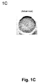
- FIG. 1C is a photograph of the actual Type I collagen matrix produced according to the method of the invention without magnification.
- FIG. 1D is a photograph of approximately 2.5 ⁇ magnified actual Type I collagen matrix showing pore distribution within the matrix.
- FIG. 1E is photograph of approximately 2.5 ⁇ magnified actual Type I collagen matrix with darkened background for better contrast showing pore distribution within the matrix.
- FIG. 2 is a scheme of an experimental set-up for preparation of a support matrix of the invention, providing means for storing and introduction of aqueous ammonia, introduction of inert atmosphere and creating reduced pressure.
- FIG. 3A is a microphotograph of a Type I collagen-based matrix prepared from 4 mg/ml of collagen in the presence of 5 ml of 3% ammonia, showing the pore size about 259 ⁇ 48 ⁇ m.
- the FIG. 3A clearly shows that the matrix has pores of substantially the same size and an equal distribution throughout the matrix.
- the ammonia volume was increased to 6 ml, the pore size increased to 334 ⁇ 89 ⁇ m, as seen in FIG. 3B .
- Scale 1000 ⁇ m, i.e. 1 cm
- FIG. 4A is a microphotograph of a Type I collagen-based matrix prepared from 6 mg/ml of collagen in the presence of 2 ml of 3% ammonia, showing the pore size 233 ⁇ 55 ⁇ m, where the pore size and distribution of pores through the matrix is substantially the same.
- the ammonia volume was increased to 4 ml, the pore size increased only slightly to 235 ⁇ 67 ⁇ m, as seen in FIG. 4B .
- Scale 1000 ⁇ m, i.e. 1 cm is shown in upper left corner.
- FIG. 5A is a microphotograph of a Type I collagen-based matrix prepared from 5 mg/ml of collagen in the presence of 1 ml of 3% ammonia prepared in an inert (nitrogen) atmosphere under reduced pressure (3 torr), showing the pore size 253 ⁇ 59 ⁇ m, wherein the pore size and distribution of pores through the matrix are substantially the same.
- the nitrogen atmosphere pressure was further decreased to 10 torr, the pore size increased to 323 ⁇ 82 ⁇ m, as seen in FIG. 5B .
- the ammonia volume was increased to 3 ml, the size of pores increased to 538 ⁇ 135 ⁇ m, as seen in FIG. 5C .
- Scale 1000 ⁇ m, i.e. 1 cm
- FIG. 6A is a microphotograph of a Type I collagen-based matrix prepared from 4 mg/ml of collagen in the presence of 5 ml of 3% ammonia prepared in the presence of non-ionic surfactant (Pluronic® F68; 0.3% weight/%).
- the pore size at the surface of the matrix was 198 ⁇ 47 ⁇ m, showing the pore size and distribution of pores through the matrix to be homogeneous and the pore sizes substantially the same.
- the amount of collagen was increased to 6 mg/ml, the pore size increased to 256 ⁇ 59 ⁇ m, as seen in FIG. 6B .
- the collagen was increased to 8 mg/ml, the size of pores rose to 380 ⁇ 100 ⁇ m, as seen in FIG. 6C .
- Scale is 500 ⁇ m shown in lower right corner.
- FIG. 7A is a graph showing results of determination of a content of S-GAG, measured by DMB assay, at day zero and day 21, obtained with seeding chondrocytes into the matrix having 200 ⁇ 50 pore sizes.
- FIG. 7B is a graph showing a content of DNA measured by DNA assay at day zero and day 21.
- FIG. 8A is a graph showing a content of S-GAG produced by chondrocytes seeded in the support matrix having small pores (153 ⁇ 39 ⁇ m) or large pores (435 ⁇ 60 ⁇ m).
- FIG. 8B is a graph showing a content of DNA in the small and large pores, measured by DNA assay at day zero and day 21.
- “Sterically stable supporting structure” means non-random vertical orientation of collagen-fibrillar structure.
- “Sterically unstable supporting structure” means random orientation mesh-like collagen-fibrillar structure.
- the matrix porosity means a pore size defined by the diameter of holes within the support matrix or substrate as well as density of the pore distribution as a function of cross-sectional area in millimeters.
- substantially homogeneous means at least 85-99% homogeneity, that is at least 85% of all pores have sizes within the given range of 200 ⁇ 100 ⁇ m. Preferable homogeneity is between 95 and 99%.
- substantially homogeneous porosity means that a pore size and diameter is within pore size range of about 200 ⁇ 100 ⁇ m, preferably 200 ⁇ 50 ⁇ m, in diameter.
- Top surface means an apical or synovial side of the matrix turned toward the joint.
- Bottom surface means basal, closest to bone surface of the matrix.
- Chondrocytes means the cells naturally residing in articular cartilage.
- “About 200 ⁇ 100 ⁇ m” means and is intended to include also pores where the mean average is within additional 10-20 ⁇ m on the upper side.
- S-GAG means sulfated glycosaminoglycan.
- the current invention is directed to a collagen-based matrix of defined porosity having substantially homogeneous pore sizes as well as to a method for preparation thereof.
- the collagen-based matrix prepared according to the method of the invention has uniformly defined vertically non-randomly organized porosity and a defined pore density.
- the support matrix or substrate is generally prepared from a biocompatible material such as collagen, particularly Type I collagen, and has vertically non-randomly oriented open pores of substantially homogeneous pore size of a narrowly defined diameter.
- the matrix is suitable for preparation of acellular implants or cellular implants seeded with chondrocytes, as well as for growth and de novo formation of hyaline or hyaline-like cartilage in vitro and in vivo.
- the invention additionally concerns a collagen-matrix composite system comprising said matrix seeded with a suspension of chondrocytes in collagen, gel, sol-gel or thermoreversible hydrogel, said system being capable of enabling formation of hyaline or hyaline-like cartilage by supporting said chondrocytes in vitro or in vivo when said system is implanted into damaged or injured cartilage in situ to produce extracellular matrix and its components Type II collagen and S-GAG.
- the Type I collagen-based matrix of the invention has substantially homogeneous pore size range of about 200 ⁇ 100 ⁇ m, preferably about 200 ⁇ 50 ⁇ m, in diameter. This pore size has been shown to provide the largest attachment of the chondrocytes to the pores of the matrix.
- the support matrix is ultimately useful for treatment of articular cartilage injuries and lesion by providing means for growing a new hyaline or hyaline-like cartilage for treatment, replacement or regeneration of the damaged or injured articular cartilage.
- Such treatment is currently difficult because of the unique properties of the articular cartilage that is not the same as and does not behave as other soft tissues.
- Articular cartilage covers the ends of bones in synovial joints. Articular cartilage is an unique tissue in that is it is avascular, aneural and alymphatic and in mature state contains a very small number of cells. These properties are the main reason why articular cartilage has such a poor intrinsic capacity to heal (Install, J. N. and Scott, W. N., Surgery of the Knee, 3 rd Edition, p. 341 (2001).
- Articular cartilage is known as a relatively acellular tissue whose extracellular space is occupied by interstitial fluid (60-80%) and organic extracellular matrix (ECM) components, primarily proteoglycans and collagens.
- interstitial fluid 60-80%
- ECM organic extracellular matrix
- Immature chondrocytes are the articular cartilage cells that are present in large numbers in cartilage of young individuals.
- the immature chondrocytes are metabolically active cells that are responsible for growth of cartilage in the young individuals.
- cartilage contains mature chondrocytes that are limited in number in mature quiescent cartilage and those present are mainly metabolically inactive.
- the mechanical function of articular cartilage is determined by its high water content and by the particular architecture of the collagen network.
- This network consists of cross-linked fibrils that extend perpendicular from the subchondral bone and curve gradually to a course parallel to the articular surface in the superficial zone (“Form and Bau der Gelenkknorpel in empueously, Z. Zellforsch ., Vol. 2, pp. 783-862 (1925)).
- Any replacement support structure should thus have orientation and organization similar to that observed naturally. Consequently, a vertical orientation of a porous structure, preferably one made of a biodegradable material used in lieu of the cartilage matrix, would seem to be an important aspect for a cartilage regeneration in vivo due to the similar structure of the native articular cartilage.
- the collagen-based matrix of the invention is an essential component of a gel-matrix composite system capable of initiating the induction of hyaline-like cartilage from chondrocytes.
- the matrix of the invention has been designed to meet requirements of properties needed for the support matrix.
- the support matrix is prepared from the biocompatible and preferably biodegradable materials that are the same or similar to those observed in the articular cartilage.
- the second requirement is that the support matrix has a spatial organization and orientation similar to that of the articular cartilage.
- the third requirement is that the support matrix has a porosity permitting the seeding of the chondrocytes into said matrix in a number of cells that are sufficient for initiation of a formation of new hyaline or hyaline-like articular cartilage in vitro and/or in vivo.
- the fourth requirement is that the support matrix has sufficient number of pores for the number of cells needed for initiation of articular cartilage formation and that the pore sizes are such that the majority of chondrocytes seeded into said support matrix are suspended within the support matrix in numbers that would result in formation of new hyaline or hyaline-like cartilage.
- the fifth requirement is that the pores have substantially the same size in a range from about 200 ⁇ 100 ⁇ m, preferably between about 200 ⁇ 50 ⁇ m in pore diameter and that such size is substantially the same from the top apical to the bottom basal surface of the pores, said pores being organized vertically from the top to the bottom.
- the support matrix preferably the collagen-based support matrix, of the invention is a three-dimensional structure made of a biocompatible and/or biodegradable material of defined density and porosity.
- the support matrix is prepared from a collagenous gel or gel solution containing Type I collagen, Type II collagen, Type IV collagen, gelatin, agarose, hyaluronin, a cell-contracted collagen containing proteoglycan, glycosaminoglycan or glycoprotein, fibronectin, laminin, a bioactive peptide growth factor, cytokine, elastin, fibrin, a synthetic polymeric fiber made of a poly-acid such as polylactic, polyglycolic or polyamino acid, polycaprolactone, polyamino acid, polypeptide gel, copolymers thereof, each alone or in a combination. Additionally, the support matrix may be prepared from the collagen precursors.
- the collagen precursors may be used to reconstitute collagen fibrillar structure for matrix protection.
- These precursors are, for example peptide monomers, such as alpha 1 (type I), and alpha 2 (type I) collagen peptide or alpha 1 (type I) alpha 2 (type I) peptides, in combination, or 2 (alpha 1, type I) and 1 (alpha 2, type I) peptides.
- the support matrix of the invention is prepared from collagen and most preferably from Type I collagen, containing a plurality of narrowly defined uniformly vertically and non-randomly organized pores.
- the pores have substantially homogeneous narrowly defined size and diameter and are uniformly distributed through the matrix dividing the matrix space into a fluid-filled column or network.
- Type I collagen-based matrix is a collagen-based sponge-like structure or honeycomb-like lattice of defined porosity having a vertically organized pores of substantially same sizes.
- the support matrix of the invention has a certain thickness and vertically organized pores of a defined diameter oriented to create an apical (top or synovial) or basal (bottom or bone) surface of the matrix for implantation.
- the diameter of said pores is chosen such that the matrix in conjunction with the chondrocyte-containing gel, preferably a sol-gel, facilitates a sterically-enhanced induction of extracellular matrix glycosaminoglycan and type II collagen deposition in ratios characteristic of hyaline articular cartilage.
- the vertically oriented cellular or acellular support matrix made of the biocompatible material having a predetermined pore size filled with chondrocytes suspension is important aspect of the quality of the repair tissue in vivo and for production of the articular cartilage in vitro.
- the size of the pores of the matrix affects the chondrocyte attachment to the matrix walls and assures the presence of chondrocytes within the matrix needed for cartilage regeneration in vitro and in vivo.
- Sizes of the pores are substantially homogeneous with homogeneity above 85%, preferably 95%, most preferably about 98-99% of pores having a diameter size of about 200 ⁇ 100 ⁇ m, preferably 200 ⁇ 50 ⁇ m.
- the pores of the collagen-based matrix are homogeneously distributed within said matrix to form a sponge-like structure able to taking in and evenly distributing the chondrocytes suspended in a gel solution and providing conditions conducive to producing extracellular matrix by the suspended chondrocytes.
- the defined and substantially homogeneous pore size diameter of the collagen-based matrix is an important aspect of the invention.
- Collagen-based matrices of defined pore sizes according to the invention having different pore size diameter permit faster or slower infiltration of the chondrocytes into said matrix, faster or slower growth and propagation of the cells and, ultimately, the higher or lower density of the cells in gel-matrix system.
- Such pore size may be adjusted by varying the pH of the gel solution, collagen concentration, lyophilization conditions, temperature, degree of cross-linking of collagen, etc.
- the pore sizes are not defined and not limited to one substantially homogeneous size.
- the collagen-based matrices known in the prior art are mostly structures containing pores having mixed pore sizes of from about 50 to about 2000 ⁇ m.
- the support matrix of the invention has the spatial organization and orientation similar to that of the articular cartilage.
- the spatial organization and orientation of support matrix is schematically depicted in FIGS. 1A and 1B .
- FIG. 1A is a schematic side view of the support matrix showing the vertical organization and orientation of support matrix pores seeded hypothetically with chondrocytes suspended in sol-gel.
- the pores are of substantially the same size in diameter and the number of cells in each pore is also substantially the same.
- the cells shown here are round and touching the walls of the pores illustrating the attachment requirement for the support matrix.
- Suspending solution for chondrocytes is any gel solution, preferably one containing collagen, gel, sol-gel or thermoreversible hydrogel that can change its state from sol to gel depending on the temperature.
- FIG. 1B is a schematic top view, showing hypothetically the ideally homogeneous distribution of pores through the support matrix, where the pores have the same sizes and chondrocytes are evenly distributed within the pores. The pores walls that separate individual pores are also seen.
- FIG. 1B is a schematic top view of a gel-matrix composite system comprising the collagen-based matrix component made of Type I collagen and embedded with chondrocytes suspended in a sol-gel showing a distribution of chondrocytes within said collagen-based matrix to be substantially homogeneous within the pores having a defined diameter of about 200 ⁇ m.
- the outer circle defines a size of the whole matrix wafer. Smaller circles are pores having a substantially the same size. Inside of the pore circles are chondrocytes deposited there in a sol-gel solution.
- FIG. 1C is the photograph of the actual Type I collagen matrix produced according to the method of the invention without magnification.
- the matrix pores are visible as a black dots.
- the matrix can be cut to order in both directions to obtain a perfect fit, if used, for example, for an articular cartilage implant.
- FIG. 1D is the photograph of 4 ⁇ magnified actual Type I collagen matrix showing the pore distribution within the matrix.
- FIG. 1E is photograph of approximately 4 ⁇ magnified actual Type I collagen matrix with darkened background for better contrast showing the pore distribution within the matrix.
- Chondrocytes suspended in a collagen, gel, sol-gel or thermoreversible hydrogel solution are introduced into narrowly defined pores of a substantially the same size and distribution within the matrix. Vertically separated columns have substantially the same diameter and defined sizes of the pores. The pores are filled with the chondrocyte suspension. This arrangement provides sterically advantageous conditions for homogeneous distribution of chondrocyte deposition within the pores that lead to enhanced production of proteoglycans and type II collagen in ratios corresponding to the ratios observed for these compounds in the healthy hyaline cartilage.
- the structural macromolecules and cartilage account for 20 to 30% of the wet weight of cartilage and includes type II collagen, large aggregating proteoglycans and non-collagenous proteins or glycoproteins. With maturation, collagen contributes over 50% and proteoglycans contribute 30-35% of the matrix dry weight.
- the matrix is typically prepared as a composite cylindrical wafer seen in FIGS. 1C-1E or as a rectangular block structure cut into a wafer-like matrix construct having from about 4 mm to about 25 mm in diameter and thickness of from 0.5 to about 5 mm.
- the seeding density of this construct is about 25,000-300,000 chondrocytes per 25 ⁇ l of collagen solution corresponding to about 1-12 millions cells/mL.
- the above density numbers are exemplary only and even lesser seeding density is possible and has been shown to lead to the production of the hyaline or hyaline-like cartilage.
- the cell density range for seeding is preferably from about 1 to about 30 million/mL.
- Chondrocytes are preferably suspended in a type I or Type II collagen or in a synthetic gel, sol-gel or hydrogel and such suspension, in combination with the support matrix, forms a gel-matrix composite system of the invention.
- the current invention additionally concerns a novel method for preparation of the support matrix that meets the requirements stated in section IIA.
- the method for preparation of the support matrix meeting such criteria involves process for standardizing the pore size, vertical orientation and organization, homogeneity and uniformity of the collagen-based matrix of the invention.
- the fabrication method for preparation of the current support matrix is based on investigation of various parameters comprising the biocompatible components and/or additives and their amounts, or reaction conditions, such as, for example, pH, temperature, pressure, reduced pressure, presence of inert gasses and an apparatus for testing these parameters all alone or in various combinations.
- Fabrication set-up for preparation of the support matrix of the invention comprises variable components depending on the conditions selected for the processing.
- It typically comprises a sealed container containing the biocompatible material or a precursor thereof, preferably Type I collagen, selected for the preparation of the support matrix wherein said material may be subjected to various conditions for testing and optimization or for final preparation of the support matrix.
- Added features may contain means for increasing or decreasing pressure, temperature, adjusting pH, adding other components, pumps, gas tanks, valves, etc.
- FIG. 2 One illustrative set-up for preparation of the support matrix is shown in FIG. 2 .
- FIG. 2 is schematic representation of one experimental set-up used for preparation of a support matrix of the invention, providing means for introduction of aqueous ammonia, means for reducing pressure and means for providing an inert atmosphere.
- the starting material in this case the collagen solution
- the collagen solution is placed into a sealed container having an outlet connected to a vacuum gauge that is further connected to a vacuum pump and a source of an inert gas.
- two sources are shown for supplying the aqueous ammonia and for storing nitrogen with a means for introducing nitrogen into the system.
- the starting material is then subjected to ammonia vapors by introducing a predetermined amount of aqueous ammonia into said container, and/or further introducing the inert gas, such as nitrogen or argon, or in one alternative, air, and optionally performing the reaction under reduced pressure, in the presence of a surfactant or under other reaction conditions. All these may be performed and/or tested in any combination to achieve the optimal expected results.
- the selected criteria for the pore size of the support matrix were set to prepare the support matrix having substantially homogeneous population of pores within said matrix wherein said pores have substantially the same size in diameter of about 200 ⁇ 100 ⁇ m.
- the process for optimization of conditions for preparation of the support matrix meeting these criteria involved testing of Type I collagen in 2, 4, 6, 8, and 10 mg per ml of solution, and its polymerization in the presence of 3% aqueous ammonia added in 1, 2, 3, 4, 5, and 6 ml volume.
- the same collagen preparations were tested with added non-ionic surfactant (Pluronic® F86, 0.3 weight/%), and the same collagen solutions were polymerized in an inert atmosphere and under reduced pressure.
- Various combinations of the above conditions were further tested to obtain the support matrix with the smallest and most homogeneous pore sizes.
- Results are summarized in Tables 1-3 and in representative FIGS. 3-6 .
- the support matrix was prepared from bovine Type I atelocollagen (2.9 mg/ml, pH 2.1), obtained from Inamed Corporation, Fremont, Calif. The collagen was concentrated by a precipitation followed by centrifugation and then dissolved in aqueous HCl in order to reach the desired concentration. The procedure for preparation of the support matrix in the presence of ammonia is described in Example 3.
- the pore sizes of the support matrix obtained under these conditions are expressed as a function of collagen concentration and volume of ammonia solution/concentration, is seen in Table 1.
- Pore size was measured within 1 mm of the surface of the formed matrix.
- FIG. 3A and FIG. 3B are representative microphotographs of support matrices prepared from 4 mg/ml collagen in the presence of 5 or 6 ml of ammonia resulting in pore sizes ranges of about 259 ⁇ 48 ⁇ m and about 334 ⁇ 89 ⁇ m, respectively. Both microphotographs are on the same scale (scale 1000 ⁇ m) and the difference in their sizes is clearly visible.
- Collagen in concentration of 6 mg/ml polymerized in the presence of 2 ml or 4 ml ammonia yielded the support matrix having pore sizes in the optimal range of about 233 ⁇ 55 ⁇ m and about 235 ⁇ 67 ⁇ m, respectively.
- FIG. 4A and FIG. 4B are representative microphotographs of support matrices prepared from 6 mg/ml collagen and 2 or 4 ml of ammonia resulting in pore sizes ranges of about 233 ⁇ 55 ⁇ m and about 235 ⁇ 67 ⁇ m. Both microphotographs are on the same scale (scale 1000 ⁇ m). Both microphotograps show the pores to be of approximately the same size. Homogeneity of their distribution is clearly visible.
- the support matrix prepared with a moderate amount of collagen between 4 and preferably 6 mg/ml, in the presence of moderate volume of ammonia, between 2 and 4 ml resulted in the matrix having the pore sizes within the optimal range of about 200 ⁇ 100 ⁇ m.
- the support matrix was prepared from bovine Type I atelocollagen, as already described above and the support matrix was prepared in the inert nitrogen atmosphere and at reduced pressure.
- the procedure conditions for preparation of the support matrix in the presence of ammonia and at inert atmosphere and reduced pressure is described in Example 5.
- Table 2 shows the pore size as a function of volume of ammonia solution/concentration and reduced pressure. Pore size is measured within 1 mm of the surface of the formed scaffold.
- FIG. 5A shows the pore size of 253 ⁇ 59 ⁇ m of the matrix made of the polymerization of 5 mg/ml collagen with 1 ml of ammonia in nitrogen atmosphere at reduced pressure to three torr.
- FIG. 5B shows the matrix having a pore size of 323 ⁇ 82 ⁇ m made of the polymerization of 5 mg/ml collagen with 1 ml of ammonia in nitrogen atmosphere at reduced pressure to 10 torr.
- FIG. 5C shows the pore size of 538 ⁇ 135 ⁇ m of the matrix made by polymerization of 5 mg/ml collagen with 3 ml of ammonia in nitrogen atmosphere at reduced pressure to 10 torr.
- the support matrix prepared with a small volume of ammonia combined with a moderate reduction of pressure performed in an inert atmosphere resulted in the matrix having the pore sizes within the optimal range of about 200 ⁇ 100 ⁇ m.
- the support matrix was prepared from bovine Type I atelocollagen, as described above.
- the procedure for preparation of the support matrix in the presence of surfactant is described in Example 4.
- the support matrix obtained under these conditions and the pore size is expressed as a function of collagen concentration in the presence of the 0.3% by weight of Pluronic® surfactant is seen in Table 3.
- Pore size seen in Table 3 is expressed as a function of collagen concentration. Pore size is measured within 1 mm of the surface of the formed matrix.
- the pore size of the support matrix was about 198 ⁇ 47 ⁇ m.
- the support matrix had pore sizes in the range from about 256 ⁇ 59 ⁇ m and about 380 ⁇ 100 ⁇ m.
- FIGS. 6A, 6B and 6C are the microphotographs of matrices resulting from polymerization of 4, 6 and 8 mg/ml of collagen in the presence of the surfactant. These figures again clearly show the uniformity and homogeneity of the resulting pores as well as their uniform sizes.
- Presence of the surfactant in the collagen pre-polymerization mixture clearly affects the formation of pore sizes within the optimal ranges, particularly when combined with lower concentration of collagen, polymerization of collagen (4 mg/ml) resulted in matrix having optimally sized pores of about 198 ⁇ 47 ⁇ m.
- the collagen precursors may be used to reconstitute collagen fibrillar structure for matrix protection.
- These precursors are, for example peptide monomers, such as alpha 1 (type I), and alpha 2 (type I) collagen peptides or in combination of 2 (alpha 1, type I) and 1 (alpha 2, type I) peptides, or 3 (alpha I, type II) peptides.
- the other factors that may be manipulated are the selective vortical shearing of the peptide monomer, the chemical composition and pH of the solubilization buffer.
- the vortical shearing step determines the dispersion and alignment of the associated collagen fibrils of alpha I (type I) and alpha II (type I) peptides. During this procedure equilibration in a nitrogen or argon atmosphere controls pH.
- Exemplary conditions for preparation of the collagen-based matrix are as follows.
- the concentration of Type I collagen for fabrication of the collagen-based matrix is about from about 2 to about 10, preferably from about 4 to about 8, most preferably about 4 to about 6 mg/ml.
- concentration of collagen is critical for the initial polymerization resulting in fibrillogenesis, that is for formation of fibrils.
- Prior and current studies show that at the concentration of collagen below 3 mg/ml there is no fibrillogenesis, and thus no pore formation, unless such is promoted by addition of some other components.
- the suitable buffers for solubilization of the Type I collagen are, for example, a formic acid containing buffer at pH 4.8, acetic acid containing buffer at pH 5.0 or a diluted hydrochloric acid at pH 3.0.
- the shearing of the collagen solution is set, for example, to be for about 10-60 seconds at 10-100 dynes/cm 2 .
- the sheared peptides are equilibrated in an inert gas atmosphere such as nitrogen or argon gas for about 30 minutes to about 2 hours at about 4° C.
- an inert gas atmosphere such as nitrogen or argon gas for about 30 minutes to about 2 hours at about 4° C.
- the use of the inert gas displaces air containing oxygen that may act as an oxidant and decrease fibrillogenesis. This factor was found to positively affect the polymerization and pore formation.
- Neutralization is typically carried out in a vapor of about 0.3% ammonia over about 12 to about 24 hour period. This factor has also been found to affect the collagen polymerization and formation of pores having homogeneous pore size.
- Freezing if there is, is carried out at ⁇ 40° C. to about ⁇ 60° C. over a period of about 2 to about 12 hours.
- Water removal is achieved by exposure to an anhydrous atmosphere from ⁇ 20° C. to about 50° C. preferably at 37° C. for about 24 to about 48 hours.
- the gradual nature of the polymerization and slow process of water removal typically maintains the architectural elements of the scaffold collagen-based to achieve the proper orientation and diameter of the longitudinal pore structure necessary for hyaline cartilage extracellular matrix deposition by the cells.
- the organization of the newly synthesized cartilage specific matrix within the porous type I collagen is visualized and quantified by, for example, ELISA/Western blot methods for determination of protein levels or quantitative RT-PCR or real-time PCR for m-RNA level.
- a main aspect of the current invention is a finding that when the pore sizes are substantially homogeneously restricted to a narrowly defined diameter, preferably to a pore size diameter of about 200 ⁇ 100, preferably 200 ⁇ 50 ⁇ m, and when such pores are vertically organized, such diameter of said pores in conjunction with the chondrocyte suspension facilitates a sterically-enhanced enablement of hyaline cartilage leading to formation of extracellular matrix.
- Such sterically-enhanced enablement leads to a deposition of collagen Type II and proteoglycans within the matrix in ratios characteristic of articular cartilage.
- the invention concerns a collagen-based matrix prepared from Type I collagen seeded with chondrocytes suspended in synthetic sol-gel, which gels at body temperature.
- the gel-matrix composite system comprises a matrix having pore diameters of about 200 ⁇ 100 ⁇ m, preferably 200 ⁇ 50 ⁇ m, that permit uniform infusion of chondrocyte/gel suspensions into the pores, and induces deposition of collagen Type II and proteoglycans (glycosaminoglycans) within the matrix in ratios characteristic of articular cartilage.
- the collagen-based matrix of the current invention acts like a porous sponge when infiltrated with the suspended chondrocytes by, for example, wicking or infusion, wherein the cells are distributed within the matrix pores.
- This arrangement permits chondrocytes to migrate and settle in the support matrix in a sterically-enhanced fashion and enables them to proliferate and secrete materials for generation of new extracellular matrix and eventually producing a hyaline cartilage.
- Suspending solution for chondrocytes is any gel solution, preferably one containing collagen, gel, sol-gel or theromoreversible hydrogel that can change its state from sol to gel depending on the temperature, and is preferably a thermo-reversible gelation hydrogel (TRGH) material in which the sol-gel transition occurs on the opposite temperature cycle of agar and gelatin gels. Consequently, the viscous fluidic phase is in a sol stage and the solid phase is in a gel stage. TRGH has very quick sol-gel transformation which requires no cure time and occurs simply as a function of temperature without hysteresis.
- TRGH thermo-reversible gelation hydrogel
- the sol-gel transition temperature for embedding chondrocytes within the collagen-based matrix is set at a temperature where the sol-gel is in a sol state whereas the temperature for stabilizing the chondrocytes within the matrix is set at a body temperature in the range of about 37° C.
- chondrocytes Healthy human articular cartilage (hAC) tissue was obtained from the Tissue Bank, The National Disease Research Interchange, Philadelphia, Pa. The cartilage tissue was digested, isolated chondrocytes were seeded on culture dishes and precultured for monolayer cell expansion. Isolated chondrocytes were seeded in a 3D culture, according to a procedure described in Examples 7 and 8.
- hAC human articular cartilage
- S-GAG total sulfated glycosaminoglycan
- Example 6 The experimental set-up is described in Example 6. Briefly, the cells were seeded into the support matrix and cultured over night and their S-GAG content and DNA (showing a number of cells) was determined at days zero and 21.
- Results are summarized in Table 4 and FIGS. 7A and 7B .
- FIG. 7A shows results of the DMB assay where the S-GAG content was determined on day zero and day 21.
- culturing of the composite for 21 days led to a substantially increased production of S-GAG.
- the production of DNA did not increase under these conditions, as seen in FIG. 7B , where DNA in ⁇ g/composite was determined.
- the average of S-GAG content in constructs with a small pore size was 183.01 ⁇ 39.78 ⁇ g (Table 5 and FIG. 8A ).
- the amount of S-GAG in constructs with a large pore size was 115.56 ⁇ 13.50 ⁇ g (Table 4 and FIG. 8A ).
- the S-GAG accumulation in constructs with small pore size was significantly greater than that with large pore size (Table 5 and FIG. 8A ).
- the DNA content in constructs with a small pore size was significantly greater than for constructs with a large pore size (Table 5 and FIG. 8A ).
- the DNA content in constructs with a small pore size was 1.5-fold greater than that with a large pore size (Table 5 and FIG. 8A ) (P ⁇ 0.05).
- FIGS. 8A and 8B Results of this study are illustrated in FIGS. 8A and 8B .
- FIG. 8A shows results obtained in DMB assay where the S-GAG content per/composite is shown for a composite having large or small pores.
- small pores composites produced approximately one half more of S-GAG than the composite having large pores.
- FIG. 8B illustrating a DNA content in composite having a large or small pores.
- the DNA content in the composite having smaller pores was significantly higher then in the composite having a large pores.
- FIG. 8A is a graph showing a content of DNA, measured by DNA assay, in composites having small pores (153 ⁇ 39 ⁇ m) or large pores (435 ⁇ 60 ⁇ m).
- FIG. 8B the production of DNA was significantly higher in the composites having smaller pores as compared to that with larger pores.
- Small pore size of the composite porous matrix shows significant cell proliferation and cartilage specific accumulation in the matrix as compared to that of large pore size.
- the matrices prepared with different collagen concentrations in the presence of surfactant readily absorbed the chondrocyte-laden gel.
- the cell counts were consistent with the pore size distribution present in FIG. 6 , with a cell viability of 98-99%.
- the matrix and a system of the invention are useful for production of hyaline cartilage in situ or in vitro.
- the collagen-based matrix is prepared as a matrix wafer.
- the matrix wafer is cut into a size of the cartilage defect and introduced into the cartilage defect or lesion or the cartilage with some bone loss such as osteochondral defects.
- the chondrocytes suspension is then introduced as a sol under colder than body temperature into said matrix emplaced in the lesion or defect thereby generating a gel-matrix composite system in situ and the temperature is raised to the body temperature whereby the sol is transitionally changed into a gel.
- the invention works in the same way for acellular implant where the collagen-based matrix filled with the sol-gel is introduced without chondrocytes.
- the implant is left in the body until the new hyaline cartilage is generated and the matrix which is biodegradable self-degrades.
- the process is similar but proceeds in the tube or Petri dish under the same conditions until the hyaline cartilage is produced. Such cartilage may then be used as an implant into the cartilage defect or lesion.
- This example describes one exemplary method for preparation of the collagen-based matrix.
- Type I collagen is dissolved in a formic acid buffer at pH 4.8 and its concentration is adjusted to about 5.2 mg/ml. The solution is subjected to a vortical shearing for 10 seconds at 10 dynes per cm. The sheared peptides is then equilibrated in nitrogen gas for 30 minutes at 4° C. to displace air. Neutralization is carried out in a vapor of 0.3% ammonia over a 24 hour period. The solution is then subjected to freezing at ⁇ 40° C. over a period of 2 hours. Water is removed by exposing the frozen solution to an anhydrous atmosphere at 37° C. for 24 hours.
- This example illustrates another exemplary method for preparation of the collagen-based matrix.
- a 1% aqueous atelocollagen solution (VITROGEN®), maintained at pH 3.0, is poured into a 10 ⁇ 20 cm tray. This tray is then placed in a 5 liter container. A 50 mL open container containing 30 mL of a 3% aqueous ammonia solution is then placed next to the tray, in the 5 liter chamber, containing 300 grams of said 1% aqueous solution of atelocollagen. The 5 liter container containing the open trays of atelocollagen and ammonia is then sealed and left to stand at room temperature for 12 hours.
- VITROGEN® 1% aqueous atelocollagen solution
- the collagenous gel is then washed with water overnight and, subsequently, freeze-dried to yield a sponge like matrix. This freeze dried matrix is then cut into squares, sterilized, and stored under a sterile wrap.
- This example illustrates a general procedure utilizing ammonia for preparation of porous honeycomb scaffold having substantially the same size and distribution of pores.
- This example illustrates preparation of porous honeycomb scaffold in the presence of a surfactant.
- This example illustrates preparation of honeycomb porous scaffold in inert atmosphere (N 2 ) and under reduced pressure.
- 15 g of bovine type 1 atelocollagen dissolved in aqueous HCl with a concentration of 5 mg/ml and pH of 3.3 was added to a 100 ml glass beaker.
- the solution was centrifuged for 5-10 minutes at 800 ⁇ g to remove air bubbles. After centrifugation the collagen solution was placed in 9.1 dm 3 container.
- the container was sealed and the air evacuated using a vacuum pump to a pressure of about 2 torr (water almost boiled). The pump was turned off and the system was filled with nitrogen to about torr. Evacuation and filling was repeated three times.
- the formed pores were vertical and cone shaped and the diameter increased with increasing distance from the surface of the collagen gel.
- the precipitated collagen was washed with deionized water for 1 day in order to remove excess ammonia and formed salts. The washed collagen was then slowly frozen and lyophilized.
- This example describes a study performed for evaluation of the importance of the pore size for induction of production of a hyaline articulate cartilage.
- chondrocytes obtained from human articulate cartilage (hACs) were suspended in hydrogel (collagen gel) and seeded into a composite of a porous sponge matrix having an average pore size 435 ⁇ m or an average pore size 153 ⁇ m. The composites were then transferred into the culture medium. After 12 hours pre-incubation, cell constructs were cultured in medium comprising 5% CO 2 , 2% O 2 and 37° C. in a multigas incubator using DMEM/F-12 medium with 10% FBS, 1% ITS, 0.1% gentamycin (Invitrogen). After 21 days of culture, constructs were harvested for biochemical evaluation.
- This example describes methods used for biochemical evaluation of collagen-based composites.
- This example describes the procedure used for cell harvesting and seeding in the collagen-based matrix according to the invention.
- chondrocytes were harvested with trypsin-EDTA (Invitrogen). Three hundred thousand human articular chondrocytes (hACs) were suspended in a collagen hydrogel and seeded into the composite of porous collagen-based matrix having a predetermined average pore size 435 ⁇ m or 153 ⁇ m. The cell constructs were incubated at 37° C. for about one hour collagen gelation and then transferred into the culture medium. After 12 hours pre-incubation, the composites comprising chondrocytes were cultivated. After zero and 21 days of culture, constructs were harvested for biochemical evaluation.
- hACs human articular chondrocytes
- This example illustrates determination of cell viability.
- the chondrocyte-contained matrices were placed in 1.5 ml microcentrifuge tubes and incubated overnight in 0.15% collagenase. The digest was spun at 2000 rpm for 5 minutes and the supernatant aspirated. An aliquot of culture medium (0.1 ml) was added to the cell pellets and an aliquot taken for counting. Cell viability and total cell count was measured using trypan blue.
Abstract
Description
| TABLE 1 | |
| Collagen | Volume of 3% aqueous ammonia |
| conc. | 2 |
3 ml | 4 ml | 5 ml | 6 ml |
| 2.8-2.9 | — | — | No pores | — | No Pores |
| mg/ml | |||||
| 4 mg/ml | — | 256 ± 68 | 274 ± 87 | 259 ± 48* | 334 ± 89 |
| 6 mg/ml | 233 ± 55 | 365 ± 107 | 235 ± 67* | — | — |
| 8 mg/ml | 284 ± 90* | 299 ± 73 | 1195 ± 294 | — | — |
| 10 mg/ml | 474 ± 108* | — | Few | — | — |
| pores | |||||
| TABLE 2 | ||
| |
Final pressure | Pore size (μm) |
| 1 |
10 torr | 323 ± 82* |
| 1 |
3 torr | 253 ± 59* |
| 3 |
10 torr | 538 ± 135 |
| 6 |
3 torr | 557 ± 148 |
| TABLE 3 | |||
| Collagen Conc. | Pores size (μm) | ||
| 4 mg/ml | 198 ± 47* | ||
| 6 mg/ml | 256 ± 59* | ||
| 8 mg/ml | 380 ± 100* | ||
| TABLE 4 |
| Results of Biochemical |
| Biochemical | Day |
| 0 | |
| Results | Mean | SD | Mean | SD |
| S-GAG accumulation | 28.53 | ±4.35 | 117.42* | ±16.55 |
| DNA contents | 1.69 | ±0.66 | 1.98 | ±0.30 |
| *P < 0.05 | ||||
| TABLE 5 |
| Results of Biochemical Evaluation |
| Biochemical | Small Pore | Large Pore |
| Results | Mean | SD | Mean | SD |
| S-GAG | 183.01* | ±39.78 | 115.56 | ±13.50 |
| accumulation | ||||
| DNA Contents | 2.78* | ±0.21 | 1.81 | ±0.10 |
| *P < 0.05 | ||||
| TABLE 6 | |||||
| Mean cell count | Mean % | ||||
| Group | ±SD | Viability | Comments | ||
| Table 6: 4 mg/ml | 100,800 ± 41,692 | 98.7 | |||
| collagen with | |||||
| Pluronic | |||||
| Table 6: 6 mg/ml | 103,500 ± 15,679 | 99% | |||
| collagen with | |||||
| Pluronic | |||||
| Table 6: 8 mg/ml | 88,350 ± 2,758 | 99.0 | |||
| collagen with | |||||
| Pluronic | |||||
Claims (19)
Priority Applications (4)
| Application Number | Priority Date | Filing Date | Title |
|---|---|---|---|
| US14/577,610 US9701940B2 (en) | 2005-09-19 | 2014-12-19 | Cell-support matrix having narrowly defined uniformly vertically and non-randomly organized porosity and pore density and a method for preparation thereof |
| US15/641,773 US20170298320A1 (en) | 2005-09-19 | 2017-07-05 | Cell-support matrix having narrowly defined uniformly vertically and non-randomly organized porosity and pore density and a method for preparation thereof |
| US16/706,372 US10900017B2 (en) | 2005-09-19 | 2019-12-06 | Cell-support matrix having narrowly defined uniformly vertically and non-randomly organized porosity and pore density and a method for preparation thereof |
| US17/156,062 US20210246418A1 (en) | 2005-09-19 | 2021-01-22 | Cell-support matrix having narrowly defined uniformly vertically and non-randomly organized porosity and pore density and a method for preparation thereof |
Applications Claiming Priority (3)
| Application Number | Priority Date | Filing Date | Title |
|---|---|---|---|
| US71871405P | 2005-09-19 | 2005-09-19 | |
| US11/523,833 US8921109B2 (en) | 2005-09-19 | 2006-09-19 | Cell-support matrix having narrowly defined uniformly vertically and non-randomly organized porosity and pore density and a method for preparation thereof |
| US14/577,610 US9701940B2 (en) | 2005-09-19 | 2014-12-19 | Cell-support matrix having narrowly defined uniformly vertically and non-randomly organized porosity and pore density and a method for preparation thereof |
Related Parent Applications (1)
| Application Number | Title | Priority Date | Filing Date |
|---|---|---|---|
| US11/523,833 Continuation US8921109B2 (en) | 2005-09-19 | 2006-09-19 | Cell-support matrix having narrowly defined uniformly vertically and non-randomly organized porosity and pore density and a method for preparation thereof |
Related Child Applications (1)
| Application Number | Title | Priority Date | Filing Date |
|---|---|---|---|
| US15/641,773 Continuation US20170298320A1 (en) | 2005-09-19 | 2017-07-05 | Cell-support matrix having narrowly defined uniformly vertically and non-randomly organized porosity and pore density and a method for preparation thereof |
Publications (2)
| Publication Number | Publication Date |
|---|---|
| US20150104872A1 US20150104872A1 (en) | 2015-04-16 |
| US9701940B2 true US9701940B2 (en) | 2017-07-11 |
Family
ID=37889483
Family Applications (5)
| Application Number | Title | Priority Date | Filing Date |
|---|---|---|---|
| US11/523,833 Active 2029-01-26 US8921109B2 (en) | 2005-09-19 | 2006-09-19 | Cell-support matrix having narrowly defined uniformly vertically and non-randomly organized porosity and pore density and a method for preparation thereof |
| US14/577,610 Active 2027-05-06 US9701940B2 (en) | 2005-09-19 | 2014-12-19 | Cell-support matrix having narrowly defined uniformly vertically and non-randomly organized porosity and pore density and a method for preparation thereof |
| US15/641,773 Abandoned US20170298320A1 (en) | 2005-09-19 | 2017-07-05 | Cell-support matrix having narrowly defined uniformly vertically and non-randomly organized porosity and pore density and a method for preparation thereof |
| US16/706,372 Active US10900017B2 (en) | 2005-09-19 | 2019-12-06 | Cell-support matrix having narrowly defined uniformly vertically and non-randomly organized porosity and pore density and a method for preparation thereof |
| US17/156,062 Pending US20210246418A1 (en) | 2005-09-19 | 2021-01-22 | Cell-support matrix having narrowly defined uniformly vertically and non-randomly organized porosity and pore density and a method for preparation thereof |
Family Applications Before (1)
| Application Number | Title | Priority Date | Filing Date |
|---|---|---|---|
| US11/523,833 Active 2029-01-26 US8921109B2 (en) | 2005-09-19 | 2006-09-19 | Cell-support matrix having narrowly defined uniformly vertically and non-randomly organized porosity and pore density and a method for preparation thereof |
Family Applications After (3)
| Application Number | Title | Priority Date | Filing Date |
|---|---|---|---|
| US15/641,773 Abandoned US20170298320A1 (en) | 2005-09-19 | 2017-07-05 | Cell-support matrix having narrowly defined uniformly vertically and non-randomly organized porosity and pore density and a method for preparation thereof |
| US16/706,372 Active US10900017B2 (en) | 2005-09-19 | 2019-12-06 | Cell-support matrix having narrowly defined uniformly vertically and non-randomly organized porosity and pore density and a method for preparation thereof |
| US17/156,062 Pending US20210246418A1 (en) | 2005-09-19 | 2021-01-22 | Cell-support matrix having narrowly defined uniformly vertically and non-randomly organized porosity and pore density and a method for preparation thereof |
Country Status (6)
| Country | Link |
|---|---|
| US (5) | US8921109B2 (en) |
| EP (1) | EP1926459B1 (en) |
| JP (1) | JP2009508596A (en) |
| AU (1) | AU2006292224B2 (en) |
| CA (1) | CA2623106C (en) |
| WO (1) | WO2007035778A2 (en) |
Families Citing this family (34)
| Publication number | Priority date | Publication date | Assignee | Title |
|---|---|---|---|---|
| US20040151705A1 (en) | 2002-03-22 | 2004-08-05 | Shuichi Mizuno | Neo-cartilage constructs and a method for preparation thereof |
| US7067123B2 (en) | 2003-04-29 | 2006-06-27 | Musculoskeletal Transplant Foundation | Glue for cartilage repair |
| US20050064042A1 (en) * | 2003-04-29 | 2005-03-24 | Musculoskeletal Transplant Foundation | Cartilage implant plug with fibrin glue and method for implantation |
| US20050222687A1 (en) * | 2004-04-02 | 2005-10-06 | Gordana Vunjak-Novakovic | Cartilage implant assembly and method for implantation |
| US7901457B2 (en) | 2003-05-16 | 2011-03-08 | Musculoskeletal Transplant Foundation | Cartilage allograft plug |
| US7488348B2 (en) * | 2003-05-16 | 2009-02-10 | Musculoskeletal Transplant Foundation | Cartilage allograft plug |
| US7837740B2 (en) | 2007-01-24 | 2010-11-23 | Musculoskeletal Transplant Foundation | Two piece cancellous construct for cartilage repair |
| US7815926B2 (en) | 2005-07-11 | 2010-10-19 | Musculoskeletal Transplant Foundation | Implant for articular cartilage repair |
| AU2006292224B2 (en) | 2005-09-19 | 2013-08-01 | Histogenics Corporation | Cell-support matrix and a method for preparation thereof |
| US9132208B2 (en) * | 2008-08-07 | 2015-09-15 | Lifenet Health | Composition for a tissue repair implant and methods of making the same |
| US9005646B2 (en) | 2005-10-12 | 2015-04-14 | Lifenet Health | Compositions for repair of defects in tissues, and methods of making the same |
| US8435551B2 (en) | 2007-03-06 | 2013-05-07 | Musculoskeletal Transplant Foundation | Cancellous construct with support ring for repair of osteochondral defects |
| US8070827B2 (en) * | 2007-07-03 | 2011-12-06 | Histogenics Corporation | Method for use of a double-structured tissue implant for treatment of tissue defects |
| US8685107B2 (en) | 2007-07-03 | 2014-04-01 | Histogenics Corporation | Double-structured tissue implant and a method for preparation and use thereof |
| US20090054984A1 (en) | 2007-08-20 | 2009-02-26 | Histogenics Corporation | Method For Use Of A Double-Structured Tissue Implant For Treatment Of Tissue Defects |
| EP2182887B1 (en) * | 2007-08-20 | 2016-12-14 | Histogenics Corporation | A method for improvement of differentiation of mesenchymal stem cells using a double-structured tissue implant |
| WO2009089340A1 (en) * | 2008-01-09 | 2009-07-16 | Innovative Health Technologies, Llc | Implant pellets and methods for performing bone augmentation and preservation |
| WO2009105820A1 (en) * | 2008-02-29 | 2009-09-03 | The University Of Sydney | Hydrogels derived from biological polymers |
| CA2717725A1 (en) | 2008-03-05 | 2009-09-11 | Musculoskeletal Transplant Foundation | Cancellous constructs, cartilage particles and combinations of cancellous constructs and cartilage particles |
| US8114156B2 (en) * | 2008-05-30 | 2012-02-14 | Edwin Burton Hatch | Flexibly compliant ceramic prosthetic meniscus for the replacement of damaged cartilage in orthopedic surgical repair or reconstruction of hip, knee, ankle, shoulder, elbow, wrist and other anatomical joints |
| DE102009018640A1 (en) * | 2009-04-17 | 2010-10-21 | Tetec Tissue Engineering Technologies Ag | Implant and therapeutic composition for the treatment of damage and / or diseases in the field of the human and / or animal musculoskeletal system |
| JP5955326B2 (en) * | 2011-08-30 | 2016-07-20 | 国立大学法人京都大学 | Porous scaffold material and method for producing the same |
| CN102716515B (en) * | 2012-07-02 | 2014-01-29 | 陕西博鸿生物科技有限公司 | Biological material for repairing meniscus tear and preparation method for biological material |
| US20140030309A1 (en) | 2012-07-11 | 2014-01-30 | Osiris Therapeutics, Inc. | Disrupted cartilage products |
| US10077420B2 (en) | 2014-12-02 | 2018-09-18 | Histogenics Corporation | Cell and tissue culture container |
| US10918767B2 (en) * | 2015-05-12 | 2021-02-16 | University Of Florida Research Foundation, Inc. | Magnetically templated tissue engineering scaffolds and methods of making and using the magnetically templated tissue engineering scaffolds |
| US11090412B2 (en) | 2018-12-21 | 2021-08-17 | Zavation Medical Products Llc | Bone repair composition and kit |
| US20230092155A1 (en) * | 2019-04-08 | 2023-03-23 | Gn Corporation Co., Ltd. | Chondrocyte culture with high tissue regeneration ability |
| JP6873436B2 (en) * | 2019-05-17 | 2021-05-19 | 学校法人大阪医科薬科大学 | Pharmaceutical composition for the treatment of joint diseases and its manufacturing method |
| CN110404110B (en) * | 2019-08-21 | 2021-10-22 | 华南理工大学 | Suture-resistant collagen-based cornea regeneration repair material and preparation method thereof |
| KR102293725B1 (en) * | 2019-11-29 | 2021-08-26 | 한국과학기술연구원 | Method for preparing bioink comprising collagen for 3d printing and method for 3d printing using bioink prepared the same |
| CN111249529B (en) * | 2020-01-16 | 2021-12-21 | 广东省人民医院(广东省医学科学院) | Bionic multilayer collagen scaffold for cartilage repair and preparation method thereof |
| WO2022065328A1 (en) * | 2020-09-23 | 2022-03-31 | 株式会社Jbm | Cell population production method |
| WO2023030435A1 (en) * | 2021-09-01 | 2023-03-09 | Shanghai Qisheng Biological Preparation Co., Ltd. | Cartilage regeneration using injectable, in situ polymerizable collagen compositions containing chondrocytes or stem cells |
Citations (688)
| Publication number | Priority date | Publication date | Assignee | Title |
|---|---|---|---|---|
| US3400199A (en) | 1965-02-26 | 1968-09-03 | Leslie L. Balassa | Wound-healing cartilage powder |
| US3476855A (en) | 1965-02-26 | 1969-11-04 | Leslie L Balassa | Sterilizing and enhancing activity of a finely divided cartilage powder |
| US3478146A (en) | 1965-02-26 | 1969-11-11 | Leslie L Balassa | Wound-healing cartilage powder extracting process |
| US3551560A (en) | 1967-10-02 | 1970-12-29 | Heinrich F Thiele | Process of reconstructing tendons,cartilage,nerve sheaths,and products |
| US3772432A (en) | 1971-01-11 | 1973-11-13 | Lescarden Ltd | Cartilage compositions for dental use |
| US3867728A (en) | 1971-12-30 | 1975-02-25 | Cutter Lab | Prosthesis for spinal repair |
| US3966908A (en) | 1973-11-29 | 1976-06-29 | Lescarden Ltd. | Method of treating degenerative joint afflictions |
| US4060081A (en) | 1975-07-15 | 1977-11-29 | Massachusetts Institute Of Technology | Multilayer membrane useful as synthetic skin |
| US4172128A (en) | 1975-03-26 | 1979-10-23 | Erhard Thiele | Process of degrading and regenerating bone and tooth material and products |
| US4182655A (en) | 1976-08-12 | 1980-01-08 | Boehringer Ingelheim Gmbh | Enzyme immobilization with a protein carrier |
| US4201845A (en) | 1976-04-12 | 1980-05-06 | Monsanto Company | Cell culture reactor |
| US4280954A (en) | 1975-07-15 | 1981-07-28 | Massachusetts Institute Of Technology | Crosslinked collagen-mucopolysaccharide composite materials |
| US4296100A (en) | 1980-06-30 | 1981-10-20 | Franco Wayne P | Method of treating the heart for myocardial infarction |
| US4350629A (en) | 1981-07-29 | 1982-09-21 | Massachusetts Institute Of Technology | Procedures for preparing composite materials from collagen and glycosaminoglycan |
| EP0068149A2 (en) | 1981-06-25 | 1983-01-05 | Serapharm GmbH & Co. KG | Dry preparation containing fibrinogene, its production and use |
| GB2102811A (en) | 1981-07-28 | 1983-02-09 | Immuno Ag | A tissue adhesive and a method of producing the same |
| US4378347A (en) | 1980-06-30 | 1983-03-29 | Franco Wayne P | Composition for treating the heart for myocardial infarction |
| EP0075444A2 (en) | 1981-09-18 | 1983-03-30 | Genentech, Inc. | Methods and products for facile microbial expression of DNA sequences |
| US4394370A (en) | 1981-09-21 | 1983-07-19 | Jefferies Steven R | Bone graft material for osseous defects and method of making same |
| US4400833A (en) | 1981-06-10 | 1983-08-30 | Kurland Kenneth Z | Means and method of implanting bioprosthetics |
| US4442655A (en) | 1981-06-25 | 1984-04-17 | Serapharm Michael Stroetmann | Fibrinogen-containing dry preparation, manufacture and use thereof |
| US4448718A (en) | 1983-09-13 | 1984-05-15 | Massachusetts Institute Of Technology | Method for the preparation of collagen-glycosaminoglycan composite materials |
| US4458678A (en) | 1981-10-26 | 1984-07-10 | Massachusetts Institute Of Technology | Cell-seeding procedures involving fibrous lattices |
| US4479271A (en) | 1981-10-26 | 1984-10-30 | Zimmer, Inc. | Prosthetic device adapted to promote bone/tissue ingrowth |
| US4501269A (en) | 1981-12-11 | 1985-02-26 | Washington State University Research Foundation, Inc. | Process for fusing bone joints |
| US4505266A (en) | 1981-10-26 | 1985-03-19 | Massachusetts Institute Of Technology | Method of using a fibrous lattice |
| US4522753A (en) | 1980-07-17 | 1985-06-11 | Massachusetts Institute Of Technology | Method for preserving porosity in porous materials |
| US4522811A (en) | 1982-07-08 | 1985-06-11 | Syntex (U.S.A.) Inc. | Serial injection of muramyldipeptides and liposomes enhances the anti-infective activity of muramyldipeptides |
| US4582865A (en) | 1984-12-06 | 1986-04-15 | Biomatrix, Inc. | Cross-linked gels of hyaluronic acid and products containing such gels |
| US4600574A (en) | 1984-03-21 | 1986-07-15 | Immuno Aktiengesellschaft Fur Chemisch-Medizinische Produkte | Method of producing a tissue adhesive |
| US4609551A (en) | 1984-03-20 | 1986-09-02 | Arnold Caplan | Process of and material for stimulating growth of cartilage and bony tissue at anatomical sites |
| US4627853A (en) | 1985-05-29 | 1986-12-09 | American Hospital Supply Corporation | Method of producing prostheses for replacement of articular cartilage and prostheses so produced |
| US4642120A (en) | 1983-03-23 | 1987-02-10 | Ramot University Authority For Applied Research And Industrial Development Ltd. | Repair of cartilage and bones |
| US4656137A (en) | 1985-09-12 | 1987-04-07 | Lescarden Inc | Method of processing animal cartilage |
| US4681763A (en) | 1985-06-11 | 1987-07-21 | University Of Medicine And Dentistry Of New Jersey | Composition for stimulating bone growth |
| US4683195A (en) | 1986-01-30 | 1987-07-28 | Cetus Corporation | Process for amplifying, detecting, and/or-cloning nucleic acid sequences |
| US4683202A (en) | 1985-03-28 | 1987-07-28 | Cetus Corporation | Process for amplifying nucleic acid sequences |
| US4713448A (en) | 1985-03-12 | 1987-12-15 | Biomatrix, Inc. | Chemically modified hyaluronic acid preparation and method of recovery thereof from animal tissues |
| US4736866A (en) | 1984-06-22 | 1988-04-12 | President And Fellows Of Harvard College | Transgenic non-human mammals |
| US4757017A (en) | 1984-09-14 | 1988-07-12 | Mcw Research Foundation, Inc. | In vitro cell culture system |
| US4776853A (en) | 1986-07-28 | 1988-10-11 | Hsc Research Development Corporation | Process for preparing biological mammalian implants |
| US4776173A (en) | 1985-12-20 | 1988-10-11 | Angio-Medical Corporation | Method for extracting a substance from animal derived material |
| US4795467A (en) | 1985-03-28 | 1989-01-03 | Collagen Corporation | Xenogeneic collagen/mineral preparations in bone repair |
| US4801299A (en) | 1983-06-10 | 1989-01-31 | University Patents, Inc. | Body implants of extracellular matrix and means and methods of making and using such implants |
| US4837379A (en) | 1988-06-02 | 1989-06-06 | Organogenesis Inc. | Fibrin-collagen tissue equivalents and methods for preparation thereof |
| US4846835A (en) | 1987-06-15 | 1989-07-11 | Grande Daniel A | Technique for healing lesions in cartilage |
| US4851354A (en) | 1987-12-07 | 1989-07-25 | Trustees Of The University Of Pennsylvania | Apparatus for mechanically stimulating cells |
| US4870009A (en) | 1982-11-22 | 1989-09-26 | The Salk Institute For Biological Studies | Method of obtaining gene product through the generation of transgenic animals |
| US4873192A (en) | 1987-02-17 | 1989-10-10 | The United States Of America As Represented By The Department Of Health And Human Services | Process for site specific mutagenesis without phenotypic selection |
| US4873191A (en) | 1981-06-12 | 1989-10-10 | Ohio University | Genetic transformation of zygotes |
| US4880610A (en) | 1988-04-20 | 1989-11-14 | Norian Corporation | In situ calcium phosphate minerals--method and composition |
| US4880429A (en) | 1987-07-20 | 1989-11-14 | Stone Kevin R | Prosthetic meniscus |
| US4902508A (en) | 1988-07-11 | 1990-02-20 | Purdue Research Foundation | Tissue graft composition |
| WO1990001342A1 (en) | 1988-08-09 | 1990-02-22 | Henkel Kommanditgesellschaft Auf Aktien | News materials for bone replacement and bone or prosthesis bonding |
| US4904259A (en) | 1988-04-29 | 1990-02-27 | Samuel Itay | Compositions and methods for repair of cartilage and bone |
| US4912032A (en) | 1986-04-17 | 1990-03-27 | Genetec Systems Corporation | Methods for selectively reacting ligands immobilized within a temperature-sensitive polymer gel |
| US4932973A (en) | 1983-09-30 | 1990-06-12 | El Gendler | Cartilage and bone induction by artificially perforated organic bone matrix |
| US4950296A (en) | 1988-04-07 | 1990-08-21 | Mcintyre Jonathan L | Bone grafting units |
| US4950483A (en) | 1988-06-30 | 1990-08-21 | Collagen Corporation | Collagen wound healing matrices and process for their production |
| US4955911A (en) | 1988-04-21 | 1990-09-11 | Sulzer Brothers Limited | Bone implant |
| WO1990011354A1 (en) | 1989-03-20 | 1990-10-04 | Institut Pasteur | Process for the specific replacement of a copy of a gene present in the receiver genome via the integration of a gene |
| US4963489A (en) | 1987-04-14 | 1990-10-16 | Marrow-Tech, Inc. | Three-dimensional cell and tissue culture system |
| US4963146A (en) | 1989-04-20 | 1990-10-16 | Colla-Tec Incorporated | Multi-layered, semi-permeable conduit for nerve regeneration |
| US4965188A (en) | 1986-08-22 | 1990-10-23 | Cetus Corporation | Process for amplifying, detecting, and/or cloning nucleic acid sequences using a thermostable enzyme |
| US4971954A (en) | 1988-11-23 | 1990-11-20 | University Of Medicine And Dentistry Of New Jersey | Collagen-based matrices ribose cross-linked |
| US4976738A (en) | 1985-01-09 | 1990-12-11 | Sulzer Brothers Limited | Porous metal overlay for an implant surface |
| US4978355A (en) | 1985-01-25 | 1990-12-18 | Sulzer Brothers Limited | Plastic bone implant having a reinforced contact surface |
| US4981783A (en) | 1986-04-16 | 1991-01-01 | Montefiore Medical Center | Method for detecting pathological conditions |
| WO1991001140A1 (en) | 1989-07-25 | 1991-02-07 | Cell Genesys, Inc. | Homologous recombination for universal donor cells and chimeric mammalian hosts |
| US4994559A (en) | 1985-12-17 | 1991-02-19 | Synergen, Inc. | Human basic fibroblast growth factor |
| US5002071A (en) | 1987-04-03 | 1991-03-26 | Research Development Foundation | Injectable soft tissue augmentation materials from the placenta and their method of manufacture |
| US5002583A (en) | 1985-08-13 | 1991-03-26 | Sandu Pitaru | Collagen implants |
| US5007934A (en) | 1987-07-20 | 1991-04-16 | Regen Corporation | Prosthetic meniscus |
| US5010892A (en) | 1988-05-04 | 1991-04-30 | Triangle Research And Development Corp. | Body lumen measuring instrument |
| WO1991009126A1 (en) | 1989-12-19 | 1991-06-27 | Takeda Chemical Industries, Ltd. | Production of human basic fgf mutein |
| US5032508A (en) | 1988-09-08 | 1991-07-16 | Marrow-Tech, Inc. | Three-dimensional cell and tissue culture system |
| FR2657352A1 (en) | 1990-01-25 | 1991-07-26 | France Etat Armement | New biological product for replacement of connective tissue, with a composite structure based on collagen, and process for its preparation |
| US5041138A (en) | 1986-11-20 | 1991-08-20 | Massachusetts Institute Of Technology | Neomorphogenesis of cartilage in vivo from cell culture |
| US5053050A (en) | 1988-04-29 | 1991-10-01 | Samuel Itay | Compositions for repair of cartilage and bone |
| US5053049A (en) | 1985-05-29 | 1991-10-01 | Baxter International | Flexible prostheses of predetermined shapes and process for making same |
| US5067964A (en) | 1989-12-13 | 1991-11-26 | Stryker Corporation | Articular surface repair |
| US5067963A (en) | 1990-08-21 | 1991-11-26 | Washington University | Method of making live autogenous skeletal replacement parts |
| US5071436A (en) | 1985-07-30 | 1991-12-10 | Societe Anonyme Bioetica | Substitute bony matrix products promoting osteogenesis |
| US5073373A (en) | 1989-09-21 | 1991-12-17 | Osteotech, Inc. | Flowable demineralized bone powder composition and its use in bone repair |
| US5084051A (en) | 1986-11-03 | 1992-01-28 | Toermaelae Pertti | Layered surgical biocomposite material |
| US5087963A (en) | 1989-10-16 | 1992-02-11 | Nec Corporation | Glass-sealed semiconductor device |
| US5092887A (en) | 1991-08-12 | 1992-03-03 | El Gendler | Artificial ligament produced from demineralized bone for the replacement and augmentation of ligaments, tendons and other fibrous connective tissue |
| US5092867A (en) | 1988-07-13 | 1992-03-03 | Harms Juergen | Correction and supporting apparatus, in particular for the spinal column |
| US5118512A (en) | 1990-01-23 | 1992-06-02 | Osteotech, Inc. (A Delaware Corp.) | Process for cryopreserving biological materials and materials prepared thereby |
| US5152791A (en) | 1989-12-07 | 1992-10-06 | Olympus Optical Co., Ltd. | Prosthetic artificial bone having ceramic layers of different porosity |
| US5155214A (en) | 1984-03-05 | 1992-10-13 | The Salk Institute For Biological Studies | Basic fibroblast growth factor |
| EP0517030A2 (en) | 1991-06-04 | 1992-12-09 | MAN Ceramics GmbH | Vertebral implant |
| EP0522569A1 (en) | 1991-07-12 | 1993-01-13 | United States Surgical Corporation | Composition for effecting bone repair |
| US5191067A (en) | 1989-04-27 | 1993-03-02 | The Salk Institute For Biological Studies | Fibroblast growth factor conjugates |
| WO1993004169A1 (en) | 1991-08-20 | 1993-03-04 | Genpharm International, Inc. | Gene targeting in animal cells using isogenic dna constructs |
| US5195892A (en) | 1990-07-23 | 1993-03-23 | Odontit S.A. | Bone-integrated dental implant system |
| US5206028A (en) | 1991-02-11 | 1993-04-27 | Li Shu Tung | Dense collagen membrane matrices for medical uses |
| US5206023A (en) | 1991-01-31 | 1993-04-27 | Robert F. Shaw | Method and compositions for the treatment and repair of defects or lesions in cartilage |
| US5227147A (en) | 1990-11-20 | 1993-07-13 | Mitsubishi Materials Corp. | Apatite whisker and method for preparation thereof |
| US5226914A (en) | 1990-11-16 | 1993-07-13 | Caplan Arnold I | Method for treating connective tissue disorders |
| US5236456A (en) | 1989-11-09 | 1993-08-17 | Osteotech, Inc. | Osteogenic composition and implant containing same |
| WO1993016739A1 (en) | 1992-02-19 | 1993-09-02 | Eberlin Jean Luc | Collagen- and fibrin adhesive-based graft for osteocartilaginous reconstruction, and preparation method therefor |
| WO1993020218A1 (en) | 1992-03-30 | 1993-10-14 | Connaught Laboratories Limited | Synthetic eukaryotic promoters containing two inducible elements |
| US5256140A (en) | 1992-03-27 | 1993-10-26 | Fallien Cosmeceuticals, Ltd. | Composition for levelling skin |
| US5256476A (en) | 1989-11-02 | 1993-10-26 | Kuraray Chemical Co., Ltd. | Fan blade comprising adsorbent particles, fine plastic particles and reinforcing fibers |
| US5260420A (en) | 1987-07-30 | 1993-11-09 | Centre Regional De Transfusion Sanguine De Lille | Concentrate of thrombin coagulable proteins, the method of obtaining same and therapeutical use thereof |
| US5270197A (en) | 1990-12-20 | 1993-12-14 | The Children's Medical Center Corporation | Cells expressing a substantial number of surface high affinity HBGF receptors but relatively few low affinity HBGF binding sites and system for assaying binding to HBGF receptor |
| US5270300A (en) | 1991-09-06 | 1993-12-14 | Robert Francis Shaw | Methods and compositions for the treatment and repair of defects or lesions in cartilage or bone |
| US5275826A (en) | 1992-11-13 | 1994-01-04 | Purdue Research Foundation | Fluidized intestinal submucosa and its use as an injectable tissue graft |
| US5281422A (en) | 1991-09-24 | 1994-01-25 | Purdue Research Foundation | Graft for promoting autogenous tissue growth |
| US5281265A (en) | 1992-02-03 | 1994-01-25 | Liu Sung Tsuen | Resorbable surgical cements |
| JPH0622744A (en) | 1992-07-09 | 1994-02-01 | Koken Co Ltd | Collagen carrier for cell culture and its production |
| US5284155A (en) | 1991-12-06 | 1994-02-08 | The General Hospital Corporation | Cartilage degradation assay system |
| WO1994003584A1 (en) | 1992-08-07 | 1994-02-17 | Tissue Engineering, Inc. | Production of graft tissue from extracellular matrix |
| US5290558A (en) | 1989-09-21 | 1994-03-01 | Osteotech, Inc. | Flowable demineralized bone powder composition and its use in bone repair |
| US5302702A (en) | 1990-11-23 | 1994-04-12 | American Cyanamid Company | Chimeric fibroblast growth factors |
| US5306500A (en) | 1988-11-21 | 1994-04-26 | Collagen Corporation | Method of augmenting tissue with collagen-polymer conjugates |
| US5306304A (en) | 1990-10-31 | 1994-04-26 | El Gendler | Flexible membranes produced from organic bone matrix for skeletal repair and reconstruction |
| US5306311A (en) | 1987-07-20 | 1994-04-26 | Regen Corporation | Prosthetic articular cartilage |
| US5314476A (en) | 1992-02-04 | 1994-05-24 | Osteotech, Inc. | Demineralized bone particles and flowable osteogenic composition containing same |
| US5326357A (en) | 1992-03-18 | 1994-07-05 | Mount Sinai Hospital Corporation | Reconstituted cartridge tissue |
| US5329846A (en) | 1991-08-12 | 1994-07-19 | Bonutti Peter M | Tissue press and system |
| EP0608546A2 (en) | 1992-12-22 | 1994-08-03 | Takeda Chemical Industries, Ltd. | Glia activating factor (GAF), antibodies against it and their uses |
| US5336616A (en) | 1990-09-12 | 1994-08-09 | Lifecell Corporation | Method for processing and preserving collagen-based tissues for transplantation |
| US5338772A (en) | 1991-06-20 | 1994-08-16 | Merck Patent Gesellschaft Mit Beschrankter Haftung | Implant material |
| US5352463A (en) | 1992-11-13 | 1994-10-04 | Badylak Steven F | Tissue graft for surgical reconstruction of a collagenous meniscus and method therefor |
| US5352589A (en) | 1988-09-16 | 1994-10-04 | Farmitalia Carlo Erba S.R.L. | Deletion mutant of basic fibroblast growth factor and production thereof |
| US5354557A (en) | 1988-04-08 | 1994-10-11 | Stryker Corporation | Osteogenic devices |
| US5356883A (en) | 1989-08-01 | 1994-10-18 | Research Foundation Of State University Of N.Y. | Water-insoluble derivatives of hyaluronic acid and their methods of preparation and use |
| WO1994029442A2 (en) | 1993-06-14 | 1994-12-22 | Basf Aktiengesellschaft | Tight control of gene expression in eucaryotic cells by tetracycline-responsive promoters |
| US5380328A (en) | 1993-08-09 | 1995-01-10 | Timesh, Inc. | Composite perforated implant structures |
| US5410016A (en) | 1990-10-15 | 1995-04-25 | Board Of Regents, The University Of Texas System | Photopolymerizable biodegradable hydrogels as tissue contacting materials and controlled-release carriers |
| US5411885A (en) | 1993-12-17 | 1995-05-02 | New York Blood Center, Inc. | Methods for tissue embedding and tissue culturing |
| US5425769A (en) | 1990-04-23 | 1995-06-20 | Snyders, Jr.; Robert V. | Composition of material for osseous repair |
| US5439818A (en) | 1985-09-12 | 1995-08-08 | Scios Nova Inc. | DNA encoding human recombinant basic fibroblast growth factor |
| US5443950A (en) | 1986-04-18 | 1995-08-22 | Advanced Tissue Sciences, Inc. | Three-dimensional cell and tissue culture system |
| WO1995025748A1 (en) | 1994-03-18 | 1995-09-28 | Baxter International Inc. | Topical fibrinogen complex |
| EP0674908A1 (en) | 1994-03-29 | 1995-10-04 | Collagen Corporation | Collagen implants having improved tensile properties |
| EP0677297A1 (en) | 1993-09-24 | 1995-10-18 | Takiron Co. Ltd. | Implantation material |
| US5466462A (en) | 1992-03-25 | 1995-11-14 | Johnson & Johnson Medical, Inc. | Heteromorphic sponges containing active agents |
| US5471893A (en) | 1992-12-11 | 1995-12-05 | Eaton Corporation | Downshift inhibitor |
| US5474987A (en) | 1991-05-02 | 1995-12-12 | Yeda Research And Development Co. Ltd. | Methods of using low molecular weight heparins treatment of pathological processes |
| US5475052A (en) | 1988-11-21 | 1995-12-12 | Collagen Corporation | Collagen-synthetic polymer matrices prepared using a multiple step reaction |
| WO1995033502A1 (en) | 1994-06-03 | 1995-12-14 | Creative Biomolecules, Inc. | Manufacture of autogenous replacement body parts |
| WO1996001313A1 (en) | 1994-07-01 | 1996-01-18 | Hermann Bujard | Tetracycline-regulated transcriptional modulators |
| WO1996003159A1 (en) | 1994-07-27 | 1996-02-08 | Minnesota Mining And Manufacturing Company | Adhesive sealant composition |
| US5491220A (en) | 1993-09-24 | 1996-02-13 | Yeda Research And Development Co., Ltd. | Surface loop structural analogues of fibroblast growth factors |
| US5496722A (en) | 1988-06-30 | 1996-03-05 | The United States Of America As Represented By The Administrator Of The National Aeronautics And Space Administration | Method for producing non-neoplastic, three dimensional, mammalian tissue and cell aggregates under microgravity culture conditions and the products produced therefrom |
| US5507813A (en) | 1993-12-09 | 1996-04-16 | Osteotech, Inc. | Shaped materials derived from elongate bone particles |
| US5512460A (en) | 1991-02-14 | 1996-04-30 | Takeda Chemical Industries, Ldt | Glia activating factor and its production |
| US5513662A (en) | 1991-12-31 | 1996-05-07 | Osteotech, Inc. | Preparation of bone for transplantation |
| US5516532A (en) | 1994-08-05 | 1996-05-14 | Children's Medical Center Corporation | Injectable non-immunogenic cartilage and bone preparation |
| WO1996015818A1 (en) | 1994-11-22 | 1996-05-30 | Tissue Engineering, Inc. | Biopolymer foams having extracellular matrix particulates |
| US5522753A (en) | 1994-01-25 | 1996-06-04 | Mcgraw; James E. | Flying device having staggered parallel airfoils |
| WO1996024310A1 (en) | 1995-02-10 | 1996-08-15 | The Hospital For Joint Diseases, Orthopaedic Institute | Multi-stage collagen-based template or implant for use in the repair of cartilage lesions |
| US5549904A (en) | 1993-06-03 | 1996-08-27 | Orthogene, Inc. | Biological adhesive composition and method of promoting adhesion between tissue surfaces |
| US5554389A (en) | 1995-04-07 | 1996-09-10 | Purdue Research Foundation | Urinary bladder submucosa derived tissue graft |
| US5565519A (en) | 1988-11-21 | 1996-10-15 | Collagen Corporation | Clear, chemically modified collagen-synthetic polymer conjugates for ophthalmic applications |
| US5569584A (en) | 1988-04-15 | 1996-10-29 | Montefiore Medical Center | Method for distinguishing or monitoring the state of premalignant or malignant transformed human colonic tissue |
| US5569272A (en) | 1991-01-31 | 1996-10-29 | Carnegie Mellon University | Tissue-connective devices with micromechanical barbs |
| EP0739631A2 (en) | 1995-04-26 | 1996-10-30 | El Gendler | Laminar bone support for cartilage growth |
| US5576288A (en) | 1989-04-27 | 1996-11-19 | The Salk Institute For Biological Studies | Fibroblast growth factor conjugates |
| WO1996040892A1 (en) | 1995-06-07 | 1996-12-19 | Basf Ag | Tetracycline regulated transcriptional modulators with altered dna binding specificities |
| US5604293A (en) | 1985-09-12 | 1997-02-18 | Scios Inc. | Recombinant human basic fibroblast growth factor |
| US5606793A (en) | 1993-05-24 | 1997-03-04 | Texas Instruments Incorporated | Multiple component assembly alignment tool |
| US5607474A (en) | 1992-02-14 | 1997-03-04 | Board Of Regents, University Of Texas System | Multi-phase bioerodible implant/carrier and method of manufacturing and using same |
| WO1997007668A1 (en) | 1995-08-31 | 1997-03-06 | Roslin Institute (Edinburgh) | Unactivated oocytes as cytoplast recipients for nuclear transfer |
| WO1997007669A1 (en) | 1995-08-31 | 1997-03-06 | Roslin Institute (Edinburgh) | Quiescent cell populations for nuclear transfer |
| US5614587A (en) | 1988-11-21 | 1997-03-25 | Collagen Corporation | Collagen-based bioadhesive compositions |
| US5614496A (en) | 1994-03-08 | 1997-03-25 | Osteosa Inc. | Use of fibroblast growth factors to stimulate bone growth |
| US5616568A (en) | 1993-11-30 | 1997-04-01 | The Research Foundation Of State University Of New York | Functionalized derivatives of hyaluronic acid |
| US5618551A (en) | 1994-01-24 | 1997-04-08 | Imedex | Biocompatible bioresorbable and non-toxic adhesive composition for surgical use |
| US5618925A (en) | 1994-04-28 | 1997-04-08 | Les Laboratories Aeterna Inc. | Extracts of shark cartilage having an anti-angiogenic activity and an effect on tumor regression; process of making thereof |
| US5629191A (en) | 1985-01-03 | 1997-05-13 | Integra Lifesciences Corporation | Method of making a porous matrix particle |
| US5631011A (en) | 1991-06-17 | 1997-05-20 | Wadstroem; Jonas | Tissue treatment composition comprising fibrin or fibrinogen and biodegradable and biocompatible polymer |
| US5630982A (en) | 1995-12-12 | 1997-05-20 | Sonoco Products Company | Method and apparatus for extruding plastic tubing |
| US5630842A (en) | 1992-07-27 | 1997-05-20 | Haemacure Biotech Inc. | Biocompatible surgical implant |
| US5632745A (en) | 1995-02-07 | 1997-05-27 | R&D Biologicals, Inc. | Surgical implantation of cartilage repair unit |
| US5650176A (en) | 1995-05-19 | 1997-07-22 | Etex Corporation | Synthesis of reactive amorphous calcium phosphates |
| EP0784985A1 (en) | 1994-09-30 | 1997-07-23 | Yamanouchi Pharmaceutical Co. Ltd. | Osteoplastic graft |
| US5653730A (en) | 1993-09-28 | 1997-08-05 | Hemodynamics, Inc. | Surface opening adhesive sealer |
| US5656492A (en) | 1993-02-12 | 1997-08-12 | Brigham And Women's Hospital, Inc. | Cell induction device |
| US5656598A (en) | 1994-03-08 | 1997-08-12 | Rhone-Poulenc Rorer Pharmaceuticals Inc. | Use of fibroblast growth factors to stimulate bone growth |
| WO1997029715A1 (en) | 1996-02-20 | 1997-08-21 | Fusion Medical Technologies, Inc. | Compositions and methods for sealing tissue and preventing post-surgical adhesions |
| US5681353A (en) | 1987-07-20 | 1997-10-28 | Regen Biologics, Inc. | Meniscal augmentation device |
| WO1997040163A1 (en) | 1996-04-19 | 1997-10-30 | Metin Colpan | Nucleic acid vaccination for parvoviral infections |
| US5695998A (en) | 1995-02-10 | 1997-12-09 | Purdue Research Foundation | Submucosa as a growth substrate for islet cells |
| US5700774A (en) | 1996-03-26 | 1997-12-23 | Genetics Institute, Inc. | Compositions comprising bone morphogenic proteins and truncated parathyroid hormone related peptide, and methods of inducing cartilage by administration of same |
| US5707962A (en) | 1994-09-28 | 1998-01-13 | Gensci Regeneration Sciences Inc. | Compositions with enhanced osteogenic potential, method for making the same and therapeutic uses thereof |
| US5713374A (en) | 1995-02-10 | 1998-02-03 | The Hospital For Joint Diseases Orthopaedic Institute | Fixation method for the attachment of wound repair materials to cartilage defects |
| US5716413A (en) | 1995-10-11 | 1998-02-10 | Osteobiologics, Inc. | Moldable, hand-shapable biodegradable implant material |
| US5723331A (en) | 1994-05-05 | 1998-03-03 | Genzyme Corporation | Methods and compositions for the repair of articular cartilage defects in mammals |
| US5728159A (en) | 1997-01-02 | 1998-03-17 | Musculoskeletal Transplant Foundation | Serrated bone graft |
| US5733337A (en) | 1995-04-07 | 1998-03-31 | Organogenesis, Inc. | Tissue repair fabric |
| US5733564A (en) | 1993-04-14 | 1998-03-31 | Leiras Oy | Method of treating endo-osteal materials with a bisphosphonate solution |
| US5736396A (en) | 1995-01-24 | 1998-04-07 | Case Western Reserve University | Lineage-directed induction of human mesenchymal stem cell differentiation |
| US5736372A (en) | 1986-11-20 | 1998-04-07 | Massachusetts Institute Of Technology | Biodegradable synthetic polymeric fibrous matrix containing chondrocyte for in vivo production of a cartilaginous structure |
| WO1998014222A1 (en) | 1996-09-30 | 1998-04-09 | Children's Medical Center Corporation | Methods and compositions for programming an organic matrix for remodeling into a target tissue |
| US5749874A (en) | 1995-02-07 | 1998-05-12 | Matrix Biotechnologies, Inc. | Cartilage repair unit and method of assembling same |
| US5755791A (en) | 1996-04-05 | 1998-05-26 | Purdue Research Foundation | Perforated submucosal tissue graft constructs |
| US5759190A (en) | 1996-08-30 | 1998-06-02 | Vts Holdings Limited | Method and kit for autologous transplantation |
| US5763416A (en) | 1994-02-18 | 1998-06-09 | The Regent Of The University Of Michigan | Gene transfer into bone cells and tissues |
| US5770417A (en) | 1986-11-20 | 1998-06-23 | Massachusetts Institute Of Technology Children's Medical Center Corporation | Three-dimensional fibrous scaffold containing attached cells for producing vascularized tissue in vivo |
| US5770229A (en) | 1994-05-13 | 1998-06-23 | Kuraray Co., Ltd. | Medical polymer gel |
| US5769899A (en) | 1994-08-12 | 1998-06-23 | Matrix Biotechnologies, Inc. | Cartilage repair unit |
| US5782915A (en) | 1995-09-15 | 1998-07-21 | Stone; Kevin R. | Articular cartilage heterografts |
| US5782835A (en) | 1995-03-07 | 1998-07-21 | Innovasive Devices, Inc. | Apparatus and methods for articular cartilage defect repair |
| US5788625A (en) | 1996-04-05 | 1998-08-04 | Depuy Orthopaedics, Inc. | Method of making reconstructive SIS structure for cartilaginous elements in situ |
| US5800537A (en) | 1992-08-07 | 1998-09-01 | Tissue Engineering, Inc. | Method and construct for producing graft tissue from an extracellular matrix |
| WO1998041246A2 (en) | 1997-03-20 | 1998-09-24 | Stryker Corporation | Osteogenic devices and methods of use thereof for repair of bone |
| US5814084A (en) | 1996-01-16 | 1998-09-29 | University Of Florida Tissue Bank, Inc. | Diaphysial cortical dowel |
| WO1998043686A1 (en) | 1997-04-03 | 1998-10-08 | California Institute Of Technology | Enzyme-mediated modification of fibrin for tissue engineering |
| WO1998044874A1 (en) | 1997-04-04 | 1998-10-15 | Barnes-Jewish Hospital | Neocartilage and methods of use |
| US5824055A (en) | 1997-03-25 | 1998-10-20 | Endotex Interventional Systems, Inc. | Stent graft delivery system and methods of use |
| US5837534A (en) | 1995-11-14 | 1998-11-17 | The Board Of Regents Of The University Of Texas System | Smooth muscle 22α promoter, gene transfer vectors containing the same, and method of use of the same to target gene expression in arterial smooth muscle cells |
| US5837458A (en) | 1994-02-17 | 1998-11-17 | Maxygen, Inc. | Methods and compositions for cellular and metabolic engineering |
| US5842477A (en) | 1996-02-21 | 1998-12-01 | Advanced Tissue Sciences, Inc. | Method for repairing cartilage |
| US5853746A (en) | 1991-01-31 | 1998-12-29 | Robert Francis Shaw | Methods and compositions for the treatment and repair of defects or lesions in cartilage or bone using functional barrier |
| US5855620A (en) | 1995-04-19 | 1999-01-05 | St. Jude Medical, Inc. | Matrix substrate for a viable body tissue-derived prosthesis and method for making the same |
| US5859208A (en) | 1988-07-06 | 1999-01-12 | Fiddes; John C. | Human basic fibroblast growth factor analog |
| US5863296A (en) | 1991-03-05 | 1999-01-26 | Colorado State University Research Foundation | Treated tissue for implantation and methods of treatment and use |
| US5866415A (en) | 1997-03-25 | 1999-02-02 | Villeneuve; Peter E. | Materials for healing cartilage and bone defects |
| US5876452A (en) | 1992-02-14 | 1999-03-02 | Board Of Regents, University Of Texas System | Biodegradable implant |
| US5876444A (en) | 1996-04-01 | 1999-03-02 | Lai; Wen-Fu | Reconstituted collagen template and the process to prepare the same |
| WO1999009914A1 (en) | 1997-08-27 | 1999-03-04 | University Of Florida Tissue Bank, Inc. | Cortical bone cervical smith-robinson fusion implant |
| WO1999011298A2 (en) | 1997-09-02 | 1999-03-11 | Gensci Regeneration Laboratories, Inc. | Reverse phase connective tissue repair composition |
| US5881733A (en) | 1996-09-13 | 1999-03-16 | Depuy Orthopaedic Technology, Inc. | Technique for osteocartilaginous transplantation in a mammalian joint |
| WO1999015209A2 (en) | 1997-09-19 | 1999-04-01 | Baxter Aktiengesellschaft | Fibrin sponge |
| US5899936A (en) | 1994-03-14 | 1999-05-04 | Cryolife, Inc. | Treated tissue for implantation and methods of preparation |
| US5899939A (en) | 1998-01-21 | 1999-05-04 | Osteotech, Inc. | Bone-derived implant for load-supporting applications |
| WO1999021497A1 (en) | 1997-10-27 | 1999-05-06 | Sulzer Orthopädie Ag | Method and instruments for repairing endochondral and osteochondral defects |
| US5902741A (en) | 1986-04-18 | 1999-05-11 | Advanced Tissue Sciences, Inc. | Three-dimensional cartilage cultures |
| WO1999022747A1 (en) | 1997-10-30 | 1999-05-14 | The General Hospital Corporation | Bonding of cartilaginous matrices using isolated chondrocytes |
| US5908924A (en) | 1996-02-27 | 1999-06-01 | Michigan State University | Cloning and expression of the gene encoding thermoanaerobacter ethanolicus 39E secondary-alcohol dehydrogenase and enzyme biochemical characterization |
| US5910315A (en) | 1997-07-18 | 1999-06-08 | Stevenson; Sharon | Allograft tissue material for filling spinal fusion cages or related surgical spaces |
| US5916557A (en) | 1993-11-12 | 1999-06-29 | The Trustees Of Columbia University In The City Of New York | Methods of repairing connective tissues |
| US5916265A (en) | 1994-03-30 | 1999-06-29 | Hu; Jie | Method of producing a biological extracellular matrix for use as a cell seeding scaffold and implant |
| US5928945A (en) | 1996-11-20 | 1999-07-27 | Advanced Tissue Sciences, Inc. | Application of shear flow stress to chondrocytes or chondrocyte stem cells to produce cartilage |
| US5942496A (en) | 1994-02-18 | 1999-08-24 | The Regent Of The University Of Michigan | Methods and compositions for multiple gene transfer into bone cells |
| US5949252A (en) | 1996-07-03 | 1999-09-07 | Fujitsu Limited | Bus configuration and input/output buffer |
| US5955438A (en) | 1994-07-19 | 1999-09-21 | Colbar R & D Ltd. | Collagen-based matrix |
| WO1999048541A1 (en) | 1998-03-26 | 1999-09-30 | University Of Pittsburgh | Assembled scaffolds for three-dimensional cell culturing and tissue generation |
| US5964805A (en) | 1997-02-12 | 1999-10-12 | Stone; Kevin R. | Method and paste for articular cartilage transplantation |
| US5965125A (en) | 1995-10-25 | 1999-10-12 | Transkaryotic Therapies, Inc. | Hybrid matrix implants and explants |
| WO1999052572A1 (en) | 1998-04-09 | 1999-10-21 | Children's Medical Center Corporation | Methods and compositions for tissue regeneration |
| US5972385A (en) | 1997-01-15 | 1999-10-26 | Orquest, Inc. | Collagen-polysaccharide matrix for bone and cartilage repair |
| US5972368A (en) | 1997-06-11 | 1999-10-26 | Sdgi Holdings, Inc. | Bone graft composites and spacers |
| US5974663A (en) | 1996-10-25 | 1999-11-02 | Honda Giken Kogya Kabushiki Kaisha | Method of manufacturing connecting rod |
| US5976524A (en) | 1997-01-06 | 1999-11-02 | Washington University | Chimeric kidney |
| WO1999056797A1 (en) | 1998-05-01 | 1999-11-11 | Zymogenetics, Inc. | Fully recombinant tissue sealant compositions |
| US5989269A (en) | 1996-08-30 | 1999-11-23 | Vts Holdings L.L.C. | Method, instruments and kit for autologous transplantation |
| US5989866A (en) | 1996-10-16 | 1999-11-23 | Zymogenetics, Inc. | FGF homologs |
| US5989289A (en) | 1995-10-16 | 1999-11-23 | Sdgi Holdings, Inc. | Bone grafts |
| US5998170A (en) | 1997-10-03 | 1999-12-07 | Amgen Inc. | Polynucleotides encoding hepatocyte-specific members of the FGF family |
| US6001352A (en) | 1997-03-31 | 1999-12-14 | Osteobiologics, Inc. | Resurfacing cartilage defects with chondrocytes proliferated without differentiation using platelet-derived growth factor |
| US6005161A (en) | 1986-01-28 | 1999-12-21 | Thm Biomedical, Inc. | Method and device for reconstruction of articular cartilage |
| US6013853A (en) | 1992-02-14 | 2000-01-11 | The University Of Texas System | Continuous release polymeric implant carrier |
| US6022744A (en) | 1994-01-13 | 2000-02-08 | Incotec International B.V. | Method for the production of desiccation tolerant plant embryoids and methods for germination of desiccation tolerant embryoids |
| US6025334A (en) | 1994-04-28 | 2000-02-15 | Les Laboratoires Aeterna Inc. | Extracts of shark cartilage having anti-collagenolytic, anti-inflammatory, anti-angiogenic and anti-tumoral activities; process of making, methods of using and compositions thereof |
| US6025538A (en) | 1998-11-20 | 2000-02-15 | Musculoskeletal Transplant Foundation | Compound bone structure fabricated from allograft tissue |
| US6027744A (en) | 1998-04-24 | 2000-02-22 | University Of Massachusetts Medical Center | Guided development and support of hydrogel-cell compositions |
| US6027742A (en) | 1995-05-19 | 2000-02-22 | Etex Corporation | Bioresorbable ceramic composites |
| US6030635A (en) | 1998-02-27 | 2000-02-29 | Musculoskeletal Transplant Foundation | Malleable paste for filling bone defects |
| US6037171A (en) | 1995-10-06 | 2000-03-14 | Microcloning Cccd Ab | Cell culture microchambers in a grid matrix sandwiched between a planar base and semipermeable membrane |
| US6039762A (en) | 1995-06-07 | 2000-03-21 | Sdgi Holdings, Inc. | Reinforced bone graft substitutes |
| US6056777A (en) | 1998-02-27 | 2000-05-02 | Mcdowell; Charles L. | Method and device for regenerating cartilage in articulating |
| US6060640A (en) | 1995-05-19 | 2000-05-09 | Baxter International Inc. | Multiple-layer, formed-in-place immunoisolation membrane structures for implantation of cells in host tissue |
| US6074663A (en) | 1995-01-16 | 2000-06-13 | Baxter International Inc. | Method of using cross-linked fibrin material |
| WO2000040177A1 (en) | 1999-01-05 | 2000-07-13 | Lifenet | Composite bone graft, method of making and using same |
| US6090996A (en) | 1997-08-04 | 2000-07-18 | Collagen Matrix, Inc. | Implant matrix |
| US6090998A (en) | 1997-10-27 | 2000-07-18 | University Of Florida | Segmentally demineralized bone implant |
| US6096347A (en) | 1996-11-05 | 2000-08-01 | Purdue Research Foundation | Myocardial graft constructs |
| WO2000044808A1 (en) | 1999-02-01 | 2000-08-03 | Hubbell Jeffrey A | Biomaterials formed by nucleophilic addition reaction to conjugated unsaturated groups |
| US6103255A (en) | 1999-04-16 | 2000-08-15 | Rutgers, The State University | Porous polymer scaffolds for tissue engineering |
| WO2000047214A1 (en) | 1999-02-09 | 2000-08-17 | Sloan-Kettering Institute For Cancer Research | Anti-resorptive bone cements and allogeneic, autografic, and xenografic bone grafts |
| WO2000047114A1 (en) | 1999-02-12 | 2000-08-17 | Collagenesis, Inc. | Injectable collagen-based delivery system for bone morphogenic proteins |
| US6110209A (en) | 1997-08-07 | 2000-08-29 | Stone; Kevin R. | Method and paste for articular cartilage transplantation |
| US6110746A (en) | 1995-06-30 | 2000-08-29 | Yeda Research And Development Co. Ltd. | Peptides derived from human heat shock protein 60 for treatment of diabetes, compositions, methods and kits |
| US6118043A (en) | 1991-06-26 | 2000-09-12 | Merck Patent Gesellschaft Mit Beschrankter Haftung | Bone replacement material with FGF |
| US6123731A (en) | 1998-02-06 | 2000-09-26 | Osteotech, Inc. | Osteoimplant and method for its manufacture |
| US6140087A (en) | 1993-06-24 | 2000-10-31 | Advec, Inc. | Adenovirus vectors for gene therapy |
| US6146385A (en) | 1997-02-11 | 2000-11-14 | Smith & Nephew, Inc. | Repairing cartilage |
| US6150163A (en) | 1996-07-25 | 2000-11-21 | Genzyme Corporation | Chondrocyte media formulations and culture procedures |
| US6156572A (en) | 1994-07-20 | 2000-12-05 | Neurotech S.A. | Bioartificial extracellular matrix containing hydrogel matrix derivatized with cell adhesive peptide fragment |
| US6159179A (en) | 1999-03-12 | 2000-12-12 | Simonson; Robert E. | Cannula and sizing and insertion method |
| US6171610B1 (en) | 1998-04-24 | 2001-01-09 | University Of Massachusetts | Guided development and support of hydrogel-cell compositions |
| WO2001002030A2 (en) | 1999-07-06 | 2001-01-11 | Ramot University Authority For Applied Research & Industrial Development Ltd. | Scaffold matrix and tissue maintaining systems |
| US6174333B1 (en) | 1994-06-06 | 2001-01-16 | Osiris Therapeutics, Inc. | Biomatrix for soft tissue regeneration using mesenchymal stem cells |
| WO2001007595A2 (en) | 1999-07-27 | 2001-02-01 | Curagen Corporation | Novel fibroblast growth factor and nucleic acids encoding same |
| US6189537B1 (en) | 1996-09-06 | 2001-02-20 | Lifenet | Process for producing osteoinductive bone, and osteoinductive bone produced thereby |
| US6197061B1 (en) | 1999-03-01 | 2001-03-06 | Koichi Masuda | In vitro production of transplantable cartilage tissue cohesive cartilage produced thereby, and method for the surgical repair of cartilage damage |
| US6197586B1 (en) | 1997-12-12 | 2001-03-06 | The Regents Of The University Of California | Chondrocyte-like cells useful for tissue engineering and methods |
| US6201165B1 (en) | 1997-10-16 | 2001-03-13 | Board Of Regents, University Of Texas System | Transgenic animal models for cardiac hypertrophy and methods of use thereof |
| US6221854B1 (en) | 1996-03-05 | 2001-04-24 | Orquest, Inc. | Method of promoting bone growth with hyaluronic acid and growth factors |
| US6231607B1 (en) | 1995-12-21 | 2001-05-15 | Hadasit Medical Research Services & Development Company Ltd. | Bone graft substitute and its preparation |
| US6231879B1 (en) | 1996-08-01 | 2001-05-15 | Neurotech S.A. | Biocompatible devices with foam scaffolds |
| WO2001038357A2 (en) | 1999-11-22 | 2001-05-31 | Millennium Pharmaceuticals, Inc. | Jaffa, a novel fibroblast growth factor family member and uses therefor |
| US6242247B1 (en) | 1996-06-04 | 2001-06-05 | Sulzer Orthopedics Ltd. | Method for making cartilage and implants |
| WO2001039788A2 (en) | 1999-12-02 | 2001-06-07 | Zymogenetics, Inc. | Methods for targeting cells that express fibroblast growth receptor-3 or-2 |
| WO2001043667A1 (en) | 1999-12-15 | 2001-06-21 | Sulzer Orthopedics Ltd. | Preparation for repairing cartilage defects or cartilage/bone defects in human or animal joints |
| US6251143B1 (en) | 1999-06-04 | 2001-06-26 | Depuy Orthopaedics, Inc. | Cartilage repair unit |
| WO2001046416A1 (en) | 1999-12-22 | 2001-06-28 | 3-Dimensional Pharmaceuticals, Inc. | Analogs of human basic fibroblast growth factor |
| US6258778B1 (en) | 1998-07-13 | 2001-07-10 | University Of Southern California | Methods for accelerating bone and cartilage growth and repair |
| US6267786B1 (en) | 1999-02-11 | 2001-07-31 | Crosscart, Inc. | Proteoglycan-reduced soft tissue xenografts |
| US20010011170A1 (en) | 1998-08-20 | 2001-08-02 | Endius Incorporated | Method for performing a surgical procedure and a cannula for use in performing the surgical procedure |
| US20010011131A1 (en) | 1997-07-28 | 2001-08-02 | Luyten Frank P. | DNA molecules encoding cartilage-derived morphogenetic proteins |
| US6270528B1 (en) | 1998-08-06 | 2001-08-07 | Sdgi Holdings, Inc. | Composited intervertebral bone spacers |
| US6274090B1 (en) | 1998-08-05 | 2001-08-14 | Thermogenesis Corp. | Apparatus and method of preparation of stable, long term thrombin from plasma and thrombin formed thereby |
| US6274663B1 (en) | 1997-10-30 | 2001-08-14 | Showa Denko K.K. | Compound resin composition |
| US6274712B1 (en) | 1997-12-23 | 2001-08-14 | 3-Dimensional Pharmaceuticals, Inc. | Analogs of human basic fibroblast growth factor mutated at one or more of the positions glutamute 89, aspartate 101 or leucine 137 |
| US20010016646A1 (en) | 1998-03-20 | 2001-08-23 | David C. Rueger | Osteogenic devices and methods of use thereof for repair of endochondral bone, osteochondral and chondral defects |
| US6280473B1 (en) | 1996-08-19 | 2001-08-28 | Macropore, Inc. | Resorbable, macro-porous, non-collapsing and flexible membrane barrier for skeletal repair and regeneration |
| US6281195B1 (en) | 1997-02-07 | 2001-08-28 | Stryker Corporation | Matrix-free osteogenic devices, implants and methods of use thereof |
| US20010018619A1 (en) | 1998-12-22 | 2001-08-30 | Robert-Jan Enzerink | Graft material convenience package |
| US6288043B1 (en) | 1999-06-18 | 2001-09-11 | Orquest, Inc. | Injectable hyaluronate-sulfated polysaccharide conjugates |
| US20010021529A1 (en) | 2000-03-02 | 2001-09-13 | Takagi Industrial Co. Ltd | Method and apparatus for cultivating a cell or tissue |
| US6294187B1 (en) | 1999-02-23 | 2001-09-25 | Osteotech, Inc. | Load-bearing osteoimplant, method for its manufacture and method of repairing bone using same |
| US6294202B1 (en) | 1994-10-06 | 2001-09-25 | Genzyme Corporation | Compositions containing polyanionic polysaccharides and hydrophobic bioabsorbable polymers |
| US6293970B1 (en) | 1998-06-30 | 2001-09-25 | Lifenet | Plasticized bone and soft tissue grafts and methods of making and using same |
| US6303585B1 (en) | 1997-07-03 | 2001-10-16 | Orquest, Inc. | Cross-linked polysaccharide drug carrier |
| US20010031254A1 (en) | 1998-11-13 | 2001-10-18 | Bianchi John R. | Assembled implant |
| US6306169B1 (en) | 1997-03-07 | 2001-10-23 | Abonetics Ltd. | Tissue implant |
| US6306174B1 (en) | 1998-12-18 | 2001-10-23 | Benoist Girard Sas | Femoral component |
| US6306424B1 (en) | 1999-06-30 | 2001-10-23 | Ethicon, Inc. | Foam composite for the repair or regeneration of tissue |
| US6310267B1 (en) | 1998-11-06 | 2001-10-30 | Aventis Behring Gmbh | Flexible wound covering based on fibrin and process for its production |
| US6312725B1 (en) | 1999-04-16 | 2001-11-06 | Cohesion Technologies, Inc. | Rapid gelling biocompatible polymer composition |
| US20010039458A1 (en) | 2000-03-22 | 2001-11-08 | Boyer Michael L. | Implants formed of coupled bone |
| US6315992B1 (en) | 1999-06-30 | 2001-11-13 | Tissuegene Co. | Generating cartilage in a mammal using fibroblasts transfected with a vector encoding TGF-β-1 |
| US6319712B1 (en) | 1998-01-30 | 2001-11-20 | Deutsche Institute Fur Textil-Und Faserforschung Stuttgart | Biohybrid articular surface replacement |
| US6322563B1 (en) | 1999-09-17 | 2001-11-27 | Genzyme Corporation | Small tissue and membrane fixation apparatus and methods for use thereof |
| US20010051834A1 (en) | 1999-03-24 | 2001-12-13 | Chondros, Inc. | Method for composite cell-based implants |
| US6333029B1 (en) | 1999-06-30 | 2001-12-25 | Ethicon, Inc. | Porous tissue scaffoldings for the repair of regeneration of tissue |
| US6334968B1 (en) | 1996-05-22 | 2002-01-01 | Ben Gurion University Of The Negev | Method of forming polysaccharide sponges for cell culture and transplantation |
| US20020009805A1 (en) | 1999-07-06 | 2002-01-24 | Ramot University Authority For Applied Research & Industrial Development Ltd. | Scaffold matrix and tissue maintaining systems |
| US20020016592A1 (en) | 1998-08-27 | 2002-02-07 | Branch Charles L. | Interbody fusion grafts and instrumentation |
| US20020022884A1 (en) | 2000-03-27 | 2002-02-21 | Mansmann Kevin A. | Meniscus-type implant with hydrogel surface reinforced by three-dimensional mesh |
| US6352558B1 (en) | 1996-02-22 | 2002-03-05 | Ed. Geistlich Soehne Ag Fuer Chemische Industrie | Method for promoting regeneration of surface cartilage in a damage joint |
| WO2002018546A2 (en) | 2000-09-01 | 2002-03-07 | Virginia Commonwealth University Intellectual Property Foundation | Plasma-derived-fibrin-based matrices and tissue |
| US20020035401A1 (en) | 2000-07-03 | 2002-03-21 | Osteotech, Inc. | Osteogenic implants derived from bone |
| WO2002022779A2 (en) | 2000-09-18 | 2002-03-21 | Prochon Biotech Ltd. | High level expression of heterologous proteins |
| US20020042373A1 (en) | 2000-07-20 | 2002-04-11 | The Board Of Regents, The University Of Texas System | Stimulation of cartilage growth with agonists of the non-proteolytically activated thrombin receptor |
| US6371958B1 (en) | 2000-03-02 | 2002-04-16 | Ethicon, Inc. | Scaffold fixation device for use in articular cartilage repair |
| US20020045940A1 (en) | 1998-08-14 | 2002-04-18 | Bruno Giannetti | Methods, instruments and materials for chondrocyte cell transplantation |
| US6376244B1 (en) | 1999-12-29 | 2002-04-23 | Children's Medical Center Corporation | Methods and compositions for organ decellularization |
| US6375935B1 (en) | 2000-04-28 | 2002-04-23 | Brent R. Constantz | Calcium phosphate cements prepared from silicate solutions |
| US6379385B1 (en) | 2000-01-06 | 2002-04-30 | Tutogen Medical Gmbh | Implant of bone matter |
| US6378527B1 (en) | 1998-04-08 | 2002-04-30 | Chondros, Inc. | Cell-culture and polymer constructs |
| US6383221B1 (en) | 1999-01-22 | 2002-05-07 | Osteotech, Inc. | Method for forming an intervertebral implant |
| US20020055783A1 (en) | 2000-05-01 | 2002-05-09 | Tallarida Steven J. | System and method for joint resurface repair |
| WO2002036732A2 (en) | 2000-10-31 | 2002-05-10 | Prochon Biotech Ltd. | Active variants of fgf with improved specificity |
| US20020062152A1 (en) | 2000-09-15 | 2002-05-23 | Martin Dauner | Medical, bioresorbable implant, process for its production and the use thereof |
| EP1208850A1 (en) | 1999-08-05 | 2002-05-29 | Takeda Chemical Industries, Ltd. | Pastes with the sustained release of osteogenesis promoter |
| WO2002041877A1 (en) | 2000-10-24 | 2002-05-30 | Clear Solutions Biotech, Inc. | Sodium hyaluronate microspheres |
| US6398972B1 (en) | 1999-04-12 | 2002-06-04 | Harvest Technologies Corporation | Method for producing platelet rich plasma and/or platelet concentrate |
| US6398816B1 (en) | 1997-09-04 | 2002-06-04 | North Shore-Long Island Jewish Research Institute | Genetic engineering of cells to enhance healing and tissue regeneration |
| US20020072806A1 (en) | 2000-01-11 | 2002-06-13 | Dayna Buskirk | Soft and calcified tissue implants |
| US6406476B1 (en) | 2000-08-15 | 2002-06-18 | Kirwan Surgical Products, Inc. | Bipolar, fluid assisted coagulator/ablator probe for arthroscopy |
| US20020082220A1 (en) | 2000-06-29 | 2002-06-27 | Hoemann Caroline D. | Composition and method for the repair and regeneration of cartilage and other tissues |
| US20020082704A1 (en) | 2000-05-15 | 2002-06-27 | Cryolife, Inc. | Osteochondral transplant techniques |
| US20020082623A1 (en) | 1999-07-28 | 2002-06-27 | Kurt Osther | Vitro repair of bone and/or cartilage defects |
| US6417247B1 (en) | 1997-10-14 | 2002-07-09 | Beth L. Armstrong | Polymer/ceramic composites |
| US20020099448A1 (en) | 1996-08-23 | 2002-07-25 | Michael C. Hiles | Multi-formed collagenous biomaterial medical device |
| WO2002058484A2 (en) | 2001-01-24 | 2002-08-01 | Collagen Ii Nutrition, Inc. | Kolla 2-desiccated avian sternal cartilage powder |
| US20020106625A1 (en) | 2002-02-07 | 2002-08-08 | Hung Clark T. | Bioreactor for generating functional cartilaginous tissue |
| US20020106393A1 (en) | 2000-02-10 | 2002-08-08 | Bianchi John R. | Assembled implant, including mixed-composition segment |
| US6432436B1 (en) | 2000-10-03 | 2002-08-13 | Musculoskeletal Transplant Foundation | Partially demineralized cortical bone constructs |
| US20020111695A1 (en) | 1995-11-06 | 2002-08-15 | Mount Sinai Hospital Corporation | Reconstituted mineralized cartilage tissue |
| US6437018B1 (en) | 1998-02-27 | 2002-08-20 | Musculoskeletal Transplant Foundation | Malleable paste with high molecular weight buffered carrier for filling bone defects |
| US6440141B1 (en) | 2000-07-24 | 2002-08-27 | Oratec Interventions, Inc. | Method and apparatus for treating osteochondral pathologies |
| US6440427B1 (en) | 1991-06-17 | 2002-08-27 | Biovitrum Ab | Tissue treatment composition comprising fibrin or fibrinogen and biodegradable and biocompatible polymer |
| US6440934B1 (en) | 1998-10-13 | 2002-08-27 | Chiron Corporation | Angiogenically effective unit dose of FGF-2 and method of use |
| EP1234552A1 (en) | 2001-02-26 | 2002-08-28 | Ethicon, Inc. | Scaffold fixation device for use in articular cartilage repair |
| EP1234555A2 (en) | 2001-02-26 | 2002-08-28 | Ethicon, Inc. | Tissue scaffold anchor for cartilage repair |
| US6443988B2 (en) | 1994-05-06 | 2002-09-03 | Disc Dynamics, Inc. | Mold apparatus and kit for in situ tissue repair |
| US6444222B1 (en) | 2001-05-08 | 2002-09-03 | Verigen Transplantation Services International Ag | Reinforced matrices |
| US6447701B1 (en) | 1997-11-19 | 2002-09-10 | Ingo Heschel | Method for producing porous structures |
| US6454811B1 (en) | 1998-10-12 | 2002-09-24 | Massachusetts Institute Of Technology | Composites for tissue regeneration and methods of manufacture thereof |
| US20020138143A1 (en) | 1997-08-27 | 2002-09-26 | Grooms Jamie M. | Cortical bone cervical Smith-Robinson fusion implant |
| US6458375B1 (en) | 1998-02-27 | 2002-10-01 | Musculoskeletal Transplant Foundation | Malleable paste with allograft bone reinforcement for filling bone defects |
| US6458144B1 (en) | 1999-12-30 | 2002-10-01 | Osteotech, Inc. | Methods for manufacturing skeletal implants |
| WO2002077199A2 (en) | 2001-03-27 | 2002-10-03 | Massachusetts Institute Of Technology | Methods and products related to fgf dimerization |
| US6475175B1 (en) | 1996-01-31 | 2002-11-05 | John Rivera | Method and apparatus for sequestering platelet rich plasma |
| US20020177224A1 (en) | 2001-03-15 | 2002-11-28 | Henning Madry | Tissue engineering enhanced by the transfer of a growth factor gene |
| WO2002095019A1 (en) | 2001-05-21 | 2002-11-28 | Omrix Biopharmaceuticals S.A. | Removal of plasmin(ogen) from protein solutions |
| US20020177859A1 (en) | 2000-06-12 | 2002-11-28 | Leonid Monassevitch | Surgical clip applicator device |
| US6489455B2 (en) | 1997-05-21 | 2002-12-03 | Clontech Laboratories, Inc. | Methods of assaying differential expression |
| EP1264607A1 (en) | 2001-06-05 | 2002-12-11 | Ethicon, Inc. | Attachment of absorbable tissue scaffolds to scaffold fixation devices |
| US20020192263A1 (en) | 1998-02-27 | 2002-12-19 | Merboth Barbara L. | Allograft bone composition having a gelatin binder |
| US6497726B1 (en) | 2000-01-11 | 2002-12-24 | Regeneration Technologies, Inc. | Materials and methods for improved bone tendon bone transplantation |
| US6504079B2 (en) | 1995-06-07 | 2003-01-07 | Stryker Corporation | Terminally sterilized osteogenic devices and preparation thereof |
| US6503277B2 (en) | 1991-08-12 | 2003-01-07 | Peter M. Bonutti | Method of transplanting human body tissue |
| US6511958B1 (en) | 1997-08-14 | 2003-01-28 | Sulzer Biologics, Inc. | Compositions for regeneration and repair of cartilage lesions |
| US6511511B1 (en) | 1997-05-30 | 2003-01-28 | Osteobiologics, Inc. | Fiber-reinforced, porous, biodegradable implant device |
| WO2003007805A2 (en) | 2001-07-16 | 2003-01-30 | Depuy Products, Inc. | Cartilage repair apparatus and method |
| WO2003007873A2 (en) | 2001-07-19 | 2003-01-30 | Prochon Biotech Ltd. | Plasma protein matrices and methods for their preparation |
| WO2003007879A2 (en) | 2001-07-16 | 2003-01-30 | Depuy Products, Inc. | Cartilage repair and regeneration scaffold and method |
| US20030021827A1 (en) | 2001-07-16 | 2003-01-30 | Prasanna Malaviya | Hybrid biologic/synthetic porous extracellular matrix scaffolds |
| US20030023316A1 (en) | 2000-08-04 | 2003-01-30 | Brown Laura Jean | Hybrid biologic-synthetic bioabsorable scaffolds |
| US6514514B1 (en) | 1997-08-14 | 2003-02-04 | Sùlzer Biologics Inc. | Device and method for regeneration and repair of cartilage lesions |
| US6517872B1 (en) | 1995-06-12 | 2003-02-11 | Yeda Research And Development Co., Ltd. | FGFR3 as a marker for mesenchymal skeletal progenitor cells |
| WO2003012053A2 (en) | 2001-08-01 | 2003-02-13 | New York University | Identification of receptor and heparin binding sites in fgf4 by structure-based mutagenesis |
| US20030032961A1 (en) | 2001-07-16 | 2003-02-13 | Pelo Mark Joseph | Devices from naturally occurring biologically derived materials |
| US20030036797A1 (en) | 2001-07-16 | 2003-02-20 | Prasanna Malaviya | Meniscus regeneration device and method |
| US20030040113A1 (en) | 2001-03-23 | 2003-02-27 | Histogenics Corporation | Composition and methods for the production of biological tissues and tissue constructs |
| US20030039695A1 (en) | 2001-08-10 | 2003-02-27 | Ed. Geistlich Soehne Ag Fuer Chemische Industrie | Collagen carrier of therapeutic genetic material, and method |
| US6528052B1 (en) | 2000-09-29 | 2003-03-04 | The Board Of Trustees Of The Leland Stanford Junior University | Method for in vivo ex vivo and in vitro repair and regeneration of cartilage and collagen and bone remodeling |
| US20030044444A1 (en) | 2001-07-16 | 2003-03-06 | Prasanna Malaviya | Porous extracellular matrix scaffold and method |
| US6530956B1 (en) | 1998-09-10 | 2003-03-11 | Kevin A. Mansmann | Resorbable scaffolds to promote cartilage regeneration |
| US20030049299A1 (en) | 2001-07-16 | 2003-03-13 | Prasanna Malaviya | Porous delivery scaffold and method |
| US20030050709A1 (en) | 2001-02-23 | 2003-03-13 | Ulrich Noth | Trabecular bone-derived human mesenchymal stem cells |
| US6533821B1 (en) | 2000-06-22 | 2003-03-18 | Thomas Lally | Bio-adhesive composition, method for adhering objects to bone |
| US20030055502A1 (en) | 2001-05-25 | 2003-03-20 | Philipp Lang | Methods and compositions for articular resurfacing |
| US6541024B1 (en) | 1996-04-19 | 2003-04-01 | Osiris Therapeutics, Inc. | Regeneration and augmentation of bone using mesenchymal stem cells |
| US6551784B2 (en) | 1989-06-07 | 2003-04-22 | Affymetrix Inc | Method of comparing nucleic acid sequences |
| US20030078617A1 (en) | 2001-07-16 | 2003-04-24 | Schwartz Herbert E. | Unitary surgical device and method |
| WO2003035851A1 (en) | 2001-10-23 | 2003-05-01 | Yissum Research Development Company Of The Hebrew University Of Jerusalem | In vitro micro-organs, and uses related thereto |
| WO2003040113A1 (en) | 2001-11-06 | 2003-05-15 | Dong Wha Pharm. Ind. Co., Ltd. | 3-amido-1,2-benzoisoxazole derivatives, process for preparation, and use thereof |
| US20030095993A1 (en) | 2000-01-28 | 2003-05-22 | Hanne Bentz | Gel-infused sponges for tissue repair and augmentation |
| US6569172B2 (en) | 1996-08-30 | 2003-05-27 | Verigen Transplantation Service International (Vtsi) | Method, instruments, and kit for autologous transplantation |
| US20030099620A1 (en) | 1997-10-30 | 2003-05-29 | The General Hospital Corporation | Bonding of cartilaginous matrices using isolated chondrocytes |
| US6576265B1 (en) | 1999-12-22 | 2003-06-10 | Acell, Inc. | Tissue regenerative composition, method of making, and method of use thereof |
| US6576285B1 (en) | 2000-11-14 | 2003-06-10 | Sunpure Ltd. | Cholesterol lowering beverage |
| US6576015B2 (en) | 2000-07-19 | 2003-06-10 | Ed. Geistlich Soehne Ag Fuer Chemische Industrie | Bone material and collagen combination for repair of injured joints |
| US6579538B1 (en) | 1999-12-22 | 2003-06-17 | Acell, Inc. | Tissue regenerative compositions for cardiac applications, method of making, and method of use thereof |
| WO2003049626A1 (en) | 2001-11-05 | 2003-06-19 | Medgenics, Inc. | Method and apparatus for production of a skin graft and the graft produced thereby |
| US6582960B1 (en) | 1998-09-18 | 2003-06-24 | Massachusetts Institute Of Technology | Use of fibroblast growth factor 2 for expansion of chondrocytes and tissue engineering |
| US6591581B2 (en) | 2000-03-08 | 2003-07-15 | Arthrex, Inc. | Method for preparing and inserting round, size specific osteochondral cores in the knee |
| US6599515B1 (en) | 1995-01-16 | 2003-07-29 | Baxter International Inc. | Fibrin porous structure |
| US20030144743A1 (en) | 2000-05-12 | 2003-07-31 | Edwards Jean T. | Osteoimplant and method for making same |
| US6607879B1 (en) | 1998-02-09 | 2003-08-19 | Incyte Corporation | Compositions for the detection of blood cell and immunological response gene expression |
| US6623963B1 (en) | 1999-12-20 | 2003-09-23 | Verigen Ag | Cellular matrix |
| US6626950B2 (en) | 2001-06-28 | 2003-09-30 | Ethicon, Inc. | Composite scaffold with post anchor for the repair and regeneration of tissue |
| WO2003079985A2 (en) | 2002-03-18 | 2003-10-02 | Carnegie Mellon University | Method and apparatus for preparing biomimetic scaffold |
| US6630457B1 (en) | 1998-09-18 | 2003-10-07 | Orthogene Llc | Functionalized derivatives of hyaluronic acid, formation of hydrogels in situ using same, and methods for making and using same |
| US20030198628A1 (en) | 2002-03-25 | 2003-10-23 | Hammerman Marc R. | Chimeric pancreas |
| WO2003087160A1 (en) | 2002-04-18 | 2003-10-23 | Nederlandse Organisatie Voor Toegepast- Natuurwetenschappeijk Onderzoek Tno | Modification of the properties of a fibrin matrix with respect to growth and ingrowth of cells |
| US6645727B2 (en) | 2000-05-26 | 2003-11-11 | Stemcell Technologies Inc. | Antibody compositions for preparing enriched mesenchymal progenitor preparations |
| WO2003094835A2 (en) | 2002-05-09 | 2003-11-20 | Prochon Biotech Ltd. | Fgf variants and methods for use thereof |
| US6652592B1 (en) | 1997-10-27 | 2003-11-25 | Regeneration Technologies, Inc. | Segmentally demineralized bone implant |
| US6652593B2 (en) | 2001-02-28 | 2003-11-25 | Synthes (Usa) | Demineralized bone implants |
| US20030225355A1 (en) | 1998-10-01 | 2003-12-04 | Butler Charles E. | Composite material for wound repair |
| US20030229400A1 (en) | 2002-05-13 | 2003-12-11 | Koichi Masuda | Tissue engineered osteochondral implant |
| US20030236573A1 (en) | 2002-06-13 | 2003-12-25 | Evans Douglas G. | Devices and methods for treating defects in the tissue of a living being |
| US6673286B2 (en) | 2001-08-03 | 2004-01-06 | Industrial Technology Research Institute | Method of making porous biodegradable polymers |
| US6686184B1 (en) | 2000-05-25 | 2004-02-03 | President And Fellows Of Harvard College | Patterning of surfaces utilizing microfluidic stamps including three-dimensionally arrayed channel networks |
| US6689747B2 (en) | 2000-03-24 | 2004-02-10 | Genentech, Inc. | Use of insulin for the treatment of cartilagenous disorders |
| US20040028717A1 (en) | 1999-11-24 | 2004-02-12 | Transtissue Technologies, Gmbh | Implantable substrates for the healing and protection of connective tissue, preferably cartilage |
| US20040033212A1 (en) | 2000-07-29 | 2004-02-19 | Thomson Brian Mark | Tissue implant |
| US6696073B2 (en) | 1999-02-23 | 2004-02-24 | Osteotech, Inc. | Shaped load-bearing osteoimplant and methods of making same |
| WO2004016276A1 (en) | 2002-08-14 | 2004-02-26 | Intercytex Limited | Kidney formation |
| US20040039447A1 (en) | 2000-03-14 | 2004-02-26 | Ultra Tec Manufacturing, Inc. | Cartilage repair plug |
| US20040044408A1 (en) | 1998-04-08 | 2004-03-04 | Hungerford David S. | Cell-culture and polymer constructs |
| US6712851B1 (en) | 1998-01-23 | 2004-03-30 | Macropore Biosurgery, Inc. | Resorbable, macro-porous non-collapsing and flexible membrane barrier for skeletal repair and regeneration |
| US20040062753A1 (en) | 2002-09-27 | 2004-04-01 | Alireza Rezania | Composite scaffolds seeded with mammalian cells |
| US20040078090A1 (en) | 2002-10-18 | 2004-04-22 | Francois Binette | Biocompatible scaffolds with tissue fragments |
| US6727224B1 (en) | 1999-02-01 | 2004-04-27 | Genetics Institute, Llc. | Methods and compositions for healing and repair of articular cartilage |
| US20040082064A1 (en) | 2001-09-07 | 2004-04-29 | Yeda Research And Development Co. Ltd. | Methods of treating disease by transplantation of developing allogeneic or xenogeneic organs or tissues |
| US6730314B2 (en) | 2000-08-29 | 2004-05-04 | Merck Patent Gesellschaft | Culturing encapsulated chondrocytes under reduced oxygen partial pressure to produce cartilage implants |
| US6734018B2 (en) | 1999-06-07 | 2004-05-11 | Lifenet | Process for decellularizing soft-tissue engineered medical implants, and decellularized soft-tissue medical implants produced |
| US6737072B1 (en) | 1998-12-03 | 2004-05-18 | Michael Nerlich | Matrix for tissue engineering formed of a hyaluronic acid ester and hydrolyzed collagen |
| US20040102850A1 (en) | 2002-11-25 | 2004-05-27 | Shepard Yolanda Denise | Cortical and cancellous allograft spacer |
| US6752834B2 (en) | 1997-10-10 | 2004-06-22 | Ed Geistlich Soehne Ag Fuer Chemische Industrie | Membrane for in guided tissue regeneration |
| US6753311B2 (en) | 2000-06-23 | 2004-06-22 | Drexel University | Collagen or collagen-like peptide containing polymeric matrices |
| US6761887B1 (en) | 1998-11-16 | 2004-07-13 | Osiris Therapeutics, Inc. | Alginate layer system for chondrogenic differentiation of human mesenchymal stem cells |
| US20040134502A1 (en) | 2002-03-22 | 2004-07-15 | Shuichi Mizuno | Method for in situ repair of injured, damaged, diseased or aged articular cartilage |
| US6764517B2 (en) | 1995-10-16 | 2004-07-20 | Depuy Acromed, Inc. | Tissue repair matrix |
| WO2004060404A1 (en) | 2002-12-27 | 2004-07-22 | Chugai Seiyaku Kabushiki Kaisha | Drug carrier |
| US20040143344A1 (en) | 2001-07-16 | 2004-07-22 | Prasanna Malaviya | Implantable tissue repair device and method |
| US20040151705A1 (en) | 2002-03-22 | 2004-08-05 | Shuichi Mizuno | Neo-cartilage constructs and a method for preparation thereof |
| US6773723B1 (en) | 2000-08-30 | 2004-08-10 | Depuy Acromed, Inc. | Collagen/polysaccharide bilayer matrix |
| WO2004067704A2 (en) | 2003-01-30 | 2004-08-12 | Prochon Biotech Ltd. | Freeze-dried fibrin matrices and methods for preparation thereof |
| WO2004069298A1 (en) | 2003-02-06 | 2004-08-19 | Institut National De La Sante Et De La Recherche Medicale (Inserm) | Fgf-2 derived proteins for the preparation of biomaterials or medical devices such as stents |
| US20040166169A1 (en) | 2002-07-15 | 2004-08-26 | Prasanna Malaviya | Porous extracellular matrix scaffold and method |
| EP1452191A2 (en) | 1999-06-30 | 2004-09-01 | Ethicon, Inc. | Porous tissue scaffoldings for the repair or regeneration of tissue |
| US20040170610A1 (en) | 2001-03-05 | 2004-09-02 | Shimon Slavin | Compositions comprising bone marrow cells together with demineralized and/or mineralized bone matrix and uses thereof in the induction of bone and cartilage formation |
| US20040175826A1 (en) | 2003-03-03 | 2004-09-09 | Technion Research & Development Foundation Ltd. | Cultured cartilage/bone cells/tissue, method of generating same and uses thereof |
| WO2004075940A1 (en) | 2003-02-26 | 2004-09-10 | Zimmer Orthobiologics, Inc. | Preparation for repairing cartilage tissue, especially articular cartilage defects |
| US6790454B1 (en) | 2000-05-26 | 2004-09-14 | Coletica | Processes for the preparation of novel collagen-based supports for tissue engineering, and biomaterials obtained |
| US20040193154A1 (en) | 2003-02-21 | 2004-09-30 | Osteobiologics, Inc. | Bone and cartilage implant delivery device |
| US20040193268A1 (en) | 2003-03-31 | 2004-09-30 | Hazebrouck Stephen A. | Intercalary prosthesis, kit and method |
| US20040197311A1 (en) | 2002-07-19 | 2004-10-07 | Brekke John H. | Device for regeneration of articular cartilage and other tissue |
| US20040197367A1 (en) | 2003-04-02 | 2004-10-07 | Alireza Rezania | Composite scaffolds seeded with mammalian cells |
| US20040197373A1 (en) | 1998-02-27 | 2004-10-07 | Gertzman Arthur A. | Composition for filling bone defects |
| US6803234B2 (en) | 2000-08-10 | 2004-10-12 | Crucell Holland B.V. | Gene delivery vectors with cell type specificity for primary human chondrocytes |
| US20040219182A1 (en) | 2003-04-29 | 2004-11-04 | Gomes Katherine A. | Novel glue for cartilage repair |
| US20040230303A1 (en) | 2003-05-16 | 2004-11-18 | Gomes Katherine A. | Cartilage allograft plug |
| US20040234549A1 (en) | 1999-04-09 | 2004-11-25 | Chiang Christina H. | Novel recombinant and mutant adenoviruses |
| US20040243242A1 (en) | 2001-02-14 | 2004-12-02 | Sybert Daryl R. | Implant derived from bone |
| US20040267277A1 (en) | 2003-06-30 | 2004-12-30 | Zannis Anthony D. | Implant delivery instrument |
| US20050004672A1 (en) | 1995-10-16 | 2005-01-06 | John Pafford | Bone grafts |
| US6841150B2 (en) | 1999-08-19 | 2005-01-11 | Artecal, Sciences, Inc. | Use of adipose tissue-derived stromal cells for chondrocyte differentiation and cartilage repair |
| US6852331B2 (en) | 2002-02-11 | 2005-02-08 | Taipei Biotechnology Ltd., Inc. | Fabrication of a cartilage implant |
| US6855167B2 (en) | 2001-12-05 | 2005-02-15 | Osteotech, Inc. | Spinal intervertebral implant, interconnections for such implant and processes for making |
| US6855189B2 (en) | 2000-01-28 | 2005-02-15 | Holcim Ltd. | Method for removing chrome and/or nickel from liquid slags |
| US20050038520A1 (en) | 2003-08-11 | 2005-02-17 | Francois Binette | Method and apparatus for resurfacing an articular surface |
| US20050042254A1 (en) | 2003-07-16 | 2005-02-24 | Toby Freyman | Aligned scaffolds for improved myocardial regeneration |
| US20050043814A1 (en) | 2003-08-20 | 2005-02-24 | Akihiko Kusanagi | Acellular matrix implanted into an articular cartilage or osteochondral lesion protected with a biodegradable polymer modified to have extended polymerization time and methods for preparation and use thereof |
| WO2005023906A1 (en) | 2003-09-08 | 2005-03-17 | Chugai Seiyaku Kabushiki Kaisha | Hyaluronic acid modification product and drug carrier therefrom |
| WO2005023321A2 (en) | 2003-09-04 | 2005-03-17 | Cook Biotech Incorporated | Extracellular matrix composite materials, and manufacture and use thereof |
| US20050064042A1 (en) | 2003-04-29 | 2005-03-24 | Musculoskeletal Transplant Foundation | Cartilage implant plug with fibrin glue and method for implantation |
| US6875442B2 (en) | 1997-11-14 | 2005-04-05 | Bonetec Corporation | Process for growing tissue in a biocompatible macroporous polymer scaffold and products therefrom |
| US20050074476A1 (en) | 2003-10-03 | 2005-04-07 | El Gendler | Cartilidge and bone induction by artificially perforated organic bone matrix augmented by undifferentiated cells suspended in bone gel |
| US6884428B2 (en) | 2000-12-21 | 2005-04-26 | Depuy Mitek, Inc. | Use of reinforced foam implants with enhanced integrity for soft tissue repair and regeneration |
| US6890354B2 (en) | 2002-03-08 | 2005-05-10 | Musculoskeletal Transplant Foundation | Bone-tendon-bone assembly with allograft bone block and method for inserting same |
| US6893466B2 (en) | 2000-08-30 | 2005-05-17 | Sdgi Holdings, Inc. | Intervertebral disc nucleus implants and methods |
| US6902584B2 (en) | 1995-10-16 | 2005-06-07 | Depuy Spine, Inc. | Bone grafting matrix |
| EP1537883A2 (en) | 2003-12-05 | 2005-06-08 | DePuy Mitek, Inc. | Implants comprising viable tissue for repairing a tissue injury or defect |
| US6911212B2 (en) | 1998-02-27 | 2005-06-28 | Musculoskeletal Transplant Foundation | Malleable putty and flowable paste with allograft bone having residual calcium for filling bone defects |
| WO2005058207A1 (en) | 2003-12-11 | 2005-06-30 | Isto Technologies, Inc. | Particulate cartilage system |
| US20050159820A1 (en) | 2002-05-13 | 2005-07-21 | Hideki Yoshikawa | Member for regenerating joint cartilage and process for producing the same, method of regenerating joint cartilage and artificial cartilage for transplantation |
| US20050159822A1 (en) | 1998-06-19 | 2005-07-21 | Lifecell Corporation, A Delaware Corporation | Particulate acellular tissue matrix |
| US20050161857A1 (en) | 2002-04-11 | 2005-07-28 | Coombes Allan Gerald A. | Polymeric fibre and method for making same |
| EP1561481A2 (en) | 2004-02-09 | 2005-08-10 | DePuy Mitek, Inc. | Scaffolds with viable tissue |
| US6932977B2 (en) | 2000-04-14 | 2005-08-23 | Depuy Spine, Inc. | Method of inducing or enhancing chondrogenesis with extracellular matrix containing BMP-4 |
| US20050191248A1 (en) | 2003-11-10 | 2005-09-01 | Angiotech International Ag | Medical implants and fibrosis-inducing agents |
| US20050196460A1 (en) | 2004-03-08 | 2005-09-08 | Malinin Theodore I. | Particulate cartilage compositions, processes for their preparation and methods for regenerating cartilage |
| US20050209705A1 (en) | 2004-03-09 | 2005-09-22 | Niederauer Gabriele G | Implant scaffold combined with autologous or allogenic tissue |
| US20050222687A1 (en) | 2004-04-02 | 2005-10-06 | Gordana Vunjak-Novakovic | Cartilage implant assembly and method for implantation |
| US20050228498A1 (en) | 2003-11-18 | 2005-10-13 | Todd Andres | Osteoconductive integrated spinal cage and method of making same |
| US20050240281A1 (en) | 1997-05-30 | 2005-10-27 | Slivka Michael A | Fiber-reinforced, porous, biodegradable implant device |
| US6962814B2 (en) | 2000-08-16 | 2005-11-08 | Duke University | Decellularized tissue engineered constructs and tissues |
| US20050251268A1 (en) | 2003-05-16 | 2005-11-10 | Musculoskeletal Transplant Foundation | Cartilage allograft plug |
| US20050255458A1 (en) | 2002-08-14 | 2005-11-17 | Hanan Polansky | Drug discovery assays based on the biology of chronic disease |
| US20050260612A1 (en) | 2003-10-02 | 2005-11-24 | Depuy Spine, Inc. | Chemical treatment for removing cellular and nuclear material from naturally occurring extracellular matrix-based biomaterials |
| US20050288796A1 (en) | 2004-06-23 | 2005-12-29 | Hani Awad | Native soft tissue matrix for therapeutic applications |
| WO2006001046A1 (en) | 2004-06-28 | 2006-01-05 | Universita' Degli Studi Di Palermo | Hydrogels of hyaluronic acid and alpha, beta-polyaspartylhydrazide and their biomedical and pharmaceutical uses |
| US6989034B2 (en) | 2002-05-31 | 2006-01-24 | Ethicon, Inc. | Attachment of absorbable tissue scaffolds to fixation devices |
| US6991652B2 (en) | 2000-06-13 | 2006-01-31 | Burg Karen J L | Tissue engineering composite |
| US6993328B1 (en) | 2000-05-08 | 2006-01-31 | Nokia Corporation | Method for over the air mobile station management |
| US6995013B2 (en) | 2002-07-08 | 2006-02-07 | Biomed Solutions, Llc | Cell-scaffold composition containing five layers |
| US20060030948A1 (en) | 2000-07-19 | 2006-02-09 | Albert Manrique | Osteoimplant and method of making same |
| EP1625832A1 (en) | 2003-05-15 | 2006-02-15 | Waseda University | Method of removing cell |
| US20060060209A1 (en) | 2004-09-23 | 2006-03-23 | Shepard David O | Osteochondral repair using plug fashioned from partial distal allograft femur or condyle |
| US7018416B2 (en) | 2000-07-06 | 2006-03-28 | Zimmer Spine, Inc. | Bone implants and methods |
| US7025916B2 (en) | 1999-09-28 | 2006-04-11 | Organogenesis, Inc. | Process of making bioengineered collagen fibrils |
| WO2006038287A1 (en) | 2004-10-06 | 2006-04-13 | Syncplus, Inc. | Synchronizing system, and synchronizing program |
| US20060099234A1 (en) | 2002-12-05 | 2006-05-11 | Heinz Winkler | Implant containing cultured cartilage cells and method for producing this implant |
| WO2006050213A2 (en) | 2004-10-29 | 2006-05-11 | Michalow Alexander E | Methods of promoting healing of cartilage defects and method of causing stem cells to differentiate by the articular chondrocyte pathway |
| US20060105015A1 (en) | 2004-11-12 | 2006-05-18 | Venu Perla | System and method for attaching soft tissue to an implant |
| US7048762B1 (en) | 1997-08-27 | 2006-05-23 | Regeneration Technologies, Inc. | Elongated cortical bone implant |
| US7070942B2 (en) | 1998-10-14 | 2006-07-04 | Depuy Spine, Inc. | Method of inducing or enhancing chondrogenesis with extracellular matrix containing GDF-5 |
| US7078232B2 (en) | 1999-08-19 | 2006-07-18 | Artecel, Inc. | Adipose tissue-derived adult stem or stromal cells for the repair of articular cartilage fractures and uses thereof |
| US7087082B2 (en) | 1998-08-03 | 2006-08-08 | Synthes (Usa) | Bone implants with central chambers |
| US20060178748A1 (en) | 2004-02-05 | 2006-08-10 | Dinger Fred B Iii | Implants and delivery system for treating defects in articulating surfaces |
| US20060204445A1 (en) | 2005-03-11 | 2006-09-14 | Anthony Atala | Cell scaffold matrices with image contrast agents |
| US7108721B2 (en) | 2000-05-11 | 2006-09-19 | Massachusetts Institute Of Technology | Tissue regrafting |
| US20060216323A1 (en) | 2003-02-04 | 2006-09-28 | David Knaack | Polyurethanes for osteoimplants |
| USRE39321E1 (en) | 1990-11-27 | 2006-10-03 | The American National Red Cross | Supplemented and unsupplemented tissue sealants, methods of their production and use |
| US20060235534A1 (en) | 2005-04-15 | 2006-10-19 | Gertzman Arthur A | Vertebral disc repair |
| US20060247791A1 (en) | 2005-04-29 | 2006-11-02 | Mckay William F | Multi-purpose medical implant devices |
| US20060247790A1 (en) | 2005-04-30 | 2006-11-02 | Mckay William F | Shaped osteochondral grafts and methods of using same |
| US7132110B2 (en) | 2001-08-30 | 2006-11-07 | Isotis Orthobiologics, Inc. | Tissue repair compositions and methods for their manufacture and use |
| EP1719463A1 (en) | 1999-12-03 | 2006-11-08 | University Of Leeds | Repair of damaged tissue |
| US20060251631A1 (en) | 2005-05-05 | 2006-11-09 | Isto Technologies, Inc. | Treatment of joint disease, methods and apparatuses therefor |
| US7141072B2 (en) | 1998-10-05 | 2006-11-28 | Ed. Geistlich Soehne Ag Fuer Chemische Industrie | Method for promoting regeneration of surface cartilage in a damaged joint using multi-layer covering |
| US20060286144A1 (en) | 2005-06-17 | 2006-12-21 | Chunlin Yang | Reinforced collagen scaffold |
| US7157428B2 (en) | 2003-11-26 | 2007-01-02 | Histogenics, Corp. | Method for treatment and repair of meniscal injuries |
| US20070009610A1 (en) | 2005-07-11 | 2007-01-11 | Carina Syring | Engineered osteochondral construct for treatment of articular cartilage defects |
| US20070014867A1 (en) | 2003-08-20 | 2007-01-18 | Histogenics Corp | Acellular matrix implants for treatment of articular cartilage, bone or osteochondral defects and injuries and a method for use thereof |
| US7169610B2 (en) | 2002-01-25 | 2007-01-30 | Genzyme Corporation | Serum-free media for chondrocytes and methods of use thereof |
| US20070026030A1 (en) | 2005-07-27 | 2007-02-01 | Berkeley Advanced Biomaterials, Inc. | Method of preparing rheological materials for bone and cartilage repair |
| US7175852B2 (en) | 2001-07-13 | 2007-02-13 | Jotec Gmbh | Implant and process for producing it |
| US20070036834A1 (en) | 2001-08-29 | 2007-02-15 | Pauletti Giovanni M | Method for augmentation of intraepithelial and systemic exposure of therapeutic agents having substrate activity for cytochrome P450 enzymes and membrane efflux systems following vaginal and oral cavity administration |
| US20070041950A1 (en) | 2005-02-01 | 2007-02-22 | Osteobiologics, Inc. | Method and device for selective addition of a bioactive agent to a multi-phase implant |
| US20070043376A1 (en) | 2003-02-21 | 2007-02-22 | Osteobiologics, Inc. | Bone and cartilage implant delivery device |
| US7182781B1 (en) | 2000-03-02 | 2007-02-27 | Regeneration Technologies, Inc. | Cervical tapered dowel |
| US20070055377A1 (en) | 2001-02-16 | 2007-03-08 | Zimmer Spine, Inc. | Bone implants and methods |
| US20070057175A1 (en) | 2005-09-13 | 2007-03-15 | Alexander Mordehai | Two dimensional ion traps with improved ion isolation and method of use |
| US7192604B2 (en) | 2000-12-22 | 2007-03-20 | Ethicon, Inc. | Implantable biodegradable devices for musculoskeletal repair or regeneration |
| US20070065943A1 (en) | 2005-09-19 | 2007-03-22 | Histogenics Corp. | Cell-support matrix having narrowly defined uniformly vertically and non-randomly organized porosity and pore density and a method for preparation thereof |
| US20070067032A1 (en) | 2003-06-27 | 2007-03-22 | Felt Jeffrey C | Meniscus preserving implant method and apparatus |
| US20070083266A1 (en) | 2001-05-25 | 2007-04-12 | Vertegen, Inc. | Devices and methods for treating facet joints, uncovertebral joints, costovertebral joints and other joints |
| US7208177B2 (en) | 1995-02-22 | 2007-04-24 | Ed. Geistlich Soehne Ag Fuer Chemische Industrie | Resorbable extracellular matrix for reconstruction of cartilage |
| US20070093912A1 (en) | 2005-10-25 | 2007-04-26 | Mark Borden | Porous and nonporous materials for tissue grafting and repair |
| US20070093896A1 (en) | 2005-10-26 | 2007-04-26 | Malinin Theodore I | Method and instrumentation for the preparation and transplantation of osteochondral allografts |
| US20070100450A1 (en) | 2005-11-02 | 2007-05-03 | Zimmer Technology, Inc. | Joint spacer implant |
| WO2007057175A2 (en) | 2005-11-17 | 2007-05-24 | Gelita Ag | Composite material, especially for medical use, and method for producing the same |
| US20070113951A1 (en) | 2005-11-07 | 2007-05-24 | National Tsing Hua University | Osteochondral composite scaffold for articular cartilage repair and preparation thereof |
| US7226482B2 (en) | 2003-09-02 | 2007-06-05 | Synthes (U.S.A.) | Multipiece allograft implant |
| US20070128155A1 (en) | 2005-12-07 | 2007-06-07 | Isto Technologies, Inc. | Cartilage repair methods |
| US20070134291A1 (en) | 1999-10-05 | 2007-06-14 | Kang Ting | Composition for cartilage |
| US20070162121A1 (en) | 2006-01-12 | 2007-07-12 | Tarrant Laurence J | Method for repair and reconstruction of ruptured ligaments or tendons and for treatment of ligament and tendon injuries |
| US20070172506A1 (en) | 2006-01-25 | 2007-07-26 | Sdgi Holdings, Inc. | Osteochondral implant procedure |
| US20070179607A1 (en) | 2006-01-31 | 2007-08-02 | Zimmer Technology, Inc. | Cartilage resurfacing implant |
| US20070178159A1 (en) | 2006-01-30 | 2007-08-02 | Alza Corporation | In-Situ Forming Porous Scaffold |
| US7252987B2 (en) | 2001-12-06 | 2007-08-07 | Islet Technology, Inc. | System for analysis and selection of encapsulated cellular materials |
| US20070185585A1 (en) | 2004-03-09 | 2007-08-09 | Brat Bracy | Implant Scaffold Combined With Autologous Tissue, Allogenic Tissue, Cultured Tissue, or combinations Thereof |
| US20070190030A1 (en) | 2001-04-17 | 2007-08-16 | Genetix Pharmaceuticals, Inc. | Method of treating arthritis using lentiviral vectors in gene therapy |
| US20070202190A1 (en) | 2006-02-27 | 2007-08-30 | Mark Borden | Bone graft materials derived from mineralized gelatin |
| US7264634B2 (en) | 2002-09-20 | 2007-09-04 | Arthrex, Inc. | Method and instrumentation for osteochondral repair using preformed implants |
| US20070219497A1 (en) | 2006-02-06 | 2007-09-20 | Johnson Royce W | System and method for purging a reduced pressure apparatus during the administration of reduced pressure treatment |
| US7291169B2 (en) | 2005-04-15 | 2007-11-06 | Zimmer Technology, Inc. | Cartilage implant |
| US7297161B2 (en) | 1999-05-10 | 2007-11-20 | Fell Barry M | Surgically implantable knee prosthesis |
| US7299805B2 (en) | 2002-06-07 | 2007-11-27 | Marctec, Llc | Scaffold and method for implanting cells |
| US20070276506A1 (en) | 2006-05-25 | 2007-11-29 | Biomet Manufacturing Corp. | Demineralized osteochondral plug |
| US7309232B2 (en) | 2003-10-10 | 2007-12-18 | Dentigenix Inc. | Methods for treating dental conditions using tissue scaffolds |
| US20070299517A1 (en) | 2006-06-21 | 2007-12-27 | Howmedica Osteonics Corp. | Articular cartilage implant |
| US7316822B2 (en) | 2003-11-26 | 2008-01-08 | Ethicon, Inc. | Conformable tissue repair implant capable of injection delivery |
| US7323011B2 (en) | 2002-10-18 | 2008-01-29 | Musculoskeletal Transplant Foundation | Cortical and cancellous allograft cervical fusion block |
| WO2008013763A2 (en) | 2006-07-25 | 2008-01-31 | Musculoskeletal Transplant Foundation | Packed demineralized cancellous tissue forms for disc nucleus augmentation, restoration, or replacement and methods of implantation |
| US7326571B2 (en) | 2003-07-17 | 2008-02-05 | Boston Scientific Scimed, Inc. | Decellularized bone marrow extracellular matrix |
| US20080031915A1 (en) | 2004-05-21 | 2008-02-07 | Jose Becerra Ratia | Cartilage and Bone Repair Composition |
| US20080038314A1 (en) | 2001-01-30 | 2008-02-14 | Hunziker Ernst B | Compositions and methods for the treatment and repair of defects or lesions in articular cartilage using synovial-derived tissue or cells |
| US20080039954A1 (en) | 2006-08-08 | 2008-02-14 | Howmedica Osteonics Corp. | Expandable cartilage implant |
| US20080039939A1 (en) | 2003-07-31 | 2008-02-14 | Yukihide Iwamoto | Method of Constructing Artificial Joint |
| US7335508B2 (en) | 2004-07-22 | 2008-02-26 | Prochon Biotech Ltd. | Porous plasma protein matrices and methods for preparation thereof |
| US7338492B2 (en) | 2002-05-15 | 2008-03-04 | Linvatec Corporation | Cross-pin graft fixation, instruments, and methods |
| US7338524B2 (en) | 1999-05-10 | 2008-03-04 | Fell Barry M | Surgically implantable knee prosthesis |
| US20080065210A1 (en) | 2006-09-11 | 2008-03-13 | Mckay William F | Multi-phase osteochondral implantable device |
| US20080077251A1 (en) | 1999-06-07 | 2008-03-27 | Chen Silvia S | Cleaning and devitalization of cartilage |
| WO2008038287A2 (en) | 2006-09-28 | 2008-04-03 | Hepacore Ltd. | N-terminal fgf variants having increased receptor selectivity and uses thereof |
| US7365051B2 (en) | 1996-11-19 | 2008-04-29 | Biopharm Gesellschaft Zur Biotechnologischen Entwicklung Von Pharmaka Mbh | Compounds with improved cartilage-inducing and/or bone-inducing activity |
| US7371400B2 (en) | 2001-01-02 | 2008-05-13 | The General Hospital Corporation | Multilayer device for tissue engineering |
| EP1923457A1 (en) | 2005-08-18 | 2008-05-21 | Koken Co., Ltd. | Cell culture support embeddable in vivo |
| US20080119947A1 (en) | 2006-11-18 | 2008-05-22 | Smith & Nephew, Inc. | Annular Ring Implant |
| US20080125868A1 (en) | 2004-12-13 | 2008-05-29 | Rickard Branemark | Implant And An Implant Member |
| US20080125863A1 (en) | 2006-11-28 | 2008-05-29 | Mckay William F | Implant designs and methods of improving cartilage repair |
| US20080138414A1 (en) | 2006-12-08 | 2008-06-12 | Smith & Nephew, Inc. | Methods of Regenerating Cartilage |
| US20080154372A1 (en) | 2006-12-21 | 2008-06-26 | Peckham Steven M | Osteochondral implant using a growth factor concentration gradient for repair of bone and cartilage tissue |
| US20080153157A1 (en) | 2006-12-20 | 2008-06-26 | Zimmer Orthobiologics, Inc. | Method of obtaining viable small tissue particles and use for tissue repair |
| WO2008081463A2 (en) | 2007-01-04 | 2008-07-10 | Hepacore Ltd. | Water soluble reactive derivatives of carboxy polysaccharides and fibrinogen conjugates thereof |
| US20080166329A1 (en) | 2005-10-24 | 2008-07-10 | Hsing-Wen Sung | Medical device and methods for living cell injection |
| WO2008106254A2 (en) | 2007-01-24 | 2008-09-04 | Musculoskeletal Transplant Foundation | Two piece cancellous construct for cartilage repair |
| US20080220044A1 (en) | 2007-03-06 | 2008-09-11 | Semler Eric J | Cancellous construct with support ring for repair of osteochondral defects |
| US7452677B2 (en) | 2003-04-14 | 2008-11-18 | Cartela R&D Ab | Monoclonal antibody capable of binding integrin alpha 10 beta 1 |
| US20080287342A1 (en) | 2004-11-23 | 2008-11-20 | The Johns Hopkins University | Compositions Comprising Modified Collagen and Uses Therefor |
| US20080306408A1 (en) | 2006-12-07 | 2008-12-11 | Arthrex, Inc. | Measuring device |
| US20080306608A1 (en) | 2005-12-07 | 2008-12-11 | Nycz Jeffrey H | Osteochondral plug graft, kit and method |
| US20080305145A1 (en) | 2007-06-01 | 2008-12-11 | Bacterin International, Inc. | Process for demineralization of bone matrix with preservation of natural growth factors |
| US7468075B2 (en) | 2001-05-25 | 2008-12-23 | Conformis, Inc. | Methods and compositions for articular repair |
| US20090001267A1 (en) | 2007-06-29 | 2009-01-01 | Hitachi High-Technologies Corporation | Charged particle beam apparatus and specimen inspection method |
| US7476257B2 (en) | 2001-09-15 | 2009-01-13 | Rush University Medical Center | Methods to engineer stratified cartilage tissue |
| US7485310B2 (en) | 2003-08-12 | 2009-02-03 | Tigenix N.V. | Use of CXCL6 chemokine in the prevention or repair of cartilage defects |
| US20090069904A1 (en) | 2007-09-12 | 2009-03-12 | Applied Medical Research | Biomaterial including micropores |
| US20090076624A1 (en) | 2007-09-14 | 2009-03-19 | Mohamed Naushad Rahaman | Synthetic osteochondral composite and method of fabrication thereof |
| US7507286B2 (en) | 2006-06-08 | 2009-03-24 | Sanatis Gmbh | Self-foaming cement for void filling and/or delivery systems |
| US20090099661A1 (en) | 2007-08-14 | 2009-04-16 | Jinia Bhattacharya | Multiple component osteoimplant |
| US7524513B2 (en) | 2001-06-15 | 2009-04-28 | Mao Hai-Quan | Biofunctional fibers |
| US20090112119A1 (en) | 2007-10-31 | 2009-04-30 | Kim Stanley I | Rotating biopsy device and biopsy robot |
| US7531503B2 (en) | 2005-03-11 | 2009-05-12 | Wake Forest University Health Sciences | Cell scaffold matrices with incorporated therapeutic agents |
| US20090131986A1 (en) | 2007-11-19 | 2009-05-21 | David Lee | Method and apparatus for spinal facet joint fusion using irregularly shaped cortical bone implants |
| US7537617B2 (en) | 2003-06-05 | 2009-05-26 | Warsaw Orthopedic, Inc. | Bone strip implants and method of making same |
| US7537780B2 (en) | 2002-03-22 | 2009-05-26 | Histogenics Corporation | Method for preparing and implanting a cartilage construct to treat cartilage lesions |
| US20090139045A1 (en) | 2007-12-03 | 2009-06-04 | William Michael Cannon | Head for a cleaning device and cleaning device usable therewith |
| US20090143867A1 (en) | 2005-08-26 | 2009-06-04 | Isto Technologies, Inc. | Implants and Methods for Repair, Replacement and Treatment of Disease |
| US20090149893A1 (en) | 2007-12-05 | 2009-06-11 | Semler Eric J | Cancellous Bone Implant for Cartilage Repair |
| US7560432B2 (en) | 2003-11-26 | 2009-07-14 | Histogenics Corporation | In situ method for treatment and repair of meniscal injuries |
| US7563455B2 (en) | 1999-02-04 | 2009-07-21 | Warsaw Orthopedic, Inc. | Highly-mineralized osteogenic sponge compositions, and uses thereof |
| US20090210057A1 (en) | 2005-12-29 | 2009-08-20 | Chun-Jan Liao | Multi-layered implant prepared by a method of tissue repair using a multi-layered matrix |
| US20090226523A1 (en) | 2007-10-19 | 2009-09-10 | Keyvan Behnam | Demineralized bone matrix compositions and methods |
| WO2009111069A1 (en) | 2008-03-05 | 2009-09-11 | Musculoskeletal Transplant Foundation | Cancellous constructs, cartilage particles and combinations of cancellous constructs and cartilage particles |
| US7595062B2 (en) | 2005-07-28 | 2009-09-29 | Depuy Products, Inc. | Joint resurfacing orthopaedic implant and associated method |
| US20090280179A1 (en) | 2006-02-09 | 2009-11-12 | Hans-Georg Neumann | Resorbable Calcium Phosphate Based Biopolymer-Cross-Linked Bone Replacement Material |
| US7618646B2 (en) | 2003-06-06 | 2009-11-17 | Humanautocell Gmbh | Matrix, cell implant and method for their production and use |
| US7622438B1 (en) | 2005-07-18 | 2009-11-24 | Acologix, Inc. | Protein formulation for promoting hard tissue formation |
| US7622562B2 (en) | 2002-06-26 | 2009-11-24 | Zimmer Orthobiologics, Inc. | Rapid isolation of osteoinductive protein mixtures from mammalian bone tissue |
| US7621963B2 (en) | 2005-04-13 | 2009-11-24 | Ebi, Llc | Composite bone graft material |
| US20090291112A1 (en) | 2003-05-16 | 2009-11-26 | Truncale Katherine G | Allograft osteochondral plug combined with cartilage particle mixture |
| US7625581B2 (en) | 2003-12-19 | 2009-12-01 | Ethicon, Inc. | Tissue scaffolds for use in muscoloskeletal repairs |
| US20090299475A1 (en) | 2008-06-03 | 2009-12-03 | Gc Corporation | Bone graft substitute |
| US7628851B2 (en) | 2003-09-05 | 2009-12-08 | Synthes Usa, Llc | Bone cement compositions having fiber-reinforcement and/or increased flowability |
| US7632311B2 (en) | 2003-10-28 | 2009-12-15 | Xiros Plc | Repair of damaged tissue on a bone site |
| US20090312842A1 (en) | 2008-06-16 | 2009-12-17 | Predrag Bursac | Assembled Cartilage Repair Graft |
| US7635592B2 (en) | 2000-08-21 | 2009-12-22 | Rice University | Tissue engineering scaffolds promoting matrix protein production |
| US7648965B2 (en) | 2004-05-14 | 2010-01-19 | Unigene Laboratories Inc. | Method for fostering bone formation and preservation |
| US7648700B2 (en) | 2004-05-14 | 2010-01-19 | Unigene Laboratories, Inc. | Method for fostering bone formation and preservation |
| US20100015202A1 (en) | 2007-03-06 | 2010-01-21 | Semler Eric J | Cancellous construct with support ring for repair of osteochondral defects |
| US20100021521A1 (en) | 2008-07-22 | 2010-01-28 | Guo-Feng Xu | Prosthesis for joint cartilage repair and method of manufacture |
| US7658768B2 (en) | 2002-06-24 | 2010-02-09 | Wright Medical Technology, Inc. | Bone graft substitute composition |
| US20100036503A1 (en) | 2008-08-07 | 2010-02-11 | Chen Silvia S | Composition for a Tissue Repair Implant and Methods of Making the Same |
| US20100036492A1 (en) | 2008-07-06 | 2010-02-11 | The Curators Of The University Of Missouri | Osteochondral implants, arthroplasty methods, devices, and systems |
| US7666230B2 (en) | 2003-12-08 | 2010-02-23 | Depuy Products, Inc. | Implant device for cartilage regeneration in load bearing articulation regions |
| US7731756B2 (en) | 2002-12-23 | 2010-06-08 | Degradable Solutions Ag | Biodegradable biocompatible implant |
| WO2010083051A2 (en) | 2009-01-15 | 2010-07-22 | ProChon Biotech, Ltd. | Cartilage particle tissue mixtures optionally combined with a cancellous construct |
| US7763272B2 (en) | 2003-03-13 | 2010-07-27 | Technische Universität Dresden | Support material for tissue engineering, for producing implants or implant materials, and an implant produced with the support material |
| US7824701B2 (en) | 2002-10-18 | 2010-11-02 | Ethicon, Inc. | Biocompatible scaffold for ligament or tendon repair |
| US20100291181A1 (en) | 2002-07-17 | 2010-11-18 | Rutgers, The State University Of New Jersey | Therapeutic devices for patterned cell growth |
| US7846466B2 (en) | 2004-06-10 | 2010-12-07 | Northwestern University | Biodegradable scaffolds and uses thereof |
| US8029992B2 (en) | 2007-04-06 | 2011-10-04 | Genzyme Corporation | Methods of evaluating cells and cell cultures |
| US8039258B2 (en) | 2004-09-28 | 2011-10-18 | Ethicon, Inc. | Tissue-engineering scaffolds containing self-assembled-peptide hydrogels |
| US8043627B2 (en) | 2004-04-15 | 2011-10-25 | Nexilis Ag | Osteogenic composite matrix, method for the production thereof and implant and scaffold for tissue engineering provided with a coating formed by said osteogenic composite matrix |
| US8062655B2 (en) | 2007-08-31 | 2011-11-22 | Phillips Plastics Corporation | Composite scaffold structure |
| US8105380B2 (en) | 2006-10-23 | 2012-01-31 | Stemedica Cell Technologies, Inc. | Cellular scaffold |
| USRE43208E1 (en) | 1998-06-16 | 2012-02-21 | Transpacific Bluetooth, Llc | Method for computing maximum traffic capacitance of a base station using a virtual call |
| US8185485B2 (en) | 2008-08-27 | 2012-05-22 | Lifeline Biotechnologies, Inc. | Device for analyzing thermal data based on breast surface temperature for the detection for use in determining cancerous conditions |
| US8420858B2 (en) | 2008-04-30 | 2013-04-16 | Lg Life Sciences Ltd. | Process for preparation of iopromide |
| US8469980B2 (en) | 2009-03-27 | 2013-06-25 | Depuy Mitek, Llc | Methods and devices for preparing and implanting tissue scaffolds |
| US8685107B2 (en) | 2007-07-03 | 2014-04-01 | Histogenics Corporation | Double-structured tissue implant and a method for preparation and use thereof |
Family Cites Families (2)
| Publication number | Priority date | Publication date | Assignee | Title |
|---|---|---|---|---|
| EP3181157B1 (en) * | 2006-11-03 | 2019-08-14 | BioMimetic Therapeutics, LLC | Compositions and methods for arthrodetic procedures |
| US8070827B2 (en) * | 2007-07-03 | 2011-12-06 | Histogenics Corporation | Method for use of a double-structured tissue implant for treatment of tissue defects |
-
2006
- 2006-09-19 AU AU2006292224A patent/AU2006292224B2/en not_active Ceased
- 2006-09-19 WO PCT/US2006/036555 patent/WO2007035778A2/en active Application Filing
- 2006-09-19 EP EP20060814983 patent/EP1926459B1/en not_active Not-in-force
- 2006-09-19 JP JP2008531443A patent/JP2009508596A/en not_active Withdrawn
- 2006-09-19 US US11/523,833 patent/US8921109B2/en active Active
- 2006-09-19 CA CA2623106A patent/CA2623106C/en active Active
-
2014
- 2014-12-19 US US14/577,610 patent/US9701940B2/en active Active
-
2017
- 2017-07-05 US US15/641,773 patent/US20170298320A1/en not_active Abandoned
-
2019
- 2019-12-06 US US16/706,372 patent/US10900017B2/en active Active
-
2021
- 2021-01-22 US US17/156,062 patent/US20210246418A1/en active Pending
Patent Citations (927)
| Publication number | Priority date | Publication date | Assignee | Title |
|---|---|---|---|---|
| US3476855A (en) | 1965-02-26 | 1969-11-04 | Leslie L Balassa | Sterilizing and enhancing activity of a finely divided cartilage powder |
| US3478146A (en) | 1965-02-26 | 1969-11-11 | Leslie L Balassa | Wound-healing cartilage powder extracting process |
| US3400199A (en) | 1965-02-26 | 1968-09-03 | Leslie L. Balassa | Wound-healing cartilage powder |
| US3551560A (en) | 1967-10-02 | 1970-12-29 | Heinrich F Thiele | Process of reconstructing tendons,cartilage,nerve sheaths,and products |
| US3772432A (en) | 1971-01-11 | 1973-11-13 | Lescarden Ltd | Cartilage compositions for dental use |
| US3867728A (en) | 1971-12-30 | 1975-02-25 | Cutter Lab | Prosthesis for spinal repair |
| US3966908A (en) | 1973-11-29 | 1976-06-29 | Lescarden Ltd. | Method of treating degenerative joint afflictions |
| US4172128A (en) | 1975-03-26 | 1979-10-23 | Erhard Thiele | Process of degrading and regenerating bone and tooth material and products |
| US4280954A (en) | 1975-07-15 | 1981-07-28 | Massachusetts Institute Of Technology | Crosslinked collagen-mucopolysaccharide composite materials |
| US4060081A (en) | 1975-07-15 | 1977-11-29 | Massachusetts Institute Of Technology | Multilayer membrane useful as synthetic skin |
| US4201845A (en) | 1976-04-12 | 1980-05-06 | Monsanto Company | Cell culture reactor |
| US4182655A (en) | 1976-08-12 | 1980-01-08 | Boehringer Ingelheim Gmbh | Enzyme immobilization with a protein carrier |
| US4296100A (en) | 1980-06-30 | 1981-10-20 | Franco Wayne P | Method of treating the heart for myocardial infarction |
| US4378347A (en) | 1980-06-30 | 1983-03-29 | Franco Wayne P | Composition for treating the heart for myocardial infarction |
| US4522753A (en) | 1980-07-17 | 1985-06-11 | Massachusetts Institute Of Technology | Method for preserving porosity in porous materials |
| US4400833A (en) | 1981-06-10 | 1983-08-30 | Kurland Kenneth Z | Means and method of implanting bioprosthetics |
| US4873191A (en) | 1981-06-12 | 1989-10-10 | Ohio University | Genetic transformation of zygotes |
| EP0068149A2 (en) | 1981-06-25 | 1983-01-05 | Serapharm GmbH & Co. KG | Dry preparation containing fibrinogene, its production and use |
| US4442655A (en) | 1981-06-25 | 1984-04-17 | Serapharm Michael Stroetmann | Fibrinogen-containing dry preparation, manufacture and use thereof |
| GB2102811A (en) | 1981-07-28 | 1983-02-09 | Immuno Ag | A tissue adhesive and a method of producing the same |
| US4350629A (en) | 1981-07-29 | 1982-09-21 | Massachusetts Institute Of Technology | Procedures for preparing composite materials from collagen and glycosaminoglycan |
| EP0075444A2 (en) | 1981-09-18 | 1983-03-30 | Genentech, Inc. | Methods and products for facile microbial expression of DNA sequences |
| US4394370A (en) | 1981-09-21 | 1983-07-19 | Jefferies Steven R | Bone graft material for osseous defects and method of making same |
| US4458678A (en) | 1981-10-26 | 1984-07-10 | Massachusetts Institute Of Technology | Cell-seeding procedures involving fibrous lattices |
| US4505266A (en) | 1981-10-26 | 1985-03-19 | Massachusetts Institute Of Technology | Method of using a fibrous lattice |
| US4479271A (en) | 1981-10-26 | 1984-10-30 | Zimmer, Inc. | Prosthetic device adapted to promote bone/tissue ingrowth |
| US4501269A (en) | 1981-12-11 | 1985-02-26 | Washington State University Research Foundation, Inc. | Process for fusing bone joints |
| US4522811A (en) | 1982-07-08 | 1985-06-11 | Syntex (U.S.A.) Inc. | Serial injection of muramyldipeptides and liposomes enhances the anti-infective activity of muramyldipeptides |
| US4870009A (en) | 1982-11-22 | 1989-09-26 | The Salk Institute For Biological Studies | Method of obtaining gene product through the generation of transgenic animals |
| US4642120A (en) | 1983-03-23 | 1987-02-10 | Ramot University Authority For Applied Research And Industrial Development Ltd. | Repair of cartilage and bones |
| US4801299A (en) | 1983-06-10 | 1989-01-31 | University Patents, Inc. | Body implants of extracellular matrix and means and methods of making and using such implants |
| US4448718A (en) | 1983-09-13 | 1984-05-15 | Massachusetts Institute Of Technology | Method for the preparation of collagen-glycosaminoglycan composite materials |
| US4932973A (en) | 1983-09-30 | 1990-06-12 | El Gendler | Cartilage and bone induction by artificially perforated organic bone matrix |
| US5155214A (en) | 1984-03-05 | 1992-10-13 | The Salk Institute For Biological Studies | Basic fibroblast growth factor |
| US4609551A (en) | 1984-03-20 | 1986-09-02 | Arnold Caplan | Process of and material for stimulating growth of cartilage and bony tissue at anatomical sites |
| US4600574A (en) | 1984-03-21 | 1986-07-15 | Immuno Aktiengesellschaft Fur Chemisch-Medizinische Produkte | Method of producing a tissue adhesive |
| US4736866A (en) | 1984-06-22 | 1988-04-12 | President And Fellows Of Harvard College | Transgenic non-human mammals |
| US4736866B1 (en) | 1984-06-22 | 1988-04-12 | Transgenic non-human mammals | |
| US4757017A (en) | 1984-09-14 | 1988-07-12 | Mcw Research Foundation, Inc. | In vitro cell culture system |
| US4582865A (en) | 1984-12-06 | 1986-04-15 | Biomatrix, Inc. | Cross-linked gels of hyaluronic acid and products containing such gels |
| US5629191A (en) | 1985-01-03 | 1997-05-13 | Integra Lifesciences Corporation | Method of making a porous matrix particle |
| US4976738A (en) | 1985-01-09 | 1990-12-11 | Sulzer Brothers Limited | Porous metal overlay for an implant surface |
| US4978355A (en) | 1985-01-25 | 1990-12-18 | Sulzer Brothers Limited | Plastic bone implant having a reinforced contact surface |
| US4713448A (en) | 1985-03-12 | 1987-12-15 | Biomatrix, Inc. | Chemically modified hyaluronic acid preparation and method of recovery thereof from animal tissues |
| US4683202A (en) | 1985-03-28 | 1987-07-28 | Cetus Corporation | Process for amplifying nucleic acid sequences |
| US4795467A (en) | 1985-03-28 | 1989-01-03 | Collagen Corporation | Xenogeneic collagen/mineral preparations in bone repair |
| US4683202B1 (en) | 1985-03-28 | 1990-11-27 | Cetus Corp | |
| US5053049A (en) | 1985-05-29 | 1991-10-01 | Baxter International | Flexible prostheses of predetermined shapes and process for making same |
| US4627853A (en) | 1985-05-29 | 1986-12-09 | American Hospital Supply Corporation | Method of producing prostheses for replacement of articular cartilage and prostheses so produced |
| US4681763A (en) | 1985-06-11 | 1987-07-21 | University Of Medicine And Dentistry Of New Jersey | Composition for stimulating bone growth |
| US5071436A (en) | 1985-07-30 | 1991-12-10 | Societe Anonyme Bioetica | Substitute bony matrix products promoting osteogenesis |
| US5002583A (en) | 1985-08-13 | 1991-03-26 | Sandu Pitaru | Collagen implants |
| US5604293A (en) | 1985-09-12 | 1997-02-18 | Scios Inc. | Recombinant human basic fibroblast growth factor |
| US5439818A (en) | 1985-09-12 | 1995-08-08 | Scios Nova Inc. | DNA encoding human recombinant basic fibroblast growth factor |
| US6294359B1 (en) | 1985-09-12 | 2001-09-25 | Scios Inc. | Human basic fibroblast growth factor analog |
| US4656137A (en) | 1985-09-12 | 1987-04-07 | Lescarden Inc | Method of processing animal cartilage |
| US4994559A (en) | 1985-12-17 | 1991-02-19 | Synergen, Inc. | Human basic fibroblast growth factor |
| US4776173A (en) | 1985-12-20 | 1988-10-11 | Angio-Medical Corporation | Method for extracting a substance from animal derived material |
| US6005161A (en) | 1986-01-28 | 1999-12-21 | Thm Biomedical, Inc. | Method and device for reconstruction of articular cartilage |
| US4683195A (en) | 1986-01-30 | 1987-07-28 | Cetus Corporation | Process for amplifying, detecting, and/or-cloning nucleic acid sequences |
| US4683195B1 (en) | 1986-01-30 | 1990-11-27 | Cetus Corp | |
| US4981783A (en) | 1986-04-16 | 1991-01-01 | Montefiore Medical Center | Method for detecting pathological conditions |
| US4912032A (en) | 1986-04-17 | 1990-03-27 | Genetec Systems Corporation | Methods for selectively reacting ligands immobilized within a temperature-sensitive polymer gel |
| US5443950A (en) | 1986-04-18 | 1995-08-22 | Advanced Tissue Sciences, Inc. | Three-dimensional cell and tissue culture system |
| US5902741A (en) | 1986-04-18 | 1999-05-11 | Advanced Tissue Sciences, Inc. | Three-dimensional cartilage cultures |
| US4776853A (en) | 1986-07-28 | 1988-10-11 | Hsc Research Development Corporation | Process for preparing biological mammalian implants |
| US4965188A (en) | 1986-08-22 | 1990-10-23 | Cetus Corporation | Process for amplifying, detecting, and/or cloning nucleic acid sequences using a thermostable enzyme |
| US5084051A (en) | 1986-11-03 | 1992-01-28 | Toermaelae Pertti | Layered surgical biocomposite material |
| US5770417A (en) | 1986-11-20 | 1998-06-23 | Massachusetts Institute Of Technology Children's Medical Center Corporation | Three-dimensional fibrous scaffold containing attached cells for producing vascularized tissue in vivo |
| US5736372A (en) | 1986-11-20 | 1998-04-07 | Massachusetts Institute Of Technology | Biodegradable synthetic polymeric fibrous matrix containing chondrocyte for in vivo production of a cartilaginous structure |
| US5041138A (en) | 1986-11-20 | 1991-08-20 | Massachusetts Institute Of Technology | Neomorphogenesis of cartilage in vivo from cell culture |
| US4873192A (en) | 1987-02-17 | 1989-10-10 | The United States Of America As Represented By The Department Of Health And Human Services | Process for site specific mutagenesis without phenotypic selection |
| US5002071A (en) | 1987-04-03 | 1991-03-26 | Research Development Foundation | Injectable soft tissue augmentation materials from the placenta and their method of manufacture |
| US4963489A (en) | 1987-04-14 | 1990-10-16 | Marrow-Tech, Inc. | Three-dimensional cell and tissue culture system |
| US4846835A (en) | 1987-06-15 | 1989-07-11 | Grande Daniel A | Technique for healing lesions in cartilage |
| US5681353A (en) | 1987-07-20 | 1997-10-28 | Regen Biologics, Inc. | Meniscal augmentation device |
| US6042610A (en) | 1987-07-20 | 2000-03-28 | Regen Biologics, Inc. | Meniscal augmentation device |
| US5007934A (en) | 1987-07-20 | 1991-04-16 | Regen Corporation | Prosthetic meniscus |
| US5306311A (en) | 1987-07-20 | 1994-04-26 | Regen Corporation | Prosthetic articular cartilage |
| US5624463A (en) | 1987-07-20 | 1997-04-29 | Regen Biologics, Inc. | Prosthetic articular cartilage |
| US4880429A (en) | 1987-07-20 | 1989-11-14 | Stone Kevin R | Prosthetic meniscus |
| US5260420A (en) | 1987-07-30 | 1993-11-09 | Centre Regional De Transfusion Sanguine De Lille | Concentrate of thrombin coagulable proteins, the method of obtaining same and therapeutical use thereof |
| US4851354A (en) | 1987-12-07 | 1989-07-25 | Trustees Of The University Of Pennsylvania | Apparatus for mechanically stimulating cells |
| US4950296A (en) | 1988-04-07 | 1990-08-21 | Mcintyre Jonathan L | Bone grafting units |
| US5354557A (en) | 1988-04-08 | 1994-10-11 | Stryker Corporation | Osteogenic devices |
| US5569584A (en) | 1988-04-15 | 1996-10-29 | Montefiore Medical Center | Method for distinguishing or monitoring the state of premalignant or malignant transformed human colonic tissue |
| US4880610A (en) | 1988-04-20 | 1989-11-14 | Norian Corporation | In situ calcium phosphate minerals--method and composition |
| US4955911A (en) | 1988-04-21 | 1990-09-11 | Sulzer Brothers Limited | Bone implant |
| US4904259A (en) | 1988-04-29 | 1990-02-27 | Samuel Itay | Compositions and methods for repair of cartilage and bone |
| US5053050A (en) | 1988-04-29 | 1991-10-01 | Samuel Itay | Compositions for repair of cartilage and bone |
| US5010892A (en) | 1988-05-04 | 1991-04-30 | Triangle Research And Development Corp. | Body lumen measuring instrument |
| US4837379A (en) | 1988-06-02 | 1989-06-06 | Organogenesis Inc. | Fibrin-collagen tissue equivalents and methods for preparation thereof |
| US4950483A (en) | 1988-06-30 | 1990-08-21 | Collagen Corporation | Collagen wound healing matrices and process for their production |
| US5496722A (en) | 1988-06-30 | 1996-03-05 | The United States Of America As Represented By The Administrator Of The National Aeronautics And Space Administration | Method for producing non-neoplastic, three dimensional, mammalian tissue and cell aggregates under microgravity culture conditions and the products produced therefrom |
| US5859208A (en) | 1988-07-06 | 1999-01-12 | Fiddes; John C. | Human basic fibroblast growth factor analog |
| US4902508A (en) | 1988-07-11 | 1990-02-20 | Purdue Research Foundation | Tissue graft composition |
| US5092867A (en) | 1988-07-13 | 1992-03-03 | Harms Juergen | Correction and supporting apparatus, in particular for the spinal column |
| WO1990001342A1 (en) | 1988-08-09 | 1990-02-22 | Henkel Kommanditgesellschaft Auf Aktien | News materials for bone replacement and bone or prosthesis bonding |
| US5032508A (en) | 1988-09-08 | 1991-07-16 | Marrow-Tech, Inc. | Three-dimensional cell and tissue culture system |
| US5352589A (en) | 1988-09-16 | 1994-10-04 | Farmitalia Carlo Erba S.R.L. | Deletion mutant of basic fibroblast growth factor and production thereof |
| US5565519A (en) | 1988-11-21 | 1996-10-15 | Collagen Corporation | Clear, chemically modified collagen-synthetic polymer conjugates for ophthalmic applications |
| US5306500A (en) | 1988-11-21 | 1994-04-26 | Collagen Corporation | Method of augmenting tissue with collagen-polymer conjugates |
| US5475052A (en) | 1988-11-21 | 1995-12-12 | Collagen Corporation | Collagen-synthetic polymer matrices prepared using a multiple step reaction |
| US5614587A (en) | 1988-11-21 | 1997-03-25 | Collagen Corporation | Collagen-based bioadhesive compositions |
| US4971954A (en) | 1988-11-23 | 1990-11-20 | University Of Medicine And Dentistry Of New Jersey | Collagen-based matrices ribose cross-linked |
| WO1990011354A1 (en) | 1989-03-20 | 1990-10-04 | Institut Pasteur | Process for the specific replacement of a copy of a gene present in the receiver genome via the integration of a gene |
| US4963146A (en) | 1989-04-20 | 1990-10-16 | Colla-Tec Incorporated | Multi-layered, semi-permeable conduit for nerve regeneration |
| US5576288A (en) | 1989-04-27 | 1996-11-19 | The Salk Institute For Biological Studies | Fibroblast growth factor conjugates |
| US5679637A (en) | 1989-04-27 | 1997-10-21 | The Salk Institute For Biological Studies | Fibroblast growth factor conjugates |
| US5191067A (en) | 1989-04-27 | 1993-03-02 | The Salk Institute For Biological Studies | Fibroblast growth factor conjugates |
| US6551784B2 (en) | 1989-06-07 | 2003-04-22 | Affymetrix Inc | Method of comparing nucleic acid sequences |
| WO1991001140A1 (en) | 1989-07-25 | 1991-02-07 | Cell Genesys, Inc. | Homologous recombination for universal donor cells and chimeric mammalian hosts |
| US5356883A (en) | 1989-08-01 | 1994-10-18 | Research Foundation Of State University Of N.Y. | Water-insoluble derivatives of hyaluronic acid and their methods of preparation and use |
| US5439684A (en) | 1989-09-21 | 1995-08-08 | Osteotech, Inc. | Shaped, swollen demineralized bone and its use in bone repair |
| US5290558A (en) | 1989-09-21 | 1994-03-01 | Osteotech, Inc. | Flowable demineralized bone powder composition and its use in bone repair |
| US5298254A (en) | 1989-09-21 | 1994-03-29 | Osteotech, Inc. | Shaped, swollen demineralized bone and its use in bone repair |
| US5073373A (en) | 1989-09-21 | 1991-12-17 | Osteotech, Inc. | Flowable demineralized bone powder composition and its use in bone repair |
| US5087963A (en) | 1989-10-16 | 1992-02-11 | Nec Corporation | Glass-sealed semiconductor device |
| US5256476A (en) | 1989-11-02 | 1993-10-26 | Kuraray Chemical Co., Ltd. | Fan blade comprising adsorbent particles, fine plastic particles and reinforcing fibers |
| US5236456A (en) | 1989-11-09 | 1993-08-17 | Osteotech, Inc. | Osteogenic composition and implant containing same |
| US5152791A (en) | 1989-12-07 | 1992-10-06 | Olympus Optical Co., Ltd. | Prosthetic artificial bone having ceramic layers of different porosity |
| US5067964A (en) | 1989-12-13 | 1991-11-26 | Stryker Corporation | Articular surface repair |
| WO1991009126A1 (en) | 1989-12-19 | 1991-06-27 | Takeda Chemical Industries, Ltd. | Production of human basic fgf mutein |
| US5118512A (en) | 1990-01-23 | 1992-06-02 | Osteotech, Inc. (A Delaware Corp.) | Process for cryopreserving biological materials and materials prepared thereby |
| FR2657352A1 (en) | 1990-01-25 | 1991-07-26 | France Etat Armement | New biological product for replacement of connective tissue, with a composite structure based on collagen, and process for its preparation |
| US5425769A (en) | 1990-04-23 | 1995-06-20 | Snyders, Jr.; Robert V. | Composition of material for osseous repair |
| US5195892A (en) | 1990-07-23 | 1993-03-23 | Odontit S.A. | Bone-integrated dental implant system |
| US5067963A (en) | 1990-08-21 | 1991-11-26 | Washington University | Method of making live autogenous skeletal replacement parts |
| US5336616A (en) | 1990-09-12 | 1994-08-09 | Lifecell Corporation | Method for processing and preserving collagen-based tissues for transplantation |
| US5410016A (en) | 1990-10-15 | 1995-04-25 | Board Of Regents, The University Of Texas System | Photopolymerizable biodegradable hydrogels as tissue contacting materials and controlled-release carriers |
| US5306304A (en) | 1990-10-31 | 1994-04-26 | El Gendler | Flexible membranes produced from organic bone matrix for skeletal repair and reconstruction |
| US5464439A (en) | 1990-10-31 | 1995-11-07 | Gendler; El | Flexible membranes produced from organic bone matrix for skeletal repair and reconstruction |
| US5556430A (en) | 1990-10-31 | 1996-09-17 | Gendler; El | Flexible membranes produced from organic bone matrix for skeletal repair and reconstruction |
| US5226914A (en) | 1990-11-16 | 1993-07-13 | Caplan Arnold I | Method for treating connective tissue disorders |
| US5227147A (en) | 1990-11-20 | 1993-07-13 | Mitsubishi Materials Corp. | Apatite whisker and method for preparation thereof |
| US5310883A (en) | 1990-11-23 | 1994-05-10 | American Cyanamid Company | Chimeric fibroblast growth factors |
| US5302702A (en) | 1990-11-23 | 1994-04-12 | American Cyanamid Company | Chimeric fibroblast growth factors |
| USRE39321E1 (en) | 1990-11-27 | 2006-10-03 | The American National Red Cross | Supplemented and unsupplemented tissue sealants, methods of their production and use |
| US5270197A (en) | 1990-12-20 | 1993-12-14 | The Children's Medical Center Corporation | Cells expressing a substantial number of surface high affinity HBGF receptors but relatively few low affinity HBGF binding sites and system for assaying binding to HBGF receptor |
| US5853746A (en) | 1991-01-31 | 1998-12-29 | Robert Francis Shaw | Methods and compositions for the treatment and repair of defects or lesions in cartilage or bone using functional barrier |
| US5206023A (en) | 1991-01-31 | 1993-04-27 | Robert F. Shaw | Method and compositions for the treatment and repair of defects or lesions in cartilage |
| US5569272A (en) | 1991-01-31 | 1996-10-29 | Carnegie Mellon University | Tissue-connective devices with micromechanical barbs |
| US5368858A (en) | 1991-01-31 | 1994-11-29 | Robert F. Shaw | Methods and compositions for the treatment and repair of defects or lesions in cartilage |
| US5206028A (en) | 1991-02-11 | 1993-04-27 | Li Shu Tung | Dense collagen membrane matrices for medical uses |
| US5512460A (en) | 1991-02-14 | 1996-04-30 | Takeda Chemical Industries, Ldt | Glia activating factor and its production |
| US5622928A (en) | 1991-02-14 | 1997-04-22 | Takeda Chemical Industries, Ltd. | Glia activation factor and its production |
| US5863296A (en) | 1991-03-05 | 1999-01-26 | Colorado State University Research Foundation | Treated tissue for implantation and methods of treatment and use |
| US5908837A (en) | 1991-05-02 | 1999-06-01 | Yeda Research And Development Co. Ltd. | Methods of using low molecular weight heparins for prevention or treatment of pathological processes |
| US5686431A (en) | 1991-05-02 | 1997-11-11 | Yeda Research And Development Co., Ltd. | Methods of using low molecular weight heparins for treatment of pathological processes |
| US5474987A (en) | 1991-05-02 | 1995-12-12 | Yeda Research And Development Co. Ltd. | Methods of using low molecular weight heparins treatment of pathological processes |
| EP0517030A2 (en) | 1991-06-04 | 1992-12-09 | MAN Ceramics GmbH | Vertebral implant |
| US6440427B1 (en) | 1991-06-17 | 2002-08-27 | Biovitrum Ab | Tissue treatment composition comprising fibrin or fibrinogen and biodegradable and biocompatible polymer |
| US5631011A (en) | 1991-06-17 | 1997-05-20 | Wadstroem; Jonas | Tissue treatment composition comprising fibrin or fibrinogen and biodegradable and biocompatible polymer |
| US5338772A (en) | 1991-06-20 | 1994-08-16 | Merck Patent Gesellschaft Mit Beschrankter Haftung | Implant material |
| US6118043A (en) | 1991-06-26 | 2000-09-12 | Merck Patent Gesellschaft Mit Beschrankter Haftung | Bone replacement material with FGF |
| EP0522569A1 (en) | 1991-07-12 | 1993-01-13 | United States Surgical Corporation | Composition for effecting bone repair |
| US5356629A (en) | 1991-07-12 | 1994-10-18 | United States Surgical Corporation | Composition for effecting bone repair |
| US6630000B1 (en) | 1991-08-12 | 2003-10-07 | Bonutti 2003 Trust-A | Method of using body tissue |
| US5662710A (en) | 1991-08-12 | 1997-09-02 | Bonutti; Peter M. | Tissue press method of use |
| US5329846A (en) | 1991-08-12 | 1994-07-19 | Bonutti Peter M | Tissue press and system |
| US6132472A (en) | 1991-08-12 | 2000-10-17 | Bonutti; Peter M. | Tissue press and system |
| US6503277B2 (en) | 1991-08-12 | 2003-01-07 | Peter M. Bonutti | Method of transplanting human body tissue |
| US5092887A (en) | 1991-08-12 | 1992-03-03 | El Gendler | Artificial ligament produced from demineralized bone for the replacement and augmentation of ligaments, tendons and other fibrous connective tissue |
| US5888219A (en) | 1991-08-12 | 1999-03-30 | Bonutti; Peter M. | Method of using human tissue with a tissue press and system |
| US6361565B1 (en) | 1991-08-12 | 2002-03-26 | Peter M. Bonutti | Expandable hip implant |
| US5545222A (en) | 1991-08-12 | 1996-08-13 | Bonutti; Peter M. | Method using human tissue |
| WO1993004169A1 (en) | 1991-08-20 | 1993-03-04 | Genpharm International, Inc. | Gene targeting in animal cells using isogenic dna constructs |
| US5270300A (en) | 1991-09-06 | 1993-12-14 | Robert Francis Shaw | Methods and compositions for the treatment and repair of defects or lesions in cartilage or bone |
| US5445833A (en) | 1991-09-24 | 1995-08-29 | Purdue Research Foundation | Tendon or ligament graft for promoting autogenous tissue growth |
| US5281422A (en) | 1991-09-24 | 1994-01-25 | Purdue Research Foundation | Graft for promoting autogenous tissue growth |
| US5372821A (en) | 1991-09-24 | 1994-12-13 | Purdue Research Foundation | Graft for promoting autogenous tissue growth |
| US5284155A (en) | 1991-12-06 | 1994-02-08 | The General Hospital Corporation | Cartilage degradation assay system |
| US5513662A (en) | 1991-12-31 | 1996-05-07 | Osteotech, Inc. | Preparation of bone for transplantation |
| US5281265A (en) | 1992-02-03 | 1994-01-25 | Liu Sung Tsuen | Resorbable surgical cements |
| US5314476A (en) | 1992-02-04 | 1994-05-24 | Osteotech, Inc. | Demineralized bone particles and flowable osteogenic composition containing same |
| US5510396A (en) | 1992-02-04 | 1996-04-23 | Osteotech, Inc. | Process for producing flowable osteogenic composition containing demineralized bone particles |
| US5876452A (en) | 1992-02-14 | 1999-03-02 | Board Of Regents, University Of Texas System | Biodegradable implant |
| US5607474A (en) | 1992-02-14 | 1997-03-04 | Board Of Regents, University Of Texas System | Multi-phase bioerodible implant/carrier and method of manufacturing and using same |
| US6013853A (en) | 1992-02-14 | 2000-01-11 | The University Of Texas System | Continuous release polymeric implant carrier |
| WO1993016739A1 (en) | 1992-02-19 | 1993-09-02 | Eberlin Jean Luc | Collagen- and fibrin adhesive-based graft for osteocartilaginous reconstruction, and preparation method therefor |
| US5326357A (en) | 1992-03-18 | 1994-07-05 | Mount Sinai Hospital Corporation | Reconstituted cartridge tissue |
| US5466462A (en) | 1992-03-25 | 1995-11-14 | Johnson & Johnson Medical, Inc. | Heteromorphic sponges containing active agents |
| US5700476A (en) | 1992-03-25 | 1997-12-23 | Johnson & Johnson Medical, Inc. | Heteromorphic sponges containing active agents |
| US5256140A (en) | 1992-03-27 | 1993-10-26 | Fallien Cosmeceuticals, Ltd. | Composition for levelling skin |
| WO1993020218A1 (en) | 1992-03-30 | 1993-10-14 | Connaught Laboratories Limited | Synthetic eukaryotic promoters containing two inducible elements |
| JPH0622744A (en) | 1992-07-09 | 1994-02-01 | Koken Co Ltd | Collagen carrier for cell culture and its production |
| US5630842A (en) | 1992-07-27 | 1997-05-20 | Haemacure Biotech Inc. | Biocompatible surgical implant |
| US5893888A (en) | 1992-08-07 | 1999-04-13 | Tissue Engineering, Inc. | Method and construct for producing graft tissue from extracellular matrix |
| US5800537A (en) | 1992-08-07 | 1998-09-01 | Tissue Engineering, Inc. | Method and construct for producing graft tissue from an extracellular matrix |
| WO1994003584A1 (en) | 1992-08-07 | 1994-02-17 | Tissue Engineering, Inc. | Production of graft tissue from extracellular matrix |
| US5352463A (en) | 1992-11-13 | 1994-10-04 | Badylak Steven F | Tissue graft for surgical reconstruction of a collagenous meniscus and method therefor |
| US5516533A (en) | 1992-11-13 | 1996-05-14 | Purdue Research Foundation | Fluidized intestinal submucosa and its use as an injectable tissue graft |
| US5275826A (en) | 1992-11-13 | 1994-01-04 | Purdue Research Foundation | Fluidized intestinal submucosa and its use as an injectable tissue graft |
| US5471893A (en) | 1992-12-11 | 1995-12-05 | Eaton Corporation | Downshift inhibitor |
| EP0608546A2 (en) | 1992-12-22 | 1994-08-03 | Takeda Chemical Industries, Ltd. | Glia activating factor (GAF), antibodies against it and their uses |
| US5571895A (en) | 1992-12-22 | 1996-11-05 | Takeda Chemical Industries, Ltd. | Modified GAF polypeptides |
| US5656492A (en) | 1993-02-12 | 1997-08-12 | Brigham And Women's Hospital, Inc. | Cell induction device |
| US5733564A (en) | 1993-04-14 | 1998-03-31 | Leiras Oy | Method of treating endo-osteal materials with a bisphosphonate solution |
| US5606793A (en) | 1993-05-24 | 1997-03-04 | Texas Instruments Incorporated | Multiple component assembly alignment tool |
| US5549904A (en) | 1993-06-03 | 1996-08-27 | Orthogene, Inc. | Biological adhesive composition and method of promoting adhesion between tissue surfaces |
| US5736132A (en) | 1993-06-03 | 1998-04-07 | Orthogene, Inc. | Method of promoting adhesion between tissue surfaces |
| WO1994029442A2 (en) | 1993-06-14 | 1994-12-22 | Basf Aktiengesellschaft | Tight control of gene expression in eucaryotic cells by tetracycline-responsive promoters |
| US6140087A (en) | 1993-06-24 | 2000-10-31 | Advec, Inc. | Adenovirus vectors for gene therapy |
| US5380328A (en) | 1993-08-09 | 1995-01-10 | Timesh, Inc. | Composite perforated implant structures |
| US5491220A (en) | 1993-09-24 | 1996-02-13 | Yeda Research And Development Co., Ltd. | Surface loop structural analogues of fibroblast growth factors |
| EP0677297A1 (en) | 1993-09-24 | 1995-10-18 | Takiron Co. Ltd. | Implantation material |
| US5653730A (en) | 1993-09-28 | 1997-08-05 | Hemodynamics, Inc. | Surface opening adhesive sealer |
| US5916557A (en) | 1993-11-12 | 1999-06-29 | The Trustees Of Columbia University In The City Of New York | Methods of repairing connective tissues |
| US5616568A (en) | 1993-11-30 | 1997-04-01 | The Research Foundation Of State University Of New York | Functionalized derivatives of hyaluronic acid |
| US5874417A (en) | 1993-11-30 | 1999-02-23 | The Research Foundation Of State University Of New York | Functionalized derivatives of hyaluronic acid |
| US5507813A (en) | 1993-12-09 | 1996-04-16 | Osteotech, Inc. | Shaped materials derived from elongate bone particles |
| US5411885A (en) | 1993-12-17 | 1995-05-02 | New York Blood Center, Inc. | Methods for tissue embedding and tissue culturing |
| US6022744A (en) | 1994-01-13 | 2000-02-08 | Incotec International B.V. | Method for the production of desiccation tolerant plant embryoids and methods for germination of desiccation tolerant embryoids |
| US5618551A (en) | 1994-01-24 | 1997-04-08 | Imedex | Biocompatible bioresorbable and non-toxic adhesive composition for surgical use |
| US5522753A (en) | 1994-01-25 | 1996-06-04 | Mcgraw; James E. | Flying device having staggered parallel airfoils |
| US5837458A (en) | 1994-02-17 | 1998-11-17 | Maxygen, Inc. | Methods and compositions for cellular and metabolic engineering |
| US5763416A (en) | 1994-02-18 | 1998-06-09 | The Regent Of The University Of Michigan | Gene transfer into bone cells and tissues |
| US5942496A (en) | 1994-02-18 | 1999-08-24 | The Regent Of The University Of Michigan | Methods and compositions for multiple gene transfer into bone cells |
| US5656598A (en) | 1994-03-08 | 1997-08-12 | Rhone-Poulenc Rorer Pharmaceuticals Inc. | Use of fibroblast growth factors to stimulate bone growth |
| US5614496A (en) | 1994-03-08 | 1997-03-25 | Osteosa Inc. | Use of fibroblast growth factors to stimulate bone growth |
| US5899936A (en) | 1994-03-14 | 1999-05-04 | Cryolife, Inc. | Treated tissue for implantation and methods of preparation |
| WO1995025748A1 (en) | 1994-03-18 | 1995-09-28 | Baxter International Inc. | Topical fibrinogen complex |
| EP0674908A1 (en) | 1994-03-29 | 1995-10-04 | Collagen Corporation | Collagen implants having improved tensile properties |
| US5916265A (en) | 1994-03-30 | 1999-06-29 | Hu; Jie | Method of producing a biological extracellular matrix for use as a cell seeding scaffold and implant |
| US5618925A (en) | 1994-04-28 | 1997-04-08 | Les Laboratories Aeterna Inc. | Extracts of shark cartilage having an anti-angiogenic activity and an effect on tumor regression; process of making thereof |
| US6025334A (en) | 1994-04-28 | 2000-02-15 | Les Laboratoires Aeterna Inc. | Extracts of shark cartilage having anti-collagenolytic, anti-inflammatory, anti-angiogenic and anti-tumoral activities; process of making, methods of using and compositions thereof |
| US5723331A (en) | 1994-05-05 | 1998-03-03 | Genzyme Corporation | Methods and compositions for the repair of articular cartilage defects in mammals |
| US5786217A (en) | 1994-05-05 | 1998-07-28 | Genzyme Corporation | Methods and compositions for the repair of articular cartilage defects in mammals |
| US6443988B2 (en) | 1994-05-06 | 2002-09-03 | Disc Dynamics, Inc. | Mold apparatus and kit for in situ tissue repair |
| US20050074481A1 (en) | 1994-05-13 | 2005-04-07 | Brekke John H. | Device for regeneration of articular cartilage and other tissue |
| US5770229A (en) | 1994-05-13 | 1998-06-23 | Kuraray Co., Ltd. | Medical polymer gel |
| US20050089544A1 (en) | 1994-06-03 | 2005-04-28 | Khouri Roger K. | Manufacture of autogenous replacement body parts |
| US6027743A (en) | 1994-06-03 | 2000-02-22 | Stryker Corporation | Manufacture of autogenous replacement body parts |
| WO1995033502A1 (en) | 1994-06-03 | 1995-12-14 | Creative Biomolecules, Inc. | Manufacture of autogenous replacement body parts |
| US5906827A (en) | 1994-06-03 | 1999-05-25 | Creative Biomolecules, Inc. | Matrix for the manufacture of autogenous replacement body parts |
| US6110482A (en) | 1994-06-03 | 2000-08-29 | Styker Corporation | Manufacture of autogenous replacement body parts |
| US6174333B1 (en) | 1994-06-06 | 2001-01-16 | Osiris Therapeutics, Inc. | Biomatrix for soft tissue regeneration using mesenchymal stem cells |
| WO1996001313A1 (en) | 1994-07-01 | 1996-01-18 | Hermann Bujard | Tetracycline-regulated transcriptional modulators |
| US5955438A (en) | 1994-07-19 | 1999-09-21 | Colbar R & D Ltd. | Collagen-based matrix |
| US6346515B1 (en) | 1994-07-19 | 2002-02-12 | Colbar R & D Ltd. | Collegan-based matrix |
| US6156572A (en) | 1994-07-20 | 2000-12-05 | Neurotech S.A. | Bioartificial extracellular matrix containing hydrogel matrix derivatized with cell adhesive peptide fragment |
| WO1996003159A1 (en) | 1994-07-27 | 1996-02-08 | Minnesota Mining And Manufacturing Company | Adhesive sealant composition |
| US5516532A (en) | 1994-08-05 | 1996-05-14 | Children's Medical Center Corporation | Injectable non-immunogenic cartilage and bone preparation |
| US5769899A (en) | 1994-08-12 | 1998-06-23 | Matrix Biotechnologies, Inc. | Cartilage repair unit |
| US5707962A (en) | 1994-09-28 | 1998-01-13 | Gensci Regeneration Sciences Inc. | Compositions with enhanced osteogenic potential, method for making the same and therapeutic uses thereof |
| US6180605B1 (en) | 1994-09-28 | 2001-01-30 | Gensci Orthobiologics, Inc. | Composition with enhanced osteogenic potential, method for making the same and therapeutic uses thereof |
| US5830493A (en) | 1994-09-30 | 1998-11-03 | Yamanouchi Pharmaceutical Co., Ltd. | Bone-forming graft |
| EP0784985A1 (en) | 1994-09-30 | 1997-07-23 | Yamanouchi Pharmaceutical Co. Ltd. | Osteoplastic graft |
| US6294202B1 (en) | 1994-10-06 | 2001-09-25 | Genzyme Corporation | Compositions containing polyanionic polysaccharides and hydrophobic bioabsorbable polymers |
| US7220558B2 (en) | 1994-11-07 | 2007-05-22 | The United States Of America As Represented By The Department Of Health And Human Services | Cartilage-derived morphogenetic proteins |
| US5948429A (en) | 1994-11-22 | 1999-09-07 | Tissue Engineering, Inc. | Methods for preparing biopolymer foams |
| WO1996015818A1 (en) | 1994-11-22 | 1996-05-30 | Tissue Engineering, Inc. | Biopolymer foams having extracellular matrix particulates |
| US6599515B1 (en) | 1995-01-16 | 2003-07-29 | Baxter International Inc. | Fibrin porous structure |
| US6074663A (en) | 1995-01-16 | 2000-06-13 | Baxter International Inc. | Method of using cross-linked fibrin material |
| US5736396A (en) | 1995-01-24 | 1998-04-07 | Case Western Reserve University | Lineage-directed induction of human mesenchymal stem cell differentiation |
| US5632745A (en) | 1995-02-07 | 1997-05-27 | R&D Biologicals, Inc. | Surgical implantation of cartilage repair unit |
| US5749874A (en) | 1995-02-07 | 1998-05-12 | Matrix Biotechnologies, Inc. | Cartilage repair unit and method of assembling same |
| US5695998A (en) | 1995-02-10 | 1997-12-09 | Purdue Research Foundation | Submucosa as a growth substrate for islet cells |
| US5713374A (en) | 1995-02-10 | 1998-02-03 | The Hospital For Joint Diseases Orthopaedic Institute | Fixation method for the attachment of wound repair materials to cartilage defects |
| WO1996024310A1 (en) | 1995-02-10 | 1996-08-15 | The Hospital For Joint Diseases, Orthopaedic Institute | Multi-stage collagen-based template or implant for use in the repair of cartilage lesions |
| US6080194A (en) | 1995-02-10 | 2000-06-27 | The Hospital For Joint Disease Orthopaedic Institute | Multi-stage collagen-based template or implant for use in the repair of cartilage lesions |
| US7208177B2 (en) | 1995-02-22 | 2007-04-24 | Ed. Geistlich Soehne Ag Fuer Chemische Industrie | Resorbable extracellular matrix for reconstruction of cartilage |
| US5782835A (en) | 1995-03-07 | 1998-07-21 | Innovasive Devices, Inc. | Apparatus and methods for articular cartilage defect repair |
| US6017348A (en) | 1995-03-07 | 2000-01-25 | Innovasive Devices, Inc. | Apparatus and methods for articular cartilage defect repair |
| US5733337A (en) | 1995-04-07 | 1998-03-31 | Organogenesis, Inc. | Tissue repair fabric |
| US5554389A (en) | 1995-04-07 | 1996-09-10 | Purdue Research Foundation | Urinary bladder submucosa derived tissue graft |
| US5855620A (en) | 1995-04-19 | 1999-01-05 | St. Jude Medical, Inc. | Matrix substrate for a viable body tissue-derived prosthesis and method for making the same |
| US5904716A (en) | 1995-04-26 | 1999-05-18 | Gendler; El | Method for reconstituting cartilage tissue using demineralized bone and product thereof |
| EP0739631A2 (en) | 1995-04-26 | 1996-10-30 | El Gendler | Laminar bone support for cartilage growth |
| US6027742A (en) | 1995-05-19 | 2000-02-22 | Etex Corporation | Bioresorbable ceramic composites |
| US5650176A (en) | 1995-05-19 | 1997-07-22 | Etex Corporation | Synthesis of reactive amorphous calcium phosphates |
| US6331312B1 (en) | 1995-05-19 | 2001-12-18 | Etex Corporation | Bioresorbable ceramic composites |
| US6060640A (en) | 1995-05-19 | 2000-05-09 | Baxter International Inc. | Multiple-layer, formed-in-place immunoisolation membrane structures for implantation of cells in host tissue |
| US5683461A (en) | 1995-05-19 | 1997-11-04 | Etex Corporation | Synthesis of reactive amorphous calcium phosphates |
| US5676976A (en) | 1995-05-19 | 1997-10-14 | Etex Corporation | Synthesis of reactive amorphous calcium phosphates |
| US6214368B1 (en) | 1995-05-19 | 2001-04-10 | Etex Corporation | Bone substitution material and a method of its manufacture |
| US6039762A (en) | 1995-06-07 | 2000-03-21 | Sdgi Holdings, Inc. | Reinforced bone graft substitutes |
| WO1996040892A1 (en) | 1995-06-07 | 1996-12-19 | Basf Ag | Tetracycline regulated transcriptional modulators with altered dna binding specificities |
| US6504079B2 (en) | 1995-06-07 | 2003-01-07 | Stryker Corporation | Terminally sterilized osteogenic devices and preparation thereof |
| US6517872B1 (en) | 1995-06-12 | 2003-02-11 | Yeda Research And Development Co., Ltd. | FGFR3 as a marker for mesenchymal skeletal progenitor cells |
| US6110746A (en) | 1995-06-30 | 2000-08-29 | Yeda Research And Development Co. Ltd. | Peptides derived from human heat shock protein 60 for treatment of diabetes, compositions, methods and kits |
| WO1997007669A1 (en) | 1995-08-31 | 1997-03-06 | Roslin Institute (Edinburgh) | Quiescent cell populations for nuclear transfer |
| WO1997007668A1 (en) | 1995-08-31 | 1997-03-06 | Roslin Institute (Edinburgh) | Unactivated oocytes as cytoplast recipients for nuclear transfer |
| US5782915A (en) | 1995-09-15 | 1998-07-21 | Stone; Kevin R. | Articular cartilage heterografts |
| US6037171A (en) | 1995-10-06 | 2000-03-14 | Microcloning Cccd Ab | Cell culture microchambers in a grid matrix sandwiched between a planar base and semipermeable membrane |
| US6156068A (en) | 1995-10-11 | 2000-12-05 | Osteobiologics, Inc. | Method of resurfacing a femoral condyle |
| US5716413A (en) | 1995-10-11 | 1998-02-10 | Osteobiologics, Inc. | Moldable, hand-shapable biodegradable implant material |
| US5863297A (en) | 1995-10-11 | 1999-01-26 | Osteobiologics, Inc. | Moldable, hand-shapable biodegradable implant material |
| US6764517B2 (en) | 1995-10-16 | 2004-07-20 | Depuy Acromed, Inc. | Tissue repair matrix |
| US20050004672A1 (en) | 1995-10-16 | 2005-01-06 | John Pafford | Bone grafts |
| US5989289A (en) | 1995-10-16 | 1999-11-23 | Sdgi Holdings, Inc. | Bone grafts |
| US6902584B2 (en) | 1995-10-16 | 2005-06-07 | Depuy Spine, Inc. | Bone grafting matrix |
| US5965125A (en) | 1995-10-25 | 1999-10-12 | Transkaryotic Therapies, Inc. | Hybrid matrix implants and explants |
| US20020111695A1 (en) | 1995-11-06 | 2002-08-15 | Mount Sinai Hospital Corporation | Reconstituted mineralized cartilage tissue |
| US5837534A (en) | 1995-11-14 | 1998-11-17 | The Board Of Regents Of The University Of Texas System | Smooth muscle 22α promoter, gene transfer vectors containing the same, and method of use of the same to target gene expression in arterial smooth muscle cells |
| US6015711A (en) | 1995-11-14 | 2000-01-18 | The Board Of Regents Of The University Of Texas System | Smooth muscle 22α promoter, gene transfer vectors containing the same, and method of use of the same to target gene expression in arterial smooth muscle cells |
| US5630982A (en) | 1995-12-12 | 1997-05-20 | Sonoco Products Company | Method and apparatus for extruding plastic tubing |
| US6231607B1 (en) | 1995-12-21 | 2001-05-15 | Hadasit Medical Research Services & Development Company Ltd. | Bone graft substitute and its preparation |
| US5814084A (en) | 1996-01-16 | 1998-09-29 | University Of Florida Tissue Bank, Inc. | Diaphysial cortical dowel |
| US6096081A (en) | 1996-01-16 | 2000-08-01 | University Of Florida Tissue Bank, Inc. | Diaphysial cortical dowel |
| US6475175B1 (en) | 1996-01-31 | 2002-11-05 | John Rivera | Method and apparatus for sequestering platelet rich plasma |
| WO1997029715A1 (en) | 1996-02-20 | 1997-08-21 | Fusion Medical Technologies, Inc. | Compositions and methods for sealing tissue and preventing post-surgical adhesions |
| US5842477A (en) | 1996-02-21 | 1998-12-01 | Advanced Tissue Sciences, Inc. | Method for repairing cartilage |
| US6352558B1 (en) | 1996-02-22 | 2002-03-05 | Ed. Geistlich Soehne Ag Fuer Chemische Industrie | Method for promoting regeneration of surface cartilage in a damage joint |
| US5908924A (en) | 1996-02-27 | 1999-06-01 | Michigan State University | Cloning and expression of the gene encoding thermoanaerobacter ethanolicus 39E secondary-alcohol dehydrogenase and enzyme biochemical characterization |
| US6221854B1 (en) | 1996-03-05 | 2001-04-24 | Orquest, Inc. | Method of promoting bone growth with hyaluronic acid and growth factors |
| US5700774A (en) | 1996-03-26 | 1997-12-23 | Genetics Institute, Inc. | Compositions comprising bone morphogenic proteins and truncated parathyroid hormone related peptide, and methods of inducing cartilage by administration of same |
| US5846931A (en) | 1996-03-26 | 1998-12-08 | Hattersley; Gary | Compositions comprising bone morphogenic proteins and truncated parathyroid hormone related peptide and methods of inducing cartilage by administration of same |
| US5876444A (en) | 1996-04-01 | 1999-03-02 | Lai; Wen-Fu | Reconstituted collagen template and the process to prepare the same |
| US6176880B1 (en) | 1996-04-05 | 2001-01-23 | Depuy Orthopaedics, Inc. | Tissue graft construct for replacement of cartilaginous structures |
| US5922028A (en) | 1996-04-05 | 1999-07-13 | Depuy Orthopaedics, Inc. | Multi-layered SIS tissue graft construct for replacement of cartilaginous elements in situ |
| US5755791A (en) | 1996-04-05 | 1998-05-26 | Purdue Research Foundation | Perforated submucosal tissue graft constructs |
| US5788625A (en) | 1996-04-05 | 1998-08-04 | Depuy Orthopaedics, Inc. | Method of making reconstructive SIS structure for cartilaginous elements in situ |
| US6541024B1 (en) | 1996-04-19 | 2003-04-01 | Osiris Therapeutics, Inc. | Regeneration and augmentation of bone using mesenchymal stem cells |
| WO1997040163A1 (en) | 1996-04-19 | 1997-10-30 | Metin Colpan | Nucleic acid vaccination for parvoviral infections |
| US6334968B1 (en) | 1996-05-22 | 2002-01-01 | Ben Gurion University Of The Negev | Method of forming polysaccharide sponges for cell culture and transplantation |
| US6425918B1 (en) | 1996-05-22 | 2002-07-30 | Ben Gurion University Of The Negev | Polysaccharide sponges for cell culture and transplantation |
| US6242247B1 (en) | 1996-06-04 | 2001-06-05 | Sulzer Orthopedics Ltd. | Method for making cartilage and implants |
| US6387693B2 (en) | 1996-06-04 | 2002-05-14 | Sulzer Orthopedics Ltd. | Method for producing cartilage tissue and implants for repairing enchondral and osteochondral defects as well as arrangement for carrying out the method |
| US5949252A (en) | 1996-07-03 | 1999-09-07 | Fujitsu Limited | Bus configuration and input/output buffer |
| US6150163A (en) | 1996-07-25 | 2000-11-21 | Genzyme Corporation | Chondrocyte media formulations and culture procedures |
| US6231879B1 (en) | 1996-08-01 | 2001-05-15 | Neurotech S.A. | Biocompatible devices with foam scaffolds |
| US6280473B1 (en) | 1996-08-19 | 2001-08-28 | Macropore, Inc. | Resorbable, macro-porous, non-collapsing and flexible membrane barrier for skeletal repair and regeneration |
| US20020099448A1 (en) | 1996-08-23 | 2002-07-25 | Michael C. Hiles | Multi-formed collagenous biomaterial medical device |
| US6666892B2 (en) | 1996-08-23 | 2003-12-23 | Cook Biotech Incorporated | Multi-formed collagenous biomaterial medical device |
| US6569172B2 (en) | 1996-08-30 | 2003-05-27 | Verigen Transplantation Service International (Vtsi) | Method, instruments, and kit for autologous transplantation |
| US6599301B2 (en) | 1996-08-30 | 2003-07-29 | Verrgen Transplantation Service International (Vtsi) | Method, instruments, and kit for autologous transplantation |
| US7048750B2 (en) | 1996-08-30 | 2006-05-23 | Verigen Ag | Method, instruments, and kits for autologous transplantation |
| US5989269A (en) | 1996-08-30 | 1999-11-23 | Vts Holdings L.L.C. | Method, instruments and kit for autologous transplantation |
| US5759190A (en) | 1996-08-30 | 1998-06-02 | Vts Holdings Limited | Method and kit for autologous transplantation |
| US6599300B2 (en) | 1996-08-30 | 2003-07-29 | Verigen Transplantation Service International (Vtsi) | Method, instruments, and kit for autologous transplantation |
| EP1181908A1 (en) | 1996-08-30 | 2002-02-27 | VTS Holdings, Ltd. | Method, instruments and kit for autologous transplantation |
| US20060167483A1 (en) | 1996-08-30 | 2006-07-27 | Verigen Ag | Method, instruments, and kit for autologous transplantation |
| EP1384452A1 (en) | 1996-08-30 | 2004-01-28 | Verigen Transplantation Service International (VTSI) AG | Method, instruments and kit for autologous transplantation |
| US6379367B1 (en) | 1996-08-30 | 2002-04-30 | Verigen Transplantation Service International (Vtsi) Ag | Method instruments and kit for autologous transplantation |
| US6592598B2 (en) | 1996-08-30 | 2003-07-15 | Verigen Transplantation Service International (Vtsi) | Method, instruments, and kit for autologous transplantation |
| US6283980B1 (en) | 1996-08-30 | 2001-09-04 | Verigen Transplantation Services Internt'l | Method, instruments, and kit for autologous transplantation |
| US6592599B2 (en) | 1996-08-30 | 2003-07-15 | Verigen Transplantation Service International (Vtsi) | Method, instruments, and kit for autologous transplantation |
| US7137989B2 (en) | 1996-08-30 | 2006-11-21 | Verigen Ag | Method, instruments, and kit for autologous transplantation |
| US6189537B1 (en) | 1996-09-06 | 2001-02-20 | Lifenet | Process for producing osteoinductive bone, and osteoinductive bone produced thereby |
| US6305379B1 (en) | 1996-09-06 | 2001-10-23 | Lifenet | Process for producing osteoinductive bone, and osteoinductive bone produced thereby |
| US5881733A (en) | 1996-09-13 | 1999-03-16 | Depuy Orthopaedic Technology, Inc. | Technique for osteocartilaginous transplantation in a mammalian joint |
| US6165487A (en) | 1996-09-30 | 2000-12-26 | Children's Medical Center Corporation | Methods and compositions for programming an organic matrix for remodeling into a target tissue |
| WO1998014222A1 (en) | 1996-09-30 | 1998-04-09 | Children's Medical Center Corporation | Methods and compositions for programming an organic matrix for remodeling into a target tissue |
| US5989866A (en) | 1996-10-16 | 1999-11-23 | Zymogenetics, Inc. | FGF homologs |
| US6352971B1 (en) | 1996-10-16 | 2002-03-05 | Zymogenetics, Inc. | FGF Homologs |
| US5974663A (en) | 1996-10-25 | 1999-11-02 | Honda Giken Kogya Kabushiki Kaisha | Method of manufacturing connecting rod |
| US6096347A (en) | 1996-11-05 | 2000-08-01 | Purdue Research Foundation | Myocardial graft constructs |
| US7365051B2 (en) | 1996-11-19 | 2008-04-29 | Biopharm Gesellschaft Zur Biotechnologischen Entwicklung Von Pharmaka Mbh | Compounds with improved cartilage-inducing and/or bone-inducing activity |
| US5928945A (en) | 1996-11-20 | 1999-07-27 | Advanced Tissue Sciences, Inc. | Application of shear flow stress to chondrocytes or chondrocyte stem cells to produce cartilage |
| US5728159A (en) | 1997-01-02 | 1998-03-17 | Musculoskeletal Transplant Foundation | Serrated bone graft |
| US5976524A (en) | 1997-01-06 | 1999-11-02 | Washington University | Chimeric kidney |
| US5972385A (en) | 1997-01-15 | 1999-10-26 | Orquest, Inc. | Collagen-polysaccharide matrix for bone and cartilage repair |
| US6281195B1 (en) | 1997-02-07 | 2001-08-28 | Stryker Corporation | Matrix-free osteogenic devices, implants and methods of use thereof |
| US6146385A (en) | 1997-02-11 | 2000-11-14 | Smith & Nephew, Inc. | Repairing cartilage |
| US5964805A (en) | 1997-02-12 | 1999-10-12 | Stone; Kevin R. | Method and paste for articular cartilage transplantation |
| US6306169B1 (en) | 1997-03-07 | 2001-10-23 | Abonetics Ltd. | Tissue implant |
| WO1998041246A2 (en) | 1997-03-20 | 1998-09-24 | Stryker Corporation | Osteogenic devices and methods of use thereof for repair of bone |
| EP1719532A2 (en) | 1997-03-20 | 2006-11-08 | Stryker Corporation | Osteogenic devices and methods of use thereof for repair of bones |
| US7041641B2 (en) | 1997-03-20 | 2006-05-09 | Stryker Corporation | Osteogenic devices and methods of use thereof for repair of endochondral bone and osteochondral defects |
| EP1719531A2 (en) | 1997-03-20 | 2006-11-08 | Stryker Corporation | Osteogenic devices and methods of use thereof for repair of bones |
| US5824055A (en) | 1997-03-25 | 1998-10-20 | Endotex Interventional Systems, Inc. | Stent graft delivery system and methods of use |
| US5866415A (en) | 1997-03-25 | 1999-02-02 | Villeneuve; Peter E. | Materials for healing cartilage and bone defects |
| US6001352A (en) | 1997-03-31 | 1999-12-14 | Osteobiologics, Inc. | Resurfacing cartilage defects with chondrocytes proliferated without differentiation using platelet-derived growth factor |
| WO1998043686A1 (en) | 1997-04-03 | 1998-10-08 | California Institute Of Technology | Enzyme-mediated modification of fibrin for tissue engineering |
| US6645764B1 (en) | 1997-04-04 | 2003-11-11 | Barnes-Jewish Hospital | Neocartilage and methods of use |
| WO1998044874A1 (en) | 1997-04-04 | 1998-10-15 | Barnes-Jewish Hospital | Neocartilage and methods of use |
| US6235316B1 (en) | 1997-04-04 | 2001-05-22 | Barnes-Jewish Hospital | Neocartilage and methods of use |
| US6489455B2 (en) | 1997-05-21 | 2002-12-03 | Clontech Laboratories, Inc. | Methods of assaying differential expression |
| US20050240281A1 (en) | 1997-05-30 | 2005-10-27 | Slivka Michael A | Fiber-reinforced, porous, biodegradable implant device |
| US6511511B1 (en) | 1997-05-30 | 2003-01-28 | Osteobiologics, Inc. | Fiber-reinforced, porous, biodegradable implant device |
| US6783712B2 (en) | 1997-05-30 | 2004-08-31 | Osteobiologics, Inc. | Fiber-reinforced, porous, biodegradable implant device |
| US5972368A (en) | 1997-06-11 | 1999-10-26 | Sdgi Holdings, Inc. | Bone graft composites and spacers |
| US6261586B1 (en) | 1997-06-11 | 2001-07-17 | Sdgi Holdings, Inc. | Bone graft composites and spacers |
| US6303585B1 (en) | 1997-07-03 | 2001-10-16 | Orquest, Inc. | Cross-linked polysaccharide drug carrier |
| US5910315A (en) | 1997-07-18 | 1999-06-08 | Stevenson; Sharon | Allograft tissue material for filling spinal fusion cages or related surgical spaces |
| US20010011131A1 (en) | 1997-07-28 | 2001-08-02 | Luyten Frank P. | DNA molecules encoding cartilage-derived morphogenetic proteins |
| US6090996A (en) | 1997-08-04 | 2000-07-18 | Collagen Matrix, Inc. | Implant matrix |
| US6110209A (en) | 1997-08-07 | 2000-08-29 | Stone; Kevin R. | Method and paste for articular cartilage transplantation |
| US6514514B1 (en) | 1997-08-14 | 2003-02-04 | Sùlzer Biologics Inc. | Device and method for regeneration and repair of cartilage lesions |
| US6511958B1 (en) | 1997-08-14 | 2003-01-28 | Sulzer Biologics, Inc. | Compositions for regeneration and repair of cartilage lesions |
| WO1999009914A1 (en) | 1997-08-27 | 1999-03-04 | University Of Florida Tissue Bank, Inc. | Cortical bone cervical smith-robinson fusion implant |
| US7048762B1 (en) | 1997-08-27 | 2006-05-23 | Regeneration Technologies, Inc. | Elongated cortical bone implant |
| US20020138143A1 (en) | 1997-08-27 | 2002-09-26 | Grooms Jamie M. | Cortical bone cervical Smith-Robinson fusion implant |
| US7048765B1 (en) | 1997-08-27 | 2006-05-23 | Regeneration Technologies, Inc. | Intervertebral spacers |
| WO1999011298A2 (en) | 1997-09-02 | 1999-03-11 | Gensci Regeneration Laboratories, Inc. | Reverse phase connective tissue repair composition |
| US6398816B1 (en) | 1997-09-04 | 2002-06-04 | North Shore-Long Island Jewish Research Institute | Genetic engineering of cells to enhance healing and tissue regeneration |
| US6548729B1 (en) | 1997-09-19 | 2003-04-15 | Baxter Aktiengesellschaft | Fibrin sponge |
| WO1999015209A2 (en) | 1997-09-19 | 1999-04-01 | Baxter Aktiengesellschaft | Fibrin sponge |
| US5998170A (en) | 1997-10-03 | 1999-12-07 | Amgen Inc. | Polynucleotides encoding hepatocyte-specific members of the FGF family |
| US6752834B2 (en) | 1997-10-10 | 2004-06-22 | Ed Geistlich Soehne Ag Fuer Chemische Industrie | Membrane for in guided tissue regeneration |
| US6417247B1 (en) | 1997-10-14 | 2002-07-09 | Beth L. Armstrong | Polymer/ceramic composites |
| US6201165B1 (en) | 1997-10-16 | 2001-03-13 | Board Of Regents, University Of Texas System | Transgenic animal models for cardiac hypertrophy and methods of use thereof |
| US6652592B1 (en) | 1997-10-27 | 2003-11-25 | Regeneration Technologies, Inc. | Segmentally demineralized bone implant |
| WO1999021497A1 (en) | 1997-10-27 | 1999-05-06 | Sulzer Orthopädie Ag | Method and instruments for repairing endochondral and osteochondral defects |
| US20010020188A1 (en) | 1997-10-27 | 2001-09-06 | Tom Sander | Selective uptake of materials by bone implants |
| US6090998A (en) | 1997-10-27 | 2000-07-18 | University Of Florida | Segmentally demineralized bone implant |
| US6183737B1 (en) | 1997-10-30 | 2001-02-06 | The General Hospital Corporation | Bonding of cartilage pieces using isolated chondrocytes and a biological gel |
| US6274663B1 (en) | 1997-10-30 | 2001-08-14 | Showa Denko K.K. | Compound resin composition |
| WO1999022747A1 (en) | 1997-10-30 | 1999-05-14 | The General Hospital Corporation | Bonding of cartilaginous matrices using isolated chondrocytes |
| US20010006634A1 (en) | 1997-10-30 | 2001-07-05 | The General Hospital Corporation | Bonding of cartilaginous matrices using isolated chondrocytes |
| US20030099620A1 (en) | 1997-10-30 | 2003-05-29 | The General Hospital Corporation | Bonding of cartilaginous matrices using isolated chondrocytes |
| US6875442B2 (en) | 1997-11-14 | 2005-04-05 | Bonetec Corporation | Process for growing tissue in a biocompatible macroporous polymer scaffold and products therefrom |
| US6447701B1 (en) | 1997-11-19 | 2002-09-10 | Ingo Heschel | Method for producing porous structures |
| US6489165B2 (en) | 1997-12-12 | 2002-12-03 | The Regents Of The University Of California | Chondrocyte-like cells useful for tissue engineering and methods |
| US20010005592A1 (en) | 1997-12-12 | 2001-06-28 | Bhatnagar Rajendra S. | Chondrocyte-like cells useful for tissue engineering and methods |
| US6197586B1 (en) | 1997-12-12 | 2001-03-06 | The Regents Of The University Of California | Chondrocyte-like cells useful for tissue engineering and methods |
| US6274712B1 (en) | 1997-12-23 | 2001-08-14 | 3-Dimensional Pharmaceuticals, Inc. | Analogs of human basic fibroblast growth factor mutated at one or more of the positions glutamute 89, aspartate 101 or leucine 137 |
| US5899939A (en) | 1998-01-21 | 1999-05-04 | Osteotech, Inc. | Bone-derived implant for load-supporting applications |
| US6712851B1 (en) | 1998-01-23 | 2004-03-30 | Macropore Biosurgery, Inc. | Resorbable, macro-porous non-collapsing and flexible membrane barrier for skeletal repair and regeneration |
| US6319712B1 (en) | 1998-01-30 | 2001-11-20 | Deutsche Institute Fur Textil-Und Faserforschung Stuttgart | Biohybrid articular surface replacement |
| US6123731A (en) | 1998-02-06 | 2000-09-26 | Osteotech, Inc. | Osteoimplant and method for its manufacture |
| US6607879B1 (en) | 1998-02-09 | 2003-08-19 | Incyte Corporation | Compositions for the detection of blood cell and immunological response gene expression |
| US6911212B2 (en) | 1998-02-27 | 2005-06-28 | Musculoskeletal Transplant Foundation | Malleable putty and flowable paste with allograft bone having residual calcium for filling bone defects |
| US7045141B2 (en) | 1998-02-27 | 2006-05-16 | Musculoskeletal Transplant Foundation | Allograft bone composition having a gelatin binder |
| US6030635A (en) | 1998-02-27 | 2000-02-29 | Musculoskeletal Transplant Foundation | Malleable paste for filling bone defects |
| US20020192263A1 (en) | 1998-02-27 | 2002-12-19 | Merboth Barbara L. | Allograft bone composition having a gelatin binder |
| US6458375B1 (en) | 1998-02-27 | 2002-10-01 | Musculoskeletal Transplant Foundation | Malleable paste with allograft bone reinforcement for filling bone defects |
| EP1127581A1 (en) | 1998-02-27 | 2001-08-29 | Musculoskeletal Transplant Foundation | Malleable paste for filling bone defects |
| US20040197373A1 (en) | 1998-02-27 | 2004-10-07 | Gertzman Arthur A. | Composition for filling bone defects |
| US6056777A (en) | 1998-02-27 | 2000-05-02 | Mcdowell; Charles L. | Method and device for regenerating cartilage in articulating |
| US6437018B1 (en) | 1998-02-27 | 2002-08-20 | Musculoskeletal Transplant Foundation | Malleable paste with high molecular weight buffered carrier for filling bone defects |
| US20010016646A1 (en) | 1998-03-20 | 2001-08-23 | David C. Rueger | Osteogenic devices and methods of use thereof for repair of endochondral bone, osteochondral and chondral defects |
| WO1999048541A1 (en) | 1998-03-26 | 1999-09-30 | University Of Pittsburgh | Assembled scaffolds for three-dimensional cell culturing and tissue generation |
| US6143293A (en) | 1998-03-26 | 2000-11-07 | Carnegie Mellon | Assembled scaffolds for three dimensional cell culturing and tissue generation |
| US20040044408A1 (en) | 1998-04-08 | 2004-03-04 | Hungerford David S. | Cell-culture and polymer constructs |
| US6378527B1 (en) | 1998-04-08 | 2002-04-30 | Chondros, Inc. | Cell-culture and polymer constructs |
| WO1999052572A1 (en) | 1998-04-09 | 1999-10-21 | Children's Medical Center Corporation | Methods and compositions for tissue regeneration |
| US6027744A (en) | 1998-04-24 | 2000-02-22 | University Of Massachusetts Medical Center | Guided development and support of hydrogel-cell compositions |
| US6171610B1 (en) | 1998-04-24 | 2001-01-09 | University Of Massachusetts | Guided development and support of hydrogel-cell compositions |
| WO1999056797A1 (en) | 1998-05-01 | 1999-11-11 | Zymogenetics, Inc. | Fully recombinant tissue sealant compositions |
| USRE43208E1 (en) | 1998-06-16 | 2012-02-21 | Transpacific Bluetooth, Llc | Method for computing maximum traffic capacitance of a base station using a virtual call |
| US6933326B1 (en) | 1998-06-19 | 2005-08-23 | Lifecell Coporation | Particulate acellular tissue matrix |
| US20050159822A1 (en) | 1998-06-19 | 2005-07-21 | Lifecell Corporation, A Delaware Corporation | Particulate acellular tissue matrix |
| US7358284B2 (en) | 1998-06-19 | 2008-04-15 | Lifecell Corporation | Particulate acellular tissue matrix |
| US6293970B1 (en) | 1998-06-30 | 2001-09-25 | Lifenet | Plasticized bone and soft tissue grafts and methods of making and using same |
| US6258778B1 (en) | 1998-07-13 | 2001-07-10 | University Of Southern California | Methods for accelerating bone and cartilage growth and repair |
| US7087082B2 (en) | 1998-08-03 | 2006-08-08 | Synthes (Usa) | Bone implants with central chambers |
| US6274090B1 (en) | 1998-08-05 | 2001-08-14 | Thermogenesis Corp. | Apparatus and method of preparation of stable, long term thrombin from plasma and thrombin formed thereby |
| US6270528B1 (en) | 1998-08-06 | 2001-08-07 | Sdgi Holdings, Inc. | Composited intervertebral bone spacers |
| US6398811B1 (en) | 1998-08-06 | 2002-06-04 | Sdgi Holdings, Inc. | Composited intervertebral bone spacers |
| US6866668B2 (en) | 1998-08-14 | 2005-03-15 | Verigen Transplantation Service International (“VTSL”) AG | Methods, instruments and materials for chondrocyte cell transplantation |
| US20020045940A1 (en) | 1998-08-14 | 2002-04-18 | Bruno Giannetti | Methods, instruments and materials for chondrocyte cell transplantation |
| US20050129668A1 (en) | 1998-08-14 | 2005-06-16 | Verigen Ag | Methods, instruments and materials for chondrocyte cell transplantation |
| US20010011170A1 (en) | 1998-08-20 | 2001-08-02 | Endius Incorporated | Method for performing a surgical procedure and a cannula for use in performing the surgical procedure |
| US20050261681A9 (en) | 1998-08-27 | 2005-11-24 | Branch Charles L | Interbody fusion grafts and instrumentation |
| US20020016592A1 (en) | 1998-08-27 | 2002-02-07 | Branch Charles L. | Interbody fusion grafts and instrumentation |
| US6530956B1 (en) | 1998-09-10 | 2003-03-11 | Kevin A. Mansmann | Resorbable scaffolds to promote cartilage regeneration |
| US6582960B1 (en) | 1998-09-18 | 2003-06-24 | Massachusetts Institute Of Technology | Use of fibroblast growth factor 2 for expansion of chondrocytes and tissue engineering |
| US6630457B1 (en) | 1998-09-18 | 2003-10-07 | Orthogene Llc | Functionalized derivatives of hyaluronic acid, formation of hydrogels in situ using same, and methods for making and using same |
| US20030225355A1 (en) | 1998-10-01 | 2003-12-04 | Butler Charles E. | Composite material for wound repair |
| US7141072B2 (en) | 1998-10-05 | 2006-11-28 | Ed. Geistlich Soehne Ag Fuer Chemische Industrie | Method for promoting regeneration of surface cartilage in a damaged joint using multi-layer covering |
| US6454811B1 (en) | 1998-10-12 | 2002-09-24 | Massachusetts Institute Of Technology | Composites for tissue regeneration and methods of manufacture thereof |
| US6440934B1 (en) | 1998-10-13 | 2002-08-27 | Chiron Corporation | Angiogenically effective unit dose of FGF-2 and method of use |
| US7070942B2 (en) | 1998-10-14 | 2006-07-04 | Depuy Spine, Inc. | Method of inducing or enhancing chondrogenesis with extracellular matrix containing GDF-5 |
| US7479160B2 (en) | 1998-10-28 | 2009-01-20 | Warsaw Orthopedic, Inc. | Interbody fusion grafts and instrumentation |
| US6486377B2 (en) | 1998-11-06 | 2002-11-26 | Aventis Behring Gmbh | Flexible wound covering based on fibrin and process for its production |
| US6310267B1 (en) | 1998-11-06 | 2001-10-30 | Aventis Behring Gmbh | Flexible wound covering based on fibrin and process for its production |
| US20010031254A1 (en) | 1998-11-13 | 2001-10-18 | Bianchi John R. | Assembled implant |
| US20040115172A1 (en) | 1998-11-13 | 2004-06-17 | Regeneration Technologies, Inc. | Assembled implant, including mixed-composition segment |
| US6761887B1 (en) | 1998-11-16 | 2004-07-13 | Osiris Therapeutics, Inc. | Alginate layer system for chondrogenic differentiation of human mesenchymal stem cells |
| US7044968B1 (en) | 1998-11-20 | 2006-05-16 | Musculoskeletal Transplant Foundation | Compound bone structure of allograft tissue with threaded fasteners |
| US6025538A (en) | 1998-11-20 | 2000-02-15 | Musculoskeletal Transplant Foundation | Compound bone structure fabricated from allograft tissue |
| US6737072B1 (en) | 1998-12-03 | 2004-05-18 | Michael Nerlich | Matrix for tissue engineering formed of a hyaluronic acid ester and hydrolyzed collagen |
| US6306174B1 (en) | 1998-12-18 | 2001-10-23 | Benoist Girard Sas | Femoral component |
| US20010018619A1 (en) | 1998-12-22 | 2001-08-30 | Robert-Jan Enzerink | Graft material convenience package |
| US20010021875A1 (en) | 1998-12-22 | 2001-09-13 | Robert-Jan Enzerink | Graft material convenience package |
| US6458158B1 (en) | 1999-01-05 | 2002-10-01 | Lifenet | Composite bone graft, method of making and using same |
| WO2000040177A1 (en) | 1999-01-05 | 2000-07-13 | Lifenet | Composite bone graft, method of making and using same |
| US20050261767A1 (en) | 1999-01-05 | 2005-11-24 | Lifenet | Composite bone graft, method of making and using same |
| US6200347B1 (en) | 1999-01-05 | 2001-03-13 | Lifenet | Composite bone graft, method of making and using same |
| US6902578B1 (en) | 1999-01-05 | 2005-06-07 | Lifenet | Composite bone graft, method of making and using same |
| US6383221B1 (en) | 1999-01-22 | 2002-05-07 | Osteotech, Inc. | Method for forming an intervertebral implant |
| US20040192605A1 (en) | 1999-02-01 | 2004-09-30 | Genetics Institute, Llc | Methods and compositions for healing and repair of articular cartilage |
| US7323445B2 (en) | 1999-02-01 | 2008-01-29 | Genetics Institute, Llc | Methods and compositions for healing and repair of articular cartilage |
| US6727224B1 (en) | 1999-02-01 | 2004-04-27 | Genetics Institute, Llc. | Methods and compositions for healing and repair of articular cartilage |
| WO2000044808A1 (en) | 1999-02-01 | 2000-08-03 | Hubbell Jeffrey A | Biomaterials formed by nucleophilic addition reaction to conjugated unsaturated groups |
| US7563455B2 (en) | 1999-02-04 | 2009-07-21 | Warsaw Orthopedic, Inc. | Highly-mineralized osteogenic sponge compositions, and uses thereof |
| US8147862B2 (en) | 1999-02-04 | 2012-04-03 | Warsaw Orthopedic, Inc. | Highly mineralized osteogenic sponge compositions and uses thereof |
| WO2000047214A1 (en) | 1999-02-09 | 2000-08-17 | Sloan-Kettering Institute For Cancer Research | Anti-resorptive bone cements and allogeneic, autografic, and xenografic bone grafts |
| US6267786B1 (en) | 1999-02-11 | 2001-07-31 | Crosscart, Inc. | Proteoglycan-reduced soft tissue xenografts |
| WO2000047114A1 (en) | 1999-02-12 | 2000-08-17 | Collagenesis, Inc. | Injectable collagen-based delivery system for bone morphogenic proteins |
| US20010043940A1 (en) | 1999-02-23 | 2001-11-22 | Boyce Todd M. | Load-bearing osteoimplant, method for its manufacture and method of repairing bone using same |
| US6294187B1 (en) | 1999-02-23 | 2001-09-25 | Osteotech, Inc. | Load-bearing osteoimplant, method for its manufacture and method of repairing bone using same |
| US6440444B2 (en) | 1999-02-23 | 2002-08-27 | Osteotech, Inc. | Load bearing osteoimplant and method of repairing bone using the same |
| US6696073B2 (en) | 1999-02-23 | 2004-02-24 | Osteotech, Inc. | Shaped load-bearing osteoimplant and methods of making same |
| US6197061B1 (en) | 1999-03-01 | 2001-03-06 | Koichi Masuda | In vitro production of transplantable cartilage tissue cohesive cartilage produced thereby, and method for the surgical repair of cartilage damage |
| US6451060B2 (en) | 1999-03-01 | 2002-09-17 | Rush-Presbyterian-St. Luke's Medical Center | Cartilage matrix and in vitro production of transplantable cartilage tissue |
| US6159179A (en) | 1999-03-12 | 2000-12-12 | Simonson; Robert E. | Cannula and sizing and insertion method |
| US20010051834A1 (en) | 1999-03-24 | 2001-12-13 | Chondros, Inc. | Method for composite cell-based implants |
| US6662805B2 (en) | 1999-03-24 | 2003-12-16 | The Johns Hopkins University | Method for composite cell-based implants |
| US20040234549A1 (en) | 1999-04-09 | 2004-11-25 | Chiang Christina H. | Novel recombinant and mutant adenoviruses |
| US6398972B1 (en) | 1999-04-12 | 2002-06-04 | Harvest Technologies Corporation | Method for producing platelet rich plasma and/or platelet concentrate |
| US6624245B2 (en) | 1999-04-16 | 2003-09-23 | Cohesion Technologies, Inc. | Rapid-gelling biocompatible polymer composition and associated methods of preparation and use |
| US20010055615A1 (en) | 1999-04-16 | 2001-12-27 | Donald G. Wallace | Rapid gelling biocompatible polymer composition |
| US6103255A (en) | 1999-04-16 | 2000-08-15 | Rutgers, The State University | Porous polymer scaffolds for tissue engineering |
| US6312725B1 (en) | 1999-04-16 | 2001-11-06 | Cohesion Technologies, Inc. | Rapid gelling biocompatible polymer composition |
| US6337198B1 (en) | 1999-04-16 | 2002-01-08 | Rutgers, The State University | Porous polymer scaffolds for tissue engineering |
| US7297161B2 (en) | 1999-05-10 | 2007-11-20 | Fell Barry M | Surgically implantable knee prosthesis |
| US7338524B2 (en) | 1999-05-10 | 2008-03-04 | Fell Barry M | Surgically implantable knee prosthesis |
| US20010010023A1 (en) | 1999-06-04 | 2001-07-26 | Schwartz Robert E. | Cartilage repair unit |
| US6251143B1 (en) | 1999-06-04 | 2001-06-26 | Depuy Orthopaedics, Inc. | Cartilage repair unit |
| US6468314B2 (en) | 1999-06-04 | 2002-10-22 | Depuy Orthopaedics, Inc. | Cartilage repair unit |
| US20080077251A1 (en) | 1999-06-07 | 2008-03-27 | Chen Silvia S | Cleaning and devitalization of cartilage |
| US6734018B2 (en) | 1999-06-07 | 2004-05-11 | Lifenet | Process for decellularizing soft-tissue engineered medical implants, and decellularized soft-tissue medical implants produced |
| US20070168030A1 (en) | 1999-06-08 | 2007-07-19 | Edwards Jean T | Osteoimplant and Method for Making Same |
| US7662184B2 (en) | 1999-06-08 | 2010-02-16 | Osteotech, Inc. | Osteoimplant and method for making same |
| US6288043B1 (en) | 1999-06-18 | 2001-09-11 | Orquest, Inc. | Injectable hyaluronate-sulfated polysaccharide conjugates |
| US6534084B1 (en) | 1999-06-30 | 2003-03-18 | Ethicon, Inc. | Porous tissue scaffoldings for the repair or regeneration of tissue |
| US6306424B1 (en) | 1999-06-30 | 2001-10-23 | Ethicon, Inc. | Foam composite for the repair or regeneration of tissue |
| US6315992B1 (en) | 1999-06-30 | 2001-11-13 | Tissuegene Co. | Generating cartilage in a mammal using fibroblasts transfected with a vector encoding TGF-β-1 |
| US6333029B1 (en) | 1999-06-30 | 2001-12-25 | Ethicon, Inc. | Porous tissue scaffoldings for the repair of regeneration of tissue |
| EP1452191A2 (en) | 1999-06-30 | 2004-09-01 | Ethicon, Inc. | Porous tissue scaffoldings for the repair or regeneration of tissue |
| WO2001002030A2 (en) | 1999-07-06 | 2001-01-11 | Ramot University Authority For Applied Research & Industrial Development Ltd. | Scaffold matrix and tissue maintaining systems |
| US6652872B2 (en) | 1999-07-06 | 2003-11-25 | Ramat At Tel Aviv University Ltd. | Scaffold formed of tissue treated to eliminate cellular and cytosolic elements |
| US20020009805A1 (en) | 1999-07-06 | 2002-01-24 | Ramot University Authority For Applied Research & Industrial Development Ltd. | Scaffold matrix and tissue maintaining systems |
| US6632651B1 (en) | 1999-07-06 | 2003-10-14 | Ramot At Tel Aviv University Ltd. | Tissue maintenance system that applies rhythmic pulses of pressure |
| WO2001007595A2 (en) | 1999-07-27 | 2001-02-01 | Curagen Corporation | Novel fibroblast growth factor and nucleic acids encoding same |
| US20020082623A1 (en) | 1999-07-28 | 2002-06-27 | Kurt Osther | Vitro repair of bone and/or cartilage defects |
| EP1208850A1 (en) | 1999-08-05 | 2002-05-29 | Takeda Chemical Industries, Ltd. | Pastes with the sustained release of osteogenesis promoter |
| US7033587B2 (en) | 1999-08-19 | 2006-04-25 | Artecel Sciences, Inc. | Use of adipose tissue-derived stromal cells for chondrocyte differentiation and cartilage repair |
| US20050112761A1 (en) | 1999-08-19 | 2005-05-26 | Artecel Sciences, Inc. | Use of adipose tissue-derived stromal cells for chondrocyte differentiation and cartilage repair |
| US7078232B2 (en) | 1999-08-19 | 2006-07-18 | Artecel, Inc. | Adipose tissue-derived adult stem or stromal cells for the repair of articular cartilage fractures and uses thereof |
| US6841150B2 (en) | 1999-08-19 | 2005-01-11 | Artecal, Sciences, Inc. | Use of adipose tissue-derived stromal cells for chondrocyte differentiation and cartilage repair |
| US6322563B1 (en) | 1999-09-17 | 2001-11-27 | Genzyme Corporation | Small tissue and membrane fixation apparatus and methods for use thereof |
| US7025916B2 (en) | 1999-09-28 | 2006-04-11 | Organogenesis, Inc. | Process of making bioengineered collagen fibrils |
| US7892799B2 (en) | 1999-10-01 | 2011-02-22 | The Board Of Trustees Of The Leland Stanford Junior University | Method for in vivo, ex vivo and in vitro repair and regeneration of cartilage |
| US20070134291A1 (en) | 1999-10-05 | 2007-06-14 | Kang Ting | Composition for cartilage |
| WO2001038357A2 (en) | 1999-11-22 | 2001-05-31 | Millennium Pharmaceuticals, Inc. | Jaffa, a novel fibroblast growth factor family member and uses therefor |
| US20040028717A1 (en) | 1999-11-24 | 2004-02-12 | Transtissue Technologies, Gmbh | Implantable substrates for the healing and protection of connective tissue, preferably cartilage |
| WO2001039788A2 (en) | 1999-12-02 | 2001-06-07 | Zymogenetics, Inc. | Methods for targeting cells that express fibroblast growth receptor-3 or-2 |
| EP1719463A1 (en) | 1999-12-03 | 2006-11-08 | University Of Leeds | Repair of damaged tissue |
| US20080183300A1 (en) | 1999-12-03 | 2008-07-31 | University Of Leeds | Fixation technology |
| WO2001043667A1 (en) | 1999-12-15 | 2001-06-21 | Sulzer Orthopedics Ltd. | Preparation for repairing cartilage defects or cartilage/bone defects in human or animal joints |
| US6858042B2 (en) | 1999-12-15 | 2005-02-22 | Zimmer Orthobiologics, Inc. | Preparation for repairing cartilage defects or cartilage/bone defects in human or animal joints |
| US6623963B1 (en) | 1999-12-20 | 2003-09-23 | Verigen Ag | Cellular matrix |
| WO2001046416A1 (en) | 1999-12-22 | 2001-06-28 | 3-Dimensional Pharmaceuticals, Inc. | Analogs of human basic fibroblast growth factor |
| US6576265B1 (en) | 1999-12-22 | 2003-06-10 | Acell, Inc. | Tissue regenerative composition, method of making, and method of use thereof |
| US6579538B1 (en) | 1999-12-22 | 2003-06-17 | Acell, Inc. | Tissue regenerative compositions for cardiac applications, method of making, and method of use thereof |
| US6376244B1 (en) | 1999-12-29 | 2002-04-23 | Children's Medical Center Corporation | Methods and compositions for organ decellularization |
| US6458144B1 (en) | 1999-12-30 | 2002-10-01 | Osteotech, Inc. | Methods for manufacturing skeletal implants |
| US6379385B1 (en) | 2000-01-06 | 2002-04-30 | Tutogen Medical Gmbh | Implant of bone matter |
| US20020072806A1 (en) | 2000-01-11 | 2002-06-13 | Dayna Buskirk | Soft and calcified tissue implants |
| US7513910B2 (en) | 2000-01-11 | 2009-04-07 | Rti Biologics, Inc. | Soft and calcified tissue implants |
| US20050101957A1 (en) | 2000-01-11 | 2005-05-12 | Regeneration Technologies, Inc. | Soft and calcified tissue implants |
| US6893462B2 (en) | 2000-01-11 | 2005-05-17 | Regeneration Technologies, Inc. | Soft and calcified tissue implants |
| US6497726B1 (en) | 2000-01-11 | 2002-12-24 | Regeneration Technologies, Inc. | Materials and methods for improved bone tendon bone transplantation |
| US20030095993A1 (en) | 2000-01-28 | 2003-05-22 | Hanne Bentz | Gel-infused sponges for tissue repair and augmentation |
| US6855189B2 (en) | 2000-01-28 | 2005-02-15 | Holcim Ltd. | Method for removing chrome and/or nickel from liquid slags |
| WO2002064180A1 (en) | 2000-02-10 | 2002-08-22 | Regeneration Technologies, Inc. | Assembled implant, including mixed-composition segment |
| US20020106393A1 (en) | 2000-02-10 | 2002-08-08 | Bianchi John R. | Assembled implant, including mixed-composition segment |
| US6432713B2 (en) | 2000-03-02 | 2002-08-13 | Takagi Industrial Co., Ltd. | Method and apparatus for cultivating a cell or tissue |
| US6371958B1 (en) | 2000-03-02 | 2002-04-16 | Ethicon, Inc. | Scaffold fixation device for use in articular cartilage repair |
| US20010021529A1 (en) | 2000-03-02 | 2001-09-13 | Takagi Industrial Co. Ltd | Method and apparatus for cultivating a cell or tissue |
| US7182781B1 (en) | 2000-03-02 | 2007-02-27 | Regeneration Technologies, Inc. | Cervical tapered dowel |
| US6591581B2 (en) | 2000-03-08 | 2003-07-15 | Arthrex, Inc. | Method for preparing and inserting round, size specific osteochondral cores in the knee |
| US6852125B2 (en) | 2000-03-14 | 2005-02-08 | Chondrosite, Inc. | Cartilage repair plug |
| US20040039447A1 (en) | 2000-03-14 | 2004-02-26 | Ultra Tec Manufacturing, Inc. | Cartilage repair plug |
| US20060276907A1 (en) | 2000-03-22 | 2006-12-07 | Boyer Michael L Ii | Multipiece implants formed of bone material |
| US6632247B2 (en) | 2000-03-22 | 2003-10-14 | Synthes (Usa) | Implants formed of coupled bone |
| US6767369B2 (en) | 2000-03-22 | 2004-07-27 | Synthes (Usa) | Plugs for filling bony defects |
| US7115146B2 (en) | 2000-03-22 | 2006-10-03 | Boyer Ii Michael L | Multipiece implants formed of bone material |
| US20040138748A1 (en) | 2000-03-22 | 2004-07-15 | Synthes (Usa) | Plugs for filling bony defects |
| US20010039457A1 (en) | 2000-03-22 | 2001-11-08 | Boyer Michael L. | Plugs for filling bony defects |
| US20010041941A1 (en) | 2000-03-22 | 2001-11-15 | Boyer Michael L. | Multipiece implants formed of bone material |
| US20010039458A1 (en) | 2000-03-22 | 2001-11-08 | Boyer Michael L. | Implants formed of coupled bone |
| US6689747B2 (en) | 2000-03-24 | 2004-02-10 | Genentech, Inc. | Use of insulin for the treatment of cartilagenous disorders |
| US20020022884A1 (en) | 2000-03-27 | 2002-02-21 | Mansmann Kevin A. | Meniscus-type implant with hydrogel surface reinforced by three-dimensional mesh |
| US6932977B2 (en) | 2000-04-14 | 2005-08-23 | Depuy Spine, Inc. | Method of inducing or enhancing chondrogenesis with extracellular matrix containing BMP-4 |
| US6375935B1 (en) | 2000-04-28 | 2002-04-23 | Brent R. Constantz | Calcium phosphate cements prepared from silicate solutions |
| US20020055783A1 (en) | 2000-05-01 | 2002-05-09 | Tallarida Steven J. | System and method for joint resurface repair |
| US6520964B2 (en) | 2000-05-01 | 2003-02-18 | Std Manufacturing, Inc. | System and method for joint resurface repair |
| US6993328B1 (en) | 2000-05-08 | 2006-01-31 | Nokia Corporation | Method for over the air mobile station management |
| US7108721B2 (en) | 2000-05-11 | 2006-09-19 | Massachusetts Institute Of Technology | Tissue regrafting |
| US7179299B2 (en) | 2000-05-12 | 2007-02-20 | Osteotech, Inc. | Osteoimplant and method for making same |
| US20030144743A1 (en) | 2000-05-12 | 2003-07-31 | Edwards Jean T. | Osteoimplant and method for making same |
| US6488033B1 (en) | 2000-05-15 | 2002-12-03 | Cryolife, Inc. | Osteochondral transplant techniques |
| US6852114B2 (en) | 2000-05-15 | 2005-02-08 | Cryolife, Inc. | Osteochondral transplant techniques |
| US20020082704A1 (en) | 2000-05-15 | 2002-06-27 | Cryolife, Inc. | Osteochondral transplant techniques |
| US6686184B1 (en) | 2000-05-25 | 2004-02-03 | President And Fellows Of Harvard College | Patterning of surfaces utilizing microfluidic stamps including three-dimensionally arrayed channel networks |
| US6790454B1 (en) | 2000-05-26 | 2004-09-14 | Coletica | Processes for the preparation of novel collagen-based supports for tissue engineering, and biomaterials obtained |
| US6645727B2 (en) | 2000-05-26 | 2003-11-11 | Stemcell Technologies Inc. | Antibody compositions for preparing enriched mesenchymal progenitor preparations |
| US20020177859A1 (en) | 2000-06-12 | 2002-11-28 | Leonid Monassevitch | Surgical clip applicator device |
| US6991652B2 (en) | 2000-06-13 | 2006-01-31 | Burg Karen J L | Tissue engineering composite |
| US6533821B1 (en) | 2000-06-22 | 2003-03-18 | Thomas Lally | Bio-adhesive composition, method for adhering objects to bone |
| US6753311B2 (en) | 2000-06-23 | 2004-06-22 | Drexel University | Collagen or collagen-like peptide containing polymeric matrices |
| US20020082220A1 (en) | 2000-06-29 | 2002-06-27 | Hoemann Caroline D. | Composition and method for the repair and regeneration of cartilage and other tissues |
| US7148209B2 (en) | 2000-06-29 | 2006-12-12 | Ecole Polytechnique | Composition and method for the repair and regeneration of cartilage and other tissues |
| US20020035401A1 (en) | 2000-07-03 | 2002-03-21 | Osteotech, Inc. | Osteogenic implants derived from bone |
| US6808585B2 (en) | 2000-07-03 | 2004-10-26 | Osteotech, Inc. | Osteogenic implants derived from bone |
| US20060200166A1 (en) | 2000-07-06 | 2006-09-07 | Zimmer Spine, Inc. | Bone implants and methods |
| US7018416B2 (en) | 2000-07-06 | 2006-03-28 | Zimmer Spine, Inc. | Bone implants and methods |
| US6576015B2 (en) | 2000-07-19 | 2003-06-10 | Ed. Geistlich Soehne Ag Fuer Chemische Industrie | Bone material and collagen combination for repair of injured joints |
| US20060030948A1 (en) | 2000-07-19 | 2006-02-09 | Albert Manrique | Osteoimplant and method of making same |
| US20020042373A1 (en) | 2000-07-20 | 2002-04-11 | The Board Of Regents, The University Of Texas System | Stimulation of cartilage growth with agonists of the non-proteolytically activated thrombin receptor |
| US6815416B2 (en) | 2000-07-20 | 2004-11-09 | The Board Of Regents, The University Of Texas System | Stimulation of cartilage growth with agonists of the non-proteolytically activated thrombin receptor |
| US6440141B1 (en) | 2000-07-24 | 2002-08-27 | Oratec Interventions, Inc. | Method and apparatus for treating osteochondral pathologies |
| US20040033212A1 (en) | 2000-07-29 | 2004-02-19 | Thomson Brian Mark | Tissue implant |
| US20030023316A1 (en) | 2000-08-04 | 2003-01-30 | Brown Laura Jean | Hybrid biologic-synthetic bioabsorable scaffolds |
| US6803234B2 (en) | 2000-08-10 | 2004-10-12 | Crucell Holland B.V. | Gene delivery vectors with cell type specificity for primary human chondrocytes |
| US6406476B1 (en) | 2000-08-15 | 2002-06-18 | Kirwan Surgical Products, Inc. | Bipolar, fluid assisted coagulator/ablator probe for arthroscopy |
| US6962814B2 (en) | 2000-08-16 | 2005-11-08 | Duke University | Decellularized tissue engineered constructs and tissues |
| US7635592B2 (en) | 2000-08-21 | 2009-12-22 | Rice University | Tissue engineering scaffolds promoting matrix protein production |
| US6730314B2 (en) | 2000-08-29 | 2004-05-04 | Merck Patent Gesellschaft | Culturing encapsulated chondrocytes under reduced oxygen partial pressure to produce cartilage implants |
| US6893466B2 (en) | 2000-08-30 | 2005-05-17 | Sdgi Holdings, Inc. | Intervertebral disc nucleus implants and methods |
| US6896904B2 (en) | 2000-08-30 | 2005-05-24 | Depuy Spine, Inc. | Collagen/polysaccharide bilayer matrix |
| US6939562B2 (en) | 2000-08-30 | 2005-09-06 | Depuy Acromed, Inc. | Collagen/polysaccharide bilayer matrix |
| US6773723B1 (en) | 2000-08-30 | 2004-08-10 | Depuy Acromed, Inc. | Collagen/polysaccharide bilayer matrix |
| WO2002018546A2 (en) | 2000-09-01 | 2002-03-07 | Virginia Commonwealth University Intellectual Property Foundation | Plasma-derived-fibrin-based matrices and tissue |
| US20020062152A1 (en) | 2000-09-15 | 2002-05-23 | Martin Dauner | Medical, bioresorbable implant, process for its production and the use thereof |
| WO2002022779A2 (en) | 2000-09-18 | 2002-03-21 | Prochon Biotech Ltd. | High level expression of heterologous proteins |
| US6528052B1 (en) | 2000-09-29 | 2003-03-04 | The Board Of Trustees Of The Leland Stanford Junior University | Method for in vivo ex vivo and in vitro repair and regeneration of cartilage and collagen and bone remodeling |
| US6432436B1 (en) | 2000-10-03 | 2002-08-13 | Musculoskeletal Transplant Foundation | Partially demineralized cortical bone constructs |
| WO2002041877A1 (en) | 2000-10-24 | 2002-05-30 | Clear Solutions Biotech, Inc. | Sodium hyaluronate microspheres |
| US7288406B2 (en) | 2000-10-31 | 2007-10-30 | Prochon Biotech Ltd. | Active variants of FGF with improved specificity |
| WO2002036732A2 (en) | 2000-10-31 | 2002-05-10 | Prochon Biotech Ltd. | Active variants of fgf with improved specificity |
| US6576285B1 (en) | 2000-11-14 | 2003-06-10 | Sunpure Ltd. | Cholesterol lowering beverage |
| US6884428B2 (en) | 2000-12-21 | 2005-04-26 | Depuy Mitek, Inc. | Use of reinforced foam implants with enhanced integrity for soft tissue repair and regeneration |
| US7192604B2 (en) | 2000-12-22 | 2007-03-20 | Ethicon, Inc. | Implantable biodegradable devices for musculoskeletal repair or regeneration |
| US7371400B2 (en) | 2001-01-02 | 2008-05-13 | The General Hospital Corporation | Multilayer device for tissue engineering |
| WO2002058484A2 (en) | 2001-01-24 | 2002-08-01 | Collagen Ii Nutrition, Inc. | Kolla 2-desiccated avian sternal cartilage powder |
| US6838440B2 (en) | 2001-01-24 | 2005-01-04 | Collagen Nutraceuticals, Inc. | Kolla2-desiccated avian sternal cartilage powder |
| US20080038314A1 (en) | 2001-01-30 | 2008-02-14 | Hunziker Ernst B | Compositions and methods for the treatment and repair of defects or lesions in articular cartilage using synovial-derived tissue or cells |
| US20080039955A1 (en) | 2001-01-30 | 2008-02-14 | Hunziker Ernst B | Compositions and methods for the treatment and repair of defects or lesions in articular cartilage using synovial-derived tissue or cells |
| US20040243242A1 (en) | 2001-02-14 | 2004-12-02 | Sybert Daryl R. | Implant derived from bone |
| US20070055377A1 (en) | 2001-02-16 | 2007-03-08 | Zimmer Spine, Inc. | Bone implants and methods |
| US20030050709A1 (en) | 2001-02-23 | 2003-03-13 | Ulrich Noth | Trabecular bone-derived human mesenchymal stem cells |
| EP1234552A1 (en) | 2001-02-26 | 2002-08-28 | Ethicon, Inc. | Scaffold fixation device for use in articular cartilage repair |
| US6743232B2 (en) | 2001-02-26 | 2004-06-01 | David W. Overaker | Tissue scaffold anchor for cartilage repair |
| US20020120274A1 (en) | 2001-02-26 | 2002-08-29 | Ethicon, Inc. | Tissue scaffold anchor for cartilage repair |
| EP1234555A2 (en) | 2001-02-26 | 2002-08-28 | Ethicon, Inc. | Tissue scaffold anchor for cartilage repair |
| US6855169B2 (en) | 2001-02-28 | 2005-02-15 | Synthes (Usa) | Demineralized bone-derived implants |
| US6652593B2 (en) | 2001-02-28 | 2003-11-25 | Synthes (Usa) | Demineralized bone implants |
| US20040107003A1 (en) | 2001-02-28 | 2004-06-03 | Synthes (Usa) | Demineralized bone implants |
| US6776800B2 (en) | 2001-02-28 | 2004-08-17 | Synthes (U.S.A.) | Implants formed with demineralized bone |
| US7608113B2 (en) | 2001-02-28 | 2009-10-27 | Synthes Usa, Llc | Demineralized bone implants |
| US20040170610A1 (en) | 2001-03-05 | 2004-09-02 | Shimon Slavin | Compositions comprising bone marrow cells together with demineralized and/or mineralized bone matrix and uses thereof in the induction of bone and cartilage formation |
| US20020177224A1 (en) | 2001-03-15 | 2002-11-28 | Henning Madry | Tissue engineering enhanced by the transfer of a growth factor gene |
| US6949252B2 (en) | 2001-03-23 | 2005-09-27 | Histogenics, Corp. | Method for preparing an implantable multilayer tissue construct |
| US20030040113A1 (en) | 2001-03-23 | 2003-02-27 | Histogenics Corporation | Composition and methods for the production of biological tissues and tissue constructs |
| WO2002077199A2 (en) | 2001-03-27 | 2002-10-03 | Massachusetts Institute Of Technology | Methods and products related to fgf dimerization |
| US20070190030A1 (en) | 2001-04-17 | 2007-08-16 | Genetix Pharmaceuticals, Inc. | Method of treating arthritis using lentiviral vectors in gene therapy |
| US6444222B1 (en) | 2001-05-08 | 2002-09-03 | Verigen Transplantation Services International Ag | Reinforced matrices |
| US7125569B2 (en) | 2001-05-21 | 2006-10-24 | Omrix Biopharmaceuticals Inc. | Removal of plasmin(ogen) from protein solutions |
| WO2002095019A1 (en) | 2001-05-21 | 2002-11-28 | Omrix Biopharmaceuticals S.A. | Removal of plasmin(ogen) from protein solutions |
| US20030055502A1 (en) | 2001-05-25 | 2003-03-20 | Philipp Lang | Methods and compositions for articular resurfacing |
| US20070083266A1 (en) | 2001-05-25 | 2007-04-12 | Vertegen, Inc. | Devices and methods for treating facet joints, uncovertebral joints, costovertebral joints and other joints |
| US20090312805A1 (en) | 2001-05-25 | 2009-12-17 | Conformis, Inc. | Methods and compositions for articular repair |
| US7468075B2 (en) | 2001-05-25 | 2008-12-23 | Conformis, Inc. | Methods and compositions for articular repair |
| EP1264607A1 (en) | 2001-06-05 | 2002-12-11 | Ethicon, Inc. | Attachment of absorbable tissue scaffolds to scaffold fixation devices |
| US7524513B2 (en) | 2001-06-15 | 2009-04-28 | Mao Hai-Quan | Biofunctional fibers |
| US6626950B2 (en) | 2001-06-28 | 2003-09-30 | Ethicon, Inc. | Composite scaffold with post anchor for the repair and regeneration of tissue |
| US7175852B2 (en) | 2001-07-13 | 2007-02-13 | Jotec Gmbh | Implant and process for producing it |
| US7163563B2 (en) | 2001-07-16 | 2007-01-16 | Depuy Products, Inc. | Unitary surgical device and method |
| US20030036797A1 (en) | 2001-07-16 | 2003-02-20 | Prasanna Malaviya | Meniscus regeneration device and method |
| US20050027307A1 (en) | 2001-07-16 | 2005-02-03 | Schwartz Herbert Eugene | Unitary surgical device and method |
| US20040220574A1 (en) | 2001-07-16 | 2004-11-04 | Pelo Mark Joseph | Device from naturally occuring biologically derived materials |
| US7361195B2 (en) | 2001-07-16 | 2008-04-22 | Depuy Products, Inc. | Cartilage repair apparatus and method |
| US7201917B2 (en) | 2001-07-16 | 2007-04-10 | Depuy Products, Inc. | Porous delivery scaffold and method |
| WO2003007805A2 (en) | 2001-07-16 | 2003-01-30 | Depuy Products, Inc. | Cartilage repair apparatus and method |
| WO2003007879A2 (en) | 2001-07-16 | 2003-01-30 | Depuy Products, Inc. | Cartilage repair and regeneration scaffold and method |
| US20030021827A1 (en) | 2001-07-16 | 2003-01-30 | Prasanna Malaviya | Hybrid biologic/synthetic porous extracellular matrix scaffolds |
| US20030032961A1 (en) | 2001-07-16 | 2003-02-13 | Pelo Mark Joseph | Devices from naturally occurring biologically derived materials |
| US20030078617A1 (en) | 2001-07-16 | 2003-04-24 | Schwartz Herbert E. | Unitary surgical device and method |
| US20030033021A1 (en) | 2001-07-16 | 2003-02-13 | Plouhar Pamela Lynn | Cartilage repair and regeneration scaffold and method |
| US20030033022A1 (en) | 2001-07-16 | 2003-02-13 | Plouhar Pamela Lynn | Cartilage repair and regeneration device and method |
| US20030049299A1 (en) | 2001-07-16 | 2003-03-13 | Prasanna Malaviya | Porous delivery scaffold and method |
| US20030036801A1 (en) | 2001-07-16 | 2003-02-20 | Schwartz Herbert E. | Cartilage repair apparatus and method |
| US20030044444A1 (en) | 2001-07-16 | 2003-03-06 | Prasanna Malaviya | Porous extracellular matrix scaffold and method |
| US20040143344A1 (en) | 2001-07-16 | 2004-07-22 | Prasanna Malaviya | Implantable tissue repair device and method |
| US20080167716A1 (en) | 2001-07-16 | 2008-07-10 | Schwartz Hebert E | Cartilage repair apparatus and method |
| WO2003007873A2 (en) | 2001-07-19 | 2003-01-30 | Prochon Biotech Ltd. | Plasma protein matrices and methods for their preparation |
| US7009039B2 (en) | 2001-07-19 | 2006-03-07 | Prochon Biotech Ltd. | Plasma protein matrices and methods for their preparation |
| WO2003012053A2 (en) | 2001-08-01 | 2003-02-13 | New York University | Identification of receptor and heparin binding sites in fgf4 by structure-based mutagenesis |
| US6673286B2 (en) | 2001-08-03 | 2004-01-06 | Industrial Technology Research Institute | Method of making porous biodegradable polymers |
| US20030039695A1 (en) | 2001-08-10 | 2003-02-27 | Ed. Geistlich Soehne Ag Fuer Chemische Industrie | Collagen carrier of therapeutic genetic material, and method |
| US20070036834A1 (en) | 2001-08-29 | 2007-02-15 | Pauletti Giovanni M | Method for augmentation of intraepithelial and systemic exposure of therapeutic agents having substrate activity for cytochrome P450 enzymes and membrane efflux systems following vaginal and oral cavity administration |
| US7132110B2 (en) | 2001-08-30 | 2006-11-07 | Isotis Orthobiologics, Inc. | Tissue repair compositions and methods for their manufacture and use |
| US20040082064A1 (en) | 2001-09-07 | 2004-04-29 | Yeda Research And Development Co. Ltd. | Methods of treating disease by transplantation of developing allogeneic or xenogeneic organs or tissues |
| US7476257B2 (en) | 2001-09-15 | 2009-01-13 | Rush University Medical Center | Methods to engineer stratified cartilage tissue |
| WO2003035851A1 (en) | 2001-10-23 | 2003-05-01 | Yissum Research Development Company Of The Hebrew University Of Jerusalem | In vitro micro-organs, and uses related thereto |
| WO2003049626A1 (en) | 2001-11-05 | 2003-06-19 | Medgenics, Inc. | Method and apparatus for production of a skin graft and the graft produced thereby |
| WO2003040113A1 (en) | 2001-11-06 | 2003-05-15 | Dong Wha Pharm. Ind. Co., Ltd. | 3-amido-1,2-benzoisoxazole derivatives, process for preparation, and use thereof |
| US6855167B2 (en) | 2001-12-05 | 2005-02-15 | Osteotech, Inc. | Spinal intervertebral implant, interconnections for such implant and processes for making |
| US7252987B2 (en) | 2001-12-06 | 2007-08-07 | Islet Technology, Inc. | System for analysis and selection of encapsulated cellular materials |
| US7169610B2 (en) | 2002-01-25 | 2007-01-30 | Genzyme Corporation | Serum-free media for chondrocytes and methods of use thereof |
| US20020106625A1 (en) | 2002-02-07 | 2002-08-08 | Hung Clark T. | Bioreactor for generating functional cartilaginous tissue |
| US6852331B2 (en) | 2002-02-11 | 2005-02-08 | Taipei Biotechnology Ltd., Inc. | Fabrication of a cartilage implant |
| US6890354B2 (en) | 2002-03-08 | 2005-05-10 | Musculoskeletal Transplant Foundation | Bone-tendon-bone assembly with allograft bone block and method for inserting same |
| WO2003079985A2 (en) | 2002-03-18 | 2003-10-02 | Carnegie Mellon University | Method and apparatus for preparing biomimetic scaffold |
| US20040151705A1 (en) | 2002-03-22 | 2004-08-05 | Shuichi Mizuno | Neo-cartilage constructs and a method for preparation thereof |
| US7537780B2 (en) | 2002-03-22 | 2009-05-26 | Histogenics Corporation | Method for preparing and implanting a cartilage construct to treat cartilage lesions |
| US20040134502A1 (en) | 2002-03-22 | 2004-07-15 | Shuichi Mizuno | Method for in situ repair of injured, damaged, diseased or aged articular cartilage |
| US7468192B2 (en) | 2002-03-22 | 2008-12-23 | Histogenics Corporation | Method for repair of cartilage lesions |
| US20060216822A1 (en) | 2002-03-22 | 2006-09-28 | Histogenics Corporation | Method for preparation of neo-cartilage constructs |
| US20130273121A1 (en) | 2002-03-22 | 2013-10-17 | Histogenics Corporation | Systems for cartilage repair |
| US20030198628A1 (en) | 2002-03-25 | 2003-10-23 | Hammerman Marc R. | Chimeric pancreas |
| US20050161857A1 (en) | 2002-04-11 | 2005-07-28 | Coombes Allan Gerald A. | Polymeric fibre and method for making same |
| WO2003087160A1 (en) | 2002-04-18 | 2003-10-23 | Nederlandse Organisatie Voor Toegepast- Natuurwetenschappeijk Onderzoek Tno | Modification of the properties of a fibrin matrix with respect to growth and ingrowth of cells |
| US7563769B2 (en) | 2002-05-09 | 2009-07-21 | ProChon Biotech, Ltd. | FGF variants and methods for use thereof |
| US8119783B2 (en) | 2002-05-09 | 2012-02-21 | Prochon Biotech Ltd. | FGF variants and methods for use thereof |
| WO2003094835A2 (en) | 2002-05-09 | 2003-11-20 | Prochon Biotech Ltd. | Fgf variants and methods for use thereof |
| US20030229400A1 (en) | 2002-05-13 | 2003-12-11 | Koichi Masuda | Tissue engineered osteochondral implant |
| US7931687B2 (en) | 2002-05-13 | 2011-04-26 | Articular Engineering, Llc | Tissue engineered osteochondral implant |
| US20050159820A1 (en) | 2002-05-13 | 2005-07-21 | Hideki Yoshikawa | Member for regenerating joint cartilage and process for producing the same, method of regenerating joint cartilage and artificial cartilage for transplantation |
| US7338492B2 (en) | 2002-05-15 | 2008-03-04 | Linvatec Corporation | Cross-pin graft fixation, instruments, and methods |
| US6989034B2 (en) | 2002-05-31 | 2006-01-24 | Ethicon, Inc. | Attachment of absorbable tissue scaffolds to fixation devices |
| US7299805B2 (en) | 2002-06-07 | 2007-11-27 | Marctec, Llc | Scaffold and method for implanting cells |
| US7156880B2 (en) | 2002-06-13 | 2007-01-02 | Kensey Nash Corporation | Devices and methods for treating defects in the tissue of a living being |
| US7241316B2 (en) | 2002-06-13 | 2007-07-10 | Douglas G Evans | Devices and methods for treating defects in the tissue of a living being |
| US20080015709A1 (en) | 2002-06-13 | 2008-01-17 | Evans Douglas G | Devices and methods for treating defects in the tissue of a living being |
| US20030236573A1 (en) | 2002-06-13 | 2003-12-25 | Evans Douglas G. | Devices and methods for treating defects in the tissue of a living being |
| US7166133B2 (en) | 2002-06-13 | 2007-01-23 | Kensey Nash Corporation | Devices and methods for treating defects in the tissue of a living being |
| US7658768B2 (en) | 2002-06-24 | 2010-02-09 | Wright Medical Technology, Inc. | Bone graft substitute composition |
| US7622562B2 (en) | 2002-06-26 | 2009-11-24 | Zimmer Orthobiologics, Inc. | Rapid isolation of osteoinductive protein mixtures from mammalian bone tissue |
| US6995013B2 (en) | 2002-07-08 | 2006-02-07 | Biomed Solutions, Llc | Cell-scaffold composition containing five layers |
| US20040166169A1 (en) | 2002-07-15 | 2004-08-26 | Prasanna Malaviya | Porous extracellular matrix scaffold and method |
| US20100291181A1 (en) | 2002-07-17 | 2010-11-18 | Rutgers, The State University Of New Jersey | Therapeutic devices for patterned cell growth |
| US20040197311A1 (en) | 2002-07-19 | 2004-10-07 | Brekke John H. | Device for regeneration of articular cartilage and other tissue |
| US20050255458A1 (en) | 2002-08-14 | 2005-11-17 | Hanan Polansky | Drug discovery assays based on the biology of chronic disease |
| WO2004016276A1 (en) | 2002-08-14 | 2004-02-26 | Intercytex Limited | Kidney formation |
| US7264634B2 (en) | 2002-09-20 | 2007-09-04 | Arthrex, Inc. | Method and instrumentation for osteochondral repair using preformed implants |
| US20070299519A1 (en) | 2002-09-20 | 2007-12-27 | Reinhold Schmieding | Preformed implants for osteochondral repair |
| US20040062753A1 (en) | 2002-09-27 | 2004-04-01 | Alireza Rezania | Composite scaffolds seeded with mammalian cells |
| US7824701B2 (en) | 2002-10-18 | 2010-11-02 | Ethicon, Inc. | Biocompatible scaffold for ligament or tendon repair |
| US20040078090A1 (en) | 2002-10-18 | 2004-04-22 | Francois Binette | Biocompatible scaffolds with tissue fragments |
| US7323011B2 (en) | 2002-10-18 | 2008-01-29 | Musculoskeletal Transplant Foundation | Cortical and cancellous allograft cervical fusion block |
| US6761739B2 (en) | 2002-11-25 | 2004-07-13 | Musculoskeletal Transplant Foundation | Cortical and cancellous allograft spacer |
| US20040102850A1 (en) | 2002-11-25 | 2004-05-27 | Shepard Yolanda Denise | Cortical and cancellous allograft spacer |
| US20060099234A1 (en) | 2002-12-05 | 2006-05-11 | Heinz Winkler | Implant containing cultured cartilage cells and method for producing this implant |
| US7731756B2 (en) | 2002-12-23 | 2010-06-08 | Degradable Solutions Ag | Biodegradable biocompatible implant |
| WO2004060404A1 (en) | 2002-12-27 | 2004-07-22 | Chugai Seiyaku Kabushiki Kaisha | Drug carrier |
| WO2004067704A2 (en) | 2003-01-30 | 2004-08-12 | Prochon Biotech Ltd. | Freeze-dried fibrin matrices and methods for preparation thereof |
| US20060216323A1 (en) | 2003-02-04 | 2006-09-28 | David Knaack | Polyurethanes for osteoimplants |
| WO2004069298A1 (en) | 2003-02-06 | 2004-08-19 | Institut National De La Sante Et De La Recherche Medicale (Inserm) | Fgf-2 derived proteins for the preparation of biomaterials or medical devices such as stents |
| US20040193154A1 (en) | 2003-02-21 | 2004-09-30 | Osteobiologics, Inc. | Bone and cartilage implant delivery device |
| US20070043376A1 (en) | 2003-02-21 | 2007-02-22 | Osteobiologics, Inc. | Bone and cartilage implant delivery device |
| US20070148242A1 (en) | 2003-02-26 | 2007-06-28 | Zimmer Orthobiologics, Inc | Preparation for repairing cartilage tissue, especially articular cartilage defects |
| WO2004075940A1 (en) | 2003-02-26 | 2004-09-10 | Zimmer Orthobiologics, Inc. | Preparation for repairing cartilage tissue, especially articular cartilage defects |
| US7642092B2 (en) | 2003-03-03 | 2010-01-05 | Technion Research & Development Foundation Ltd. | Cultured cartilage/bone cells/tissue, method of generating same and uses thereof |
| US20040175826A1 (en) | 2003-03-03 | 2004-09-09 | Technion Research & Development Foundation Ltd. | Cultured cartilage/bone cells/tissue, method of generating same and uses thereof |
| US7763272B2 (en) | 2003-03-13 | 2010-07-27 | Technische Universität Dresden | Support material for tissue engineering, for producing implants or implant materials, and an implant produced with the support material |
| US20040193268A1 (en) | 2003-03-31 | 2004-09-30 | Hazebrouck Stephen A. | Intercalary prosthesis, kit and method |
| US7125423B2 (en) | 2003-03-31 | 2006-10-24 | Depuy Products, Inc. | Intercalary prosthesis, kit and method |
| US20040197367A1 (en) | 2003-04-02 | 2004-10-07 | Alireza Rezania | Composite scaffolds seeded with mammalian cells |
| US20040197375A1 (en) | 2003-04-02 | 2004-10-07 | Alireza Rezania | Composite scaffolds seeded with mammalian cells |
| US7452677B2 (en) | 2003-04-14 | 2008-11-18 | Cartela R&D Ab | Monoclonal antibody capable of binding integrin alpha 10 beta 1 |
| WO2004096983A2 (en) | 2003-04-29 | 2004-11-11 | Musculoskeletal Transplant Foundation | Novel glue for cartilage repair |
| US20080133008A1 (en) | 2003-04-29 | 2008-06-05 | Muscuoskeletal Transplant Foundation | Cartilage repair mixture containing allograft chondrocytes |
| US7067123B2 (en) | 2003-04-29 | 2006-06-27 | Musculoskeletal Transplant Foundation | Glue for cartilage repair |
| US20060210643A1 (en) | 2003-04-29 | 2006-09-21 | Truncale Katherine A G | Cartilage repair mixture containing allograft chondrocytes |
| USRE42208E1 (en) | 2003-04-29 | 2011-03-08 | Musculoskeletal Transplant Foundation | Glue for cartilage repair |
| WO2005110278A2 (en) | 2003-04-29 | 2005-11-24 | Musculoskeletal Transplant Foundation | Cartilage repair mixture containing allograft chondrocytes |
| US20050064042A1 (en) | 2003-04-29 | 2005-03-24 | Musculoskeletal Transplant Foundation | Cartilage implant plug with fibrin glue and method for implantation |
| US20040219182A1 (en) | 2003-04-29 | 2004-11-04 | Gomes Katherine A. | Novel glue for cartilage repair |
| US20080274157A1 (en) | 2003-04-29 | 2008-11-06 | Gordana Vunjak-Novakovic | Cartilage implant plug with fibrin glue and method for implantation |
| US20110196508A1 (en) | 2003-04-29 | 2011-08-11 | Katherine Gomes Truncale | Cartilage repair mixture containing allograft chondrocytes |
| EP1625832A1 (en) | 2003-05-15 | 2006-02-15 | Waseda University | Method of removing cell |
| US7901457B2 (en) | 2003-05-16 | 2011-03-08 | Musculoskeletal Transplant Foundation | Cartilage allograft plug |
| US20090069901A1 (en) | 2003-05-16 | 2009-03-12 | Katherine Gomes Truncale | Cartilage allograft plug |
| US20040230303A1 (en) | 2003-05-16 | 2004-11-18 | Gomes Katherine A. | Cartilage allograft plug |
| US7488348B2 (en) | 2003-05-16 | 2009-02-10 | Musculoskeletal Transplant Foundation | Cartilage allograft plug |
| WO2004103224A1 (en) | 2003-05-16 | 2004-12-02 | Musculoskeletal Transplant Foundation | Cartilage allograft plug |
| US20090291112A1 (en) | 2003-05-16 | 2009-11-26 | Truncale Katherine G | Allograft osteochondral plug combined with cartilage particle mixture |
| US20050251268A1 (en) | 2003-05-16 | 2005-11-10 | Musculoskeletal Transplant Foundation | Cartilage allograft plug |
| US7537617B2 (en) | 2003-06-05 | 2009-05-26 | Warsaw Orthopedic, Inc. | Bone strip implants and method of making same |
| US7618646B2 (en) | 2003-06-06 | 2009-11-17 | Humanautocell Gmbh | Matrix, cell implant and method for their production and use |
| US20070067032A1 (en) | 2003-06-27 | 2007-03-22 | Felt Jeffrey C | Meniscus preserving implant method and apparatus |
| US20040267277A1 (en) | 2003-06-30 | 2004-12-30 | Zannis Anthony D. | Implant delivery instrument |
| US20050042254A1 (en) | 2003-07-16 | 2005-02-24 | Toby Freyman | Aligned scaffolds for improved myocardial regeneration |
| US7326571B2 (en) | 2003-07-17 | 2008-02-05 | Boston Scientific Scimed, Inc. | Decellularized bone marrow extracellular matrix |
| US20080039939A1 (en) | 2003-07-31 | 2008-02-14 | Yukihide Iwamoto | Method of Constructing Artificial Joint |
| US20050038520A1 (en) | 2003-08-11 | 2005-02-17 | Francois Binette | Method and apparatus for resurfacing an articular surface |
| US7485310B2 (en) | 2003-08-12 | 2009-02-03 | Tigenix N.V. | Use of CXCL6 chemokine in the prevention or repair of cartilage defects |
| US20090117652A1 (en) | 2003-08-12 | 2009-05-07 | Tigenix N.V. | Use of cxcl6 chemokine in the prevention or repair of cartilage defects |
| US7217294B2 (en) | 2003-08-20 | 2007-05-15 | Histogenics Corp. | Acellular matrix implants for treatment of articular cartilage, bone or osteochondral defects and injuries and method for use thereof |
| US20050043814A1 (en) | 2003-08-20 | 2005-02-24 | Akihiko Kusanagi | Acellular matrix implanted into an articular cartilage or osteochondral lesion protected with a biodegradable polymer modified to have extended polymerization time and methods for preparation and use thereof |
| US20070014867A1 (en) | 2003-08-20 | 2007-01-18 | Histogenics Corp | Acellular matrix implants for treatment of articular cartilage, bone or osteochondral defects and injuries and a method for use thereof |
| US7601173B2 (en) | 2003-09-02 | 2009-10-13 | Synthes Usa, Llc | Multipiece allograft implant |
| US7226482B2 (en) | 2003-09-02 | 2007-06-05 | Synthes (U.S.A.) | Multipiece allograft implant |
| WO2005023321A2 (en) | 2003-09-04 | 2005-03-17 | Cook Biotech Incorporated | Extracellular matrix composite materials, and manufacture and use thereof |
| US7628851B2 (en) | 2003-09-05 | 2009-12-08 | Synthes Usa, Llc | Bone cement compositions having fiber-reinforcement and/or increased flowability |
| US7767806B2 (en) | 2003-09-08 | 2010-08-03 | Chugai Seiyaku Kabushiki Kaisha | Hyaluronic acid modification products and drug carriers using them |
| WO2005023906A1 (en) | 2003-09-08 | 2005-03-17 | Chugai Seiyaku Kabushiki Kaisha | Hyaluronic acid modification product and drug carrier therefrom |
| US20050260612A1 (en) | 2003-10-02 | 2005-11-24 | Depuy Spine, Inc. | Chemical treatment for removing cellular and nuclear material from naturally occurring extracellular matrix-based biomaterials |
| US20050074476A1 (en) | 2003-10-03 | 2005-04-07 | El Gendler | Cartilidge and bone induction by artificially perforated organic bone matrix augmented by undifferentiated cells suspended in bone gel |
| US7309232B2 (en) | 2003-10-10 | 2007-12-18 | Dentigenix Inc. | Methods for treating dental conditions using tissue scaffolds |
| US7632311B2 (en) | 2003-10-28 | 2009-12-15 | Xiros Plc | Repair of damaged tissue on a bone site |
| US20050191248A1 (en) | 2003-11-10 | 2005-09-01 | Angiotech International Ag | Medical implants and fibrosis-inducing agents |
| US20050228498A1 (en) | 2003-11-18 | 2005-10-13 | Todd Andres | Osteoconductive integrated spinal cage and method of making same |
| US7157428B2 (en) | 2003-11-26 | 2007-01-02 | Histogenics, Corp. | Method for treatment and repair of meniscal injuries |
| US7316822B2 (en) | 2003-11-26 | 2008-01-08 | Ethicon, Inc. | Conformable tissue repair implant capable of injection delivery |
| US7560432B2 (en) | 2003-11-26 | 2009-07-14 | Histogenics Corporation | In situ method for treatment and repair of meniscal injuries |
| US7875296B2 (en) | 2003-11-26 | 2011-01-25 | Depuy Mitek, Inc. | Conformable tissue repair implant capable of injection delivery |
| US7901461B2 (en) | 2003-12-05 | 2011-03-08 | Ethicon, Inc. | Viable tissue repair implants and methods of use |
| US20050125077A1 (en) | 2003-12-05 | 2005-06-09 | Harmon Alexander M. | Viable tissue repair implants and methods of use |
| EP1537883A2 (en) | 2003-12-05 | 2005-06-08 | DePuy Mitek, Inc. | Implants comprising viable tissue for repairing a tissue injury or defect |
| US7666230B2 (en) | 2003-12-08 | 2010-02-23 | Depuy Products, Inc. | Implant device for cartilage regeneration in load bearing articulation regions |
| WO2005058207A1 (en) | 2003-12-11 | 2005-06-30 | Isto Technologies, Inc. | Particulate cartilage system |
| US20050152882A1 (en) | 2003-12-11 | 2005-07-14 | Isto Technologies | Particulate cartilage system |
| US7625581B2 (en) | 2003-12-19 | 2009-12-01 | Ethicon, Inc. | Tissue scaffolds for use in muscoloskeletal repairs |
| US20060178748A1 (en) | 2004-02-05 | 2006-08-10 | Dinger Fred B Iii | Implants and delivery system for treating defects in articulating surfaces |
| EP1561481A2 (en) | 2004-02-09 | 2005-08-10 | DePuy Mitek, Inc. | Scaffolds with viable tissue |
| US20070098759A1 (en) | 2004-03-08 | 2007-05-03 | Malinin Theodore I | Method for regenerating cartilage |
| US20050196460A1 (en) | 2004-03-08 | 2005-09-08 | Malinin Theodore I. | Particulate cartilage compositions, processes for their preparation and methods for regenerating cartilage |
| US20050209705A1 (en) | 2004-03-09 | 2005-09-22 | Niederauer Gabriele G | Implant scaffold combined with autologous or allogenic tissue |
| US20070185585A1 (en) | 2004-03-09 | 2007-08-09 | Brat Bracy | Implant Scaffold Combined With Autologous Tissue, Allogenic Tissue, Cultured Tissue, or combinations Thereof |
| US20050222687A1 (en) | 2004-04-02 | 2005-10-06 | Gordana Vunjak-Novakovic | Cartilage implant assembly and method for implantation |
| WO2007024238A1 (en) | 2004-04-02 | 2007-03-01 | Musculoskeletal Transplant Foundation | Cartilage implant assembly and method for implantation |
| US20090043389A1 (en) | 2004-04-02 | 2009-02-12 | Gordana Vunjak-Novakovic | Cartilage implant plug with fibrin glue and method for implantation |
| US8043627B2 (en) | 2004-04-15 | 2011-10-25 | Nexilis Ag | Osteogenic composite matrix, method for the production thereof and implant and scaffold for tissue engineering provided with a coating formed by said osteogenic composite matrix |
| US7648700B2 (en) | 2004-05-14 | 2010-01-19 | Unigene Laboratories, Inc. | Method for fostering bone formation and preservation |
| US7648965B2 (en) | 2004-05-14 | 2010-01-19 | Unigene Laboratories Inc. | Method for fostering bone formation and preservation |
| US20080031915A1 (en) | 2004-05-21 | 2008-02-07 | Jose Becerra Ratia | Cartilage and Bone Repair Composition |
| US7846466B2 (en) | 2004-06-10 | 2010-12-07 | Northwestern University | Biodegradable scaffolds and uses thereof |
| US20050288796A1 (en) | 2004-06-23 | 2005-12-29 | Hani Awad | Native soft tissue matrix for therapeutic applications |
| WO2006001046A1 (en) | 2004-06-28 | 2006-01-05 | Universita' Degli Studi Di Palermo | Hydrogels of hyaluronic acid and alpha, beta-polyaspartylhydrazide and their biomedical and pharmaceutical uses |
| US7335508B2 (en) | 2004-07-22 | 2008-02-26 | Prochon Biotech Ltd. | Porous plasma protein matrices and methods for preparation thereof |
| US20060060209A1 (en) | 2004-09-23 | 2006-03-23 | Shepard David O | Osteochondral repair using plug fashioned from partial distal allograft femur or condyle |
| US8039258B2 (en) | 2004-09-28 | 2011-10-18 | Ethicon, Inc. | Tissue-engineering scaffolds containing self-assembled-peptide hydrogels |
| WO2006038287A1 (en) | 2004-10-06 | 2006-04-13 | Syncplus, Inc. | Synchronizing system, and synchronizing program |
| US8292968B2 (en) | 2004-10-12 | 2012-10-23 | Musculoskeletal Transplant Foundation | Cancellous constructs, cartilage particles and combinations of cancellous constructs and cartilage particles |
| WO2006042311A2 (en) | 2004-10-12 | 2006-04-20 | Musculoskeletal Transplant Foundation | Cartilage implant plug with fibrin glue and method for implantation |
| US20110070271A1 (en) | 2004-10-12 | 2011-03-24 | Truncale Katherine G | Cancellous constructs, cartilage particles and combinations of cancellous constructs and cartilage particles |
| WO2006050213A2 (en) | 2004-10-29 | 2006-05-11 | Michalow Alexander E | Methods of promoting healing of cartilage defects and method of causing stem cells to differentiate by the articular chondrocyte pathway |
| US20060111778A1 (en) | 2004-10-29 | 2006-05-25 | Michalow Alexander E | Methods of promoting healing of cartilage defects and method of causing stem cells to differentiate by the articular chondrocyte pathway |
| US20060105015A1 (en) | 2004-11-12 | 2006-05-18 | Venu Perla | System and method for attaching soft tissue to an implant |
| US20080287342A1 (en) | 2004-11-23 | 2008-11-20 | The Johns Hopkins University | Compositions Comprising Modified Collagen and Uses Therefor |
| US20080125868A1 (en) | 2004-12-13 | 2008-05-29 | Rickard Branemark | Implant And An Implant Member |
| US20070041950A1 (en) | 2005-02-01 | 2007-02-22 | Osteobiologics, Inc. | Method and device for selective addition of a bioactive agent to a multi-phase implant |
| US7531503B2 (en) | 2005-03-11 | 2009-05-12 | Wake Forest University Health Sciences | Cell scaffold matrices with incorporated therapeutic agents |
| US20060204445A1 (en) | 2005-03-11 | 2006-09-14 | Anthony Atala | Cell scaffold matrices with image contrast agents |
| US7621963B2 (en) | 2005-04-13 | 2009-11-24 | Ebi, Llc | Composite bone graft material |
| US20060235534A1 (en) | 2005-04-15 | 2006-10-19 | Gertzman Arthur A | Vertebral disc repair |
| US7291169B2 (en) | 2005-04-15 | 2007-11-06 | Zimmer Technology, Inc. | Cartilage implant |
| US7531000B2 (en) | 2005-04-15 | 2009-05-12 | Zimmer, Inc. | Cartilage implant |
| WO2006113586A2 (en) | 2005-04-15 | 2006-10-26 | Musculoskeletal Transplant Foundation | Vertebral disc repair |
| US20080051889A1 (en) | 2005-04-15 | 2008-02-28 | Zimmer, Inc. | Cartilage implant |
| US20060247791A1 (en) | 2005-04-29 | 2006-11-02 | Mckay William F | Multi-purpose medical implant devices |
| US20060247790A1 (en) | 2005-04-30 | 2006-11-02 | Mckay William F | Shaped osteochondral grafts and methods of using same |
| US20060251631A1 (en) | 2005-05-05 | 2006-11-09 | Isto Technologies, Inc. | Treatment of joint disease, methods and apparatuses therefor |
| US20060286144A1 (en) | 2005-06-17 | 2006-12-21 | Chunlin Yang | Reinforced collagen scaffold |
| US20070009610A1 (en) | 2005-07-11 | 2007-01-11 | Carina Syring | Engineered osteochondral construct for treatment of articular cartilage defects |
| US7622438B1 (en) | 2005-07-18 | 2009-11-24 | Acologix, Inc. | Protein formulation for promoting hard tissue formation |
| US7638486B2 (en) | 2005-07-18 | 2009-12-29 | Acologix, Inc. | Method for promoting hard tissue formation |
| US20070026030A1 (en) | 2005-07-27 | 2007-02-01 | Berkeley Advanced Biomaterials, Inc. | Method of preparing rheological materials for bone and cartilage repair |
| US7595062B2 (en) | 2005-07-28 | 2009-09-29 | Depuy Products, Inc. | Joint resurfacing orthopaedic implant and associated method |
| EP1923457A1 (en) | 2005-08-18 | 2008-05-21 | Koken Co., Ltd. | Cell culture support embeddable in vivo |
| US8030361B2 (en) | 2005-08-18 | 2011-10-04 | Koken Co., Ltd. | Cell culture carrier implantable in vivo |
| US20090143867A1 (en) | 2005-08-26 | 2009-06-04 | Isto Technologies, Inc. | Implants and Methods for Repair, Replacement and Treatment of Disease |
| US20070057175A1 (en) | 2005-09-13 | 2007-03-15 | Alexander Mordehai | Two dimensional ion traps with improved ion isolation and method of use |
| WO2007035778A2 (en) | 2005-09-19 | 2007-03-29 | Histogenics Corporation | Cell-support matrix and a method for preparation thereof |
| US20070065943A1 (en) | 2005-09-19 | 2007-03-22 | Histogenics Corp. | Cell-support matrix having narrowly defined uniformly vertically and non-randomly organized porosity and pore density and a method for preparation thereof |
| US8921109B2 (en) * | 2005-09-19 | 2014-12-30 | Histogenics Corporation | Cell-support matrix having narrowly defined uniformly vertically and non-randomly organized porosity and pore density and a method for preparation thereof |
| US20080166329A1 (en) | 2005-10-24 | 2008-07-10 | Hsing-Wen Sung | Medical device and methods for living cell injection |
| US20070093912A1 (en) | 2005-10-25 | 2007-04-26 | Mark Borden | Porous and nonporous materials for tissue grafting and repair |
| US8110007B2 (en) | 2005-10-25 | 2012-02-07 | Globus Medical, Inc | Porous and nonporous materials for tissue grafting and repair |
| US20070093896A1 (en) | 2005-10-26 | 2007-04-26 | Malinin Theodore I | Method and instrumentation for the preparation and transplantation of osteochondral allografts |
| US20070135917A1 (en) | 2005-10-26 | 2007-06-14 | Malinin Theodore I | Instrumentation for the preparation and transplantation of osteochondral allografts |
| US7550007B2 (en) | 2005-10-26 | 2009-06-23 | Biomet Sports Medicine, Llc | Osteochondral allografts |
| US20070135918A1 (en) | 2005-10-26 | 2007-06-14 | Malinin Theodore I | Instrumentation for the preparation and transplantation of osteochondral allografts |
| US20070135928A1 (en) | 2005-10-26 | 2007-06-14 | Malinin Theodore I | Osteochondral allografts |
| US20070100450A1 (en) | 2005-11-02 | 2007-05-03 | Zimmer Technology, Inc. | Joint spacer implant |
| US20070113951A1 (en) | 2005-11-07 | 2007-05-24 | National Tsing Hua University | Osteochondral composite scaffold for articular cartilage repair and preparation thereof |
| WO2007057175A2 (en) | 2005-11-17 | 2007-05-24 | Gelita Ag | Composite material, especially for medical use, and method for producing the same |
| US20080260801A1 (en) | 2005-11-17 | 2008-10-23 | Gelita Ag | Composite material, especially for medical use, and method for producing the material |
| US20070128155A1 (en) | 2005-12-07 | 2007-06-07 | Isto Technologies, Inc. | Cartilage repair methods |
| US20080306608A1 (en) | 2005-12-07 | 2008-12-11 | Nycz Jeffrey H | Osteochondral plug graft, kit and method |
| US20090319051A9 (en) | 2005-12-07 | 2009-12-24 | Nycz Jeffrey H | Osteochondral plug graft, kit and method |
| US20090210057A1 (en) | 2005-12-29 | 2009-08-20 | Chun-Jan Liao | Multi-layered implant prepared by a method of tissue repair using a multi-layered matrix |
| US20070162121A1 (en) | 2006-01-12 | 2007-07-12 | Tarrant Laurence J | Method for repair and reconstruction of ruptured ligaments or tendons and for treatment of ligament and tendon injuries |
| US20070172506A1 (en) | 2006-01-25 | 2007-07-26 | Sdgi Holdings, Inc. | Osteochondral implant procedure |
| US20070178159A1 (en) | 2006-01-30 | 2007-08-02 | Alza Corporation | In-Situ Forming Porous Scaffold |
| US20070179607A1 (en) | 2006-01-31 | 2007-08-02 | Zimmer Technology, Inc. | Cartilage resurfacing implant |
| US20070219497A1 (en) | 2006-02-06 | 2007-09-20 | Johnson Royce W | System and method for purging a reduced pressure apparatus during the administration of reduced pressure treatment |
| US20090280179A1 (en) | 2006-02-09 | 2009-11-12 | Hans-Georg Neumann | Resorbable Calcium Phosphate Based Biopolymer-Cross-Linked Bone Replacement Material |
| US20070202190A1 (en) | 2006-02-27 | 2007-08-30 | Mark Borden | Bone graft materials derived from mineralized gelatin |
| US20070276506A1 (en) | 2006-05-25 | 2007-11-29 | Biomet Manufacturing Corp. | Demineralized osteochondral plug |
| US7507286B2 (en) | 2006-06-08 | 2009-03-24 | Sanatis Gmbh | Self-foaming cement for void filling and/or delivery systems |
| US20070299517A1 (en) | 2006-06-21 | 2007-12-27 | Howmedica Osteonics Corp. | Articular cartilage implant |
| US20100241228A1 (en) | 2006-07-07 | 2010-09-23 | Carina Syring | Engineered osteochondral construct for treatment of articular cartilage defects |
| US20080027546A1 (en) | 2006-07-25 | 2008-01-31 | Semler Eric J | Packed demineralized cancellous tissue forms for disc nucleus augmentation, restoration, or replacement and methods of implantation |
| WO2008013763A2 (en) | 2006-07-25 | 2008-01-31 | Musculoskeletal Transplant Foundation | Packed demineralized cancellous tissue forms for disc nucleus augmentation, restoration, or replacement and methods of implantation |
| US20080039954A1 (en) | 2006-08-08 | 2008-02-14 | Howmedica Osteonics Corp. | Expandable cartilage implant |
| WO2008021127A2 (en) | 2006-08-08 | 2008-02-21 | Howmedica Osteonics Corp. | Expandable cartilage implant |
| US20080065210A1 (en) | 2006-09-11 | 2008-03-13 | Mckay William F | Multi-phase osteochondral implantable device |
| US20110053841A1 (en) | 2006-09-28 | 2011-03-03 | Avner Yayon | N-terminal fgf variants having increased receptor selectivity and uses thereof |
| WO2008038287A2 (en) | 2006-09-28 | 2008-04-03 | Hepacore Ltd. | N-terminal fgf variants having increased receptor selectivity and uses thereof |
| US8105380B2 (en) | 2006-10-23 | 2012-01-31 | Stemedica Cell Technologies, Inc. | Cellular scaffold |
| US20080119947A1 (en) | 2006-11-18 | 2008-05-22 | Smith & Nephew, Inc. | Annular Ring Implant |
| US20080125863A1 (en) | 2006-11-28 | 2008-05-29 | Mckay William F | Implant designs and methods of improving cartilage repair |
| US20080306408A1 (en) | 2006-12-07 | 2008-12-11 | Arthrex, Inc. | Measuring device |
| US20080138414A1 (en) | 2006-12-08 | 2008-06-12 | Smith & Nephew, Inc. | Methods of Regenerating Cartilage |
| US20080153157A1 (en) | 2006-12-20 | 2008-06-26 | Zimmer Orthobiologics, Inc. | Method of obtaining viable small tissue particles and use for tissue repair |
| US20080154372A1 (en) | 2006-12-21 | 2008-06-26 | Peckham Steven M | Osteochondral implant using a growth factor concentration gradient for repair of bone and cartilage tissue |
| WO2008081463A2 (en) | 2007-01-04 | 2008-07-10 | Hepacore Ltd. | Water soluble reactive derivatives of carboxy polysaccharides and fibrinogen conjugates thereof |
| US20110224797A1 (en) | 2007-01-24 | 2011-09-15 | Semler Eric J | Two piece cancellous construct for cartilage repair |
| US20080255676A1 (en) | 2007-01-24 | 2008-10-16 | Musculoskeletal Transplant Foundation | Two piece cancellous construct for cartilage repair |
| US7837740B2 (en) | 2007-01-24 | 2010-11-23 | Musculoskeletal Transplant Foundation | Two piece cancellous construct for cartilage repair |
| WO2008106254A2 (en) | 2007-01-24 | 2008-09-04 | Musculoskeletal Transplant Foundation | Two piece cancellous construct for cartilage repair |
| US20080220044A1 (en) | 2007-03-06 | 2008-09-11 | Semler Eric J | Cancellous construct with support ring for repair of osteochondral defects |
| US20100015202A1 (en) | 2007-03-06 | 2010-01-21 | Semler Eric J | Cancellous construct with support ring for repair of osteochondral defects |
| US8029992B2 (en) | 2007-04-06 | 2011-10-04 | Genzyme Corporation | Methods of evaluating cells and cell cultures |
| US20080305145A1 (en) | 2007-06-01 | 2008-12-11 | Bacterin International, Inc. | Process for demineralization of bone matrix with preservation of natural growth factors |
| US20090001267A1 (en) | 2007-06-29 | 2009-01-01 | Hitachi High-Technologies Corporation | Charged particle beam apparatus and specimen inspection method |
| US8685107B2 (en) | 2007-07-03 | 2014-04-01 | Histogenics Corporation | Double-structured tissue implant and a method for preparation and use thereof |
| US20090099661A1 (en) | 2007-08-14 | 2009-04-16 | Jinia Bhattacharya | Multiple component osteoimplant |
| US8062655B2 (en) | 2007-08-31 | 2011-11-22 | Phillips Plastics Corporation | Composite scaffold structure |
| US20090069904A1 (en) | 2007-09-12 | 2009-03-12 | Applied Medical Research | Biomaterial including micropores |
| US20090076624A1 (en) | 2007-09-14 | 2009-03-19 | Mohamed Naushad Rahaman | Synthetic osteochondral composite and method of fabrication thereof |
| US20090226523A1 (en) | 2007-10-19 | 2009-09-10 | Keyvan Behnam | Demineralized bone matrix compositions and methods |
| US20090112119A1 (en) | 2007-10-31 | 2009-04-30 | Kim Stanley I | Rotating biopsy device and biopsy robot |
| US20090131986A1 (en) | 2007-11-19 | 2009-05-21 | David Lee | Method and apparatus for spinal facet joint fusion using irregularly shaped cortical bone implants |
| US20090139045A1 (en) | 2007-12-03 | 2009-06-04 | William Michael Cannon | Head for a cleaning device and cleaning device usable therewith |
| US20090149893A1 (en) | 2007-12-05 | 2009-06-11 | Semler Eric J | Cancellous Bone Implant for Cartilage Repair |
| WO2009076164A2 (en) | 2007-12-05 | 2009-06-18 | Musculoskeletal Transplant Foundation | Cancellous bone implant for cartilage repair |
| WO2009111069A1 (en) | 2008-03-05 | 2009-09-11 | Musculoskeletal Transplant Foundation | Cancellous constructs, cartilage particles and combinations of cancellous constructs and cartilage particles |
| US8420858B2 (en) | 2008-04-30 | 2013-04-16 | Lg Life Sciences Ltd. | Process for preparation of iopromide |
| US20090299475A1 (en) | 2008-06-03 | 2009-12-03 | Gc Corporation | Bone graft substitute |
| US20090312842A1 (en) | 2008-06-16 | 2009-12-17 | Predrag Bursac | Assembled Cartilage Repair Graft |
| US20100036492A1 (en) | 2008-07-06 | 2010-02-11 | The Curators Of The University Of Missouri | Osteochondral implants, arthroplasty methods, devices, and systems |
| US20100021521A1 (en) | 2008-07-22 | 2010-01-28 | Guo-Feng Xu | Prosthesis for joint cartilage repair and method of manufacture |
| US20100036503A1 (en) | 2008-08-07 | 2010-02-11 | Chen Silvia S | Composition for a Tissue Repair Implant and Methods of Making the Same |
| US8185485B2 (en) | 2008-08-27 | 2012-05-22 | Lifeline Biotechnologies, Inc. | Device for analyzing thermal data based on breast surface temperature for the detection for use in determining cancerous conditions |
| WO2010083051A2 (en) | 2009-01-15 | 2010-07-22 | ProChon Biotech, Ltd. | Cartilage particle tissue mixtures optionally combined with a cancellous construct |
| US20100274362A1 (en) | 2009-01-15 | 2010-10-28 | Avner Yayon | Cartilage particle tissue mixtures optionally combined with a cancellous construct |
| US8469980B2 (en) | 2009-03-27 | 2013-06-25 | Depuy Mitek, Llc | Methods and devices for preparing and implanting tissue scaffolds |
Non-Patent Citations (307)
| Title |
|---|
| Abbate et. al., 2001, Biotechniques 3 1:336-40. |
| Abraham et. al., 1996, Human Basic Fibroblast Growth Factor: Nucleotide Sequence and Genomic Organization, EMBO Journal., 5(10):2523-2528. |
| Agrawal et. al., 1991, Pharmacokinetics, Biodistribution, and Stability of Oligodeoxynucleotide Phosphorothioates in Mice, PNAS, 88:7595-7599. |
| Altschul et. al., 1990, Basic Local Alignment Search Tool, J Mol Biol 215:403-410. |
| Andres et. al., 2008, A Pro-Inflammatory Signature Mediates FGF2-induced Angiogenesis, Journal of Cellular and Molecular Medicine, 13(8B):2083-2108. |
| Arai et. al., 2000, Gene delivery to human chondrocytes by an adeno associated virus vector J Rheumatol 27(4):979-82. |
| Arai et. al., 2004, Effect of adenovirus-mediated overexpression of bovine ADAMTS-4 and human ADAMTS-5 in primary bovine articular chondrocyte pellet culture system Osteoarthritis Cartilage, 8:599-613. |
| Arakawa et. al., 1993, Production and Characterization of an Analog of Acidic Fibroblast Growth Factor With Enhanced Stability and Biological Activity, Protein Engineering, 6(5):541-546. |
| Aston et. al., 1986, Repair of Articular Surfaces by Allografts of Articular and Growth-Plate Cartilage, Journal of Bone and Joint Surgery, 68-B(1):29-35. |
| Attwood, 2000, The Babel of bioinformatics, Science, 290:471-473. |
| Aviezer et. al., 1994, Differential Structural Requirements of Heparin and Heparan Sulfate Proteoglycans That Promote Binding of Basic Fibroblast Growth Factor to Its Receptor, The Journal of Biological Chemistry, 269(1):114-121. |
| Aviles et. al., 2003, Testing clinical therapeutic angiogenesis using basic fibroblast growth factor (FGF-2), British Journal of Pharmacology, 140:637-646. |
| Bailly et. al., 2000, Uncoupling of Cell Proliferation and Differentiation Activities of Basic Fibroblast Growth Factor, FASEB Journal., 14:333-344. |
| Baird, 1994, Fibroblast growth factors: activities and significance of non-neurotrophin neurotrophic growth factors, Current Opinions in Neurobiology, 4:78-86. |
| Bange et. al., 2002, Cancer Progression and Tumor Cell Motility are Associated with the FGFR4 Arg 388 Allele, Cancer Research, 62:840-846. |
| Baragi et. al., 1995, Transplantation of transduced chondrocytes protects articular cartilage from interleukin 1-induced extracellular matrix degradation, J. Clin. Invest. 96(5):2454-60. |
| Barralet et. al., 2000, Dissolution of dense carbonate apatite subcutaneously implanted in Wistar rats, J Biomed Mater Res, 49(2):176-82. |
| Bartholomew et. al., 1997, The Evi-1 proto-oncogene encodes a transcriptional repressor activity associated with transformation, Oncogene, 14:569-577. |
| Behr et. al., 2010, Fgf-9 is required for angiogenesis and osteogenesis in long bone repair, PNAS, 107(26):11853-11858. |
| Bellosta et. al., 2001, Identification of Receptor and Heparin Binding Sites in Fibroblast Growth Factor 4 by Structure-Based Mutagenesis, Molecular and Cellular Biology 21(17):5946-5957. |
| Ben-Bassat et. al., 2000, in Biomaterials Engineering and Devices: Human Applications v2:I55-169. |
| Berclaz et. al., 2002, regulates alveolar macrophage Fc?R-mediated phagocytosis and the IL-18/IFN?-mediated molecular connection between innate and adaptive immunity in the lung, Blood, 100:4193-4200. |
| Beynnon et. al., 2005, Treatment of Anterior Cruciate Ligament Injuries Part 2, The American Journal of Sports Medicine, 33(11):1751-1767. |
| Beynnon, et. al., 2005, Treatment of Anterior Cruciate Ligament Injuries Part 1, The American Journal of Sports Medicine, 33(10):1579-1602. |
| Bikfalvi et. al., 1997, Biological Roles of Fibroblast Growth Factor-2, Endocrine Reviews, 18(I):26-45. |
| Blessing et. al., 1993, Transgenic mice as a model to study the role of TGF-beta-related molecules in hair follicles, Genes Dev, 7:204-215. |
| Bolander, 1992, Regulation of fracture repair by growth factors, Proc Soc Exp Biol Med., 200(2):165-170. |
| Bork et. al., 1996, Go hunting in sequence databases but watch out for the traps, Trends in Genetics 12(10):425-427. |
| Bork, 2000, Powers and pitfalls in sequence analysis: The 70% hurdle, Genome Res 10(4):398-400. |
| Borok et. al., 2000, Differential regulation of rat aquaporin-5 promoter/enhancer activities in lung and salivary epithelial cells, J Biol Chem, 275:26507-14. |
| Bradford, 1976, A Rapid and Sensitive Method for the Quantitation of Micro-gram Quantities of Protein Utilizing the Principle of Protein-Dye Binding, Analytical Biochemistry, 72(1-2):248-254. |
| Bradley, 1987, Production and analysis of chimaeric mice. In Teratocarcinomas and Embryonic Stem Cells-A Practica Approach:113-152. |
| Bradley, 1987, Production and analysis of chimaeric mice. In Teratocarcinomas and Embryonic Stem Cells—A Practica Approach:113-152. |
| Breinan et. al., 1997, Effect of Cultured Autologous Chondrocytes on Repair of Chondral Defects in a Canine Model, Journal of Bone and Joint Surgery [Am], 79-A(10):1439-1451. |
| Breinan et. al., 2001, Autologous Chondrocyte Implantation in a Canine Model: Change in Composition of Reparative Tissue with Time, Journal of Orthopaedic Research, 19:482-492. |
| Brenner, 1999, Errors in genome annotation, Trends in Genetics 15(4):132-133. |
| Brittberg et. al., 1994, Treatment of Deep Cartilage Defects in the Knee with Autologous Chondrocyte Transplantation, New England Journal of Medicine, 331(14):889-895. |
| Brittberg et. al., 1996, Rabbit Articular Cartilage Defects Treated with Autologous Cultured Chondrocytes, Clinical Orthopaedics and Related Research, 326:270-283. |
| Brittberg et. al., 2001, Autologous Chondrocytes Used for Articular Cartilage Repair: An Update, Clinical Orthopaedics and Related Research, 391 Suppl: S337-S348. |
| Brown et. al., 2005, Hyaluronic acid: a unique topical vehicle for the localized delivery of drugs to the skin, JEADV, 19(3):308-318. |
| Buckwalter et. al., 1998, Articular Cartilage: Degeneration and Osteoarthritis, Repair, Regeneration, and Transplantation, AAOS Instructional Course Lectures, 47:487-504. |
| Bugbee, 2000, Fresh Osteochondral Allografting, Operative Techniques in Sports Medicine, 8(2):158-162. |
| Bujard, 1999, Controlling genes with tetracyclines, J Gene Med, 1:372-374. |
| Bulpitt et al., 1999, New strategy for chemical modification of hyaluronic acid: Preparation of functionalized derivatives and their use in the formation of novel biocompatible hydrogels, J Biomed Mater Res, 47:152-169. |
| Burdette et. al., 1996, Cloning and expression of the gene encoding the Thermoanaerobacter ethanolicus 39E secondary-alcohol dehydrogenase and biochemical characterization of the enzyme, Biochem J, 316:115-122. |
| Burger et. al., 2002, Fibroblast growth factor receptor-1 is expressed by endothelial progenitor cells, Blood, 100(10):3527-35. |
| Bursac, 2002, Collagen Network Contributions to Structure-Function Relationships in Cartilaginous Tissues in Compression (Dissertation), Boston University College of Engineering. |
| Bystricky et. al., 2001, Nonbiodegradable hyaluronan derivative prepared by reaction with a water-soluble carbodiimide Chem Paper, 1:49-52. |
| Cappellen et. al., 1999, Frequent Activating Mutations of FGFR3 in Human Bladder and Cervix Carcinomas, Nature Genetics, 23:18-20. |
| Carr, 1988, Fibrin formed in plasma is composed of fibers more massive than those formed from purified fibrinogen, Thromb Haemost., 59(3):535-539. |
| Chalfie et. al., 1994, Green fluorescent protein as a marker for gene expression, Science 263:802-805. |
| Charron et. al., 1999, Cooperative Interaction between GATA-4 and GATA-6 Regulates Myocardial Gene Expression, Molecular & Cellular Biology 19(6):4355-4365. |
| Chellaiah et. al., 1994, Fibroblast Growth Factor Receptor (FGFR) 3, The Journal of Biological Chemistry 269(15):11620-11627. |
| Chen et. al., 1999, Repair of Articular Cartilage Defects: Part I Basic Science of Cartilage Healing, The American Journal of Orthopedics:31-33. |
| Chole et. al., 2001, JARO 2:65-71. |
| Chusho et. al., 2001, Dwarfism and Early Death in Mice Lacking C-Type Natriuretic Peptide, PNAS, 98(7):4016-4021. |
| Coffin et. al., 1997, Retroviruses Cold Spring Harbor Laboratory Press:758-763. |
| Colombier et. al., 1999, Cells Tissues Organs 164:131-140. |
| Communication pursuant to Article 94(3) EPC for European Patent Application No. 06814983.0, dated Sep. 23, 2013, 6 pages. |
| Cook et. al., 2003, Biocompatibility of three-dimensional chondrocyte grafts in large tibial defects of rabbits, Am J Vet. Res 64(1):12-20. |
| Corpet, 1988, Multiple sequence alignment with hierarchical clustering, Nucleic Acids Res, 16(22):10881-90. |
| Coughlin et. al., 1988, Acidic and Basic Fibroblast Growth Factors Stimulate Tyrosine Kinase Activity in Vivo, Journal of Biological Chemistry, 263(2):988-993. |
| Coulson et. al., 1999, Collagen and a thermally reversible poloxamer deliver demineralized bone matrix (DBM) and biologically active proteins to sites of bone regeneration In: Portland Bone Symposium, Jeffrey O Hollinger: Proceedings from Portland Bone Symposium, Oregon Health Sciences University, US, 619-637. |
| Crameri et. al.,1997, Molecular evolution of an arsenate detoxification pathway by DNA shuffling, Nature Biotech, 15:436-438. |
| Cuenco et. al., 2001, Cooperation of BCR-ABL and AML1/MDS1/EVI1 in blocking myeloid differentiation and rapid induction of an acute myelogenous leukemia, Oncogene, 20:8236-8248. |
| Dahlberg et. al., 1991, Demineralized Allogeneic Bone Matrix for Cartilage Repair, Journal of Orthopaedic Research, 9:11-19. |
| Danilenko et. al., 1999, Recombinant rat fibroblast growth factor-16: structure and biological activity, Arch. Biochem. Biophys, 361:34-46. |
| De Jagereta et. al., 2003, Simultaneous detection of 15 human cytokines in a single sample of stimulated peripheral blood mononuclear cells, Clin & Diagn Lab Immunol, 10:133-139. |
| DeKoter et. al.,1998, EMBO 17:4456-4468. |
| Delezoide et. al., 1998, Spatio-temporal expression of FGFR 1, 2 and 3 genes during human embryo-fetal ossification, Mech Dev 77(1):19-30. |
| Dell' Accio et. al., 2001, Molecular markers predictive of the capacity of expanded human articular chondrocytes to form stable cartilage in vivo, Arthritis Rheum, 44(7):1608-19. |
| Dellow, et. al., 2001, Cardiovasc Res 50:3-6. |
| Deng et. al., 1996, Fibroblast Growth Factor Receptor 3 is a Negative Regulator of Bone Growth, Cell 84:911-921. |
| Denissen et. al., 1994, Bone Miner, 25:123-134. |
| Denissen, et. al., 2000, J Periodontol, 71:279-86. |
| Diduch et. al., 2002, Joint Repair: Treatment Options for Articular Cartilage Injury Orthopedic Technology Review, 4:24-27. |
| Dinser et. al., 2001, Comparison of long-term transgene expression after non-viral and adenoviral gene transfer into primary articular chondrocytes, Histochem Cell Biol, 116(I):69-77. |
| Dionne et. al., 1990, Cloning and expression of two distinct high-affinity receptors cross-reacting with acidic and basic fibroblast growth factors, The EMBO Journal, 9(9):2685-2692. |
| Doerks, 1998, Protein annotation: detective work for function prediction, Trends Genet., 14(6):248-250. |
| Dreyfus et. al., 1995, Expression of the Evi-1 gene in myelodysplastic syndromes, Leukemia, 9:203-205. |
| Dvorakova et. al., 2001, Changes in the Expression of FGFR3 in Patients With Chronic Myeloid Leukaemia Receiving Transplants of Allogeneic Peripheral Blood Stem Cells, British Journal of Haematology, 113:832-835. |
| Eliopoulos et. al., 2002, Human cytidine deaminase as an ex vivo drug selectable marker in gene-modified primary bone marrow stromal cells, Gene Ther, 9:452-462. |
| Elroy-Stein et. al., 1989, Cap-independent translation of mRNA conferred by encephalomyocarditis virus 5′ sequence improves the performance of the vaccinia virus/bacteriophage T7 hybrid expression system, Proc. Natl. Acad. Sci., 86:6126-6130. |
| Eriksson et. al., 1991, Three-Dimensional structure of human basic fibroblast growth factor Proceedings of the National Academy of Science of USA, National Academy of Science, Washington, DC, US, 88:3441-3445. |
| Erlebacher et. al., 1995, Toward a Molecular Understanding of Skelet.al Development, Cell, 80:371-378. |
| European Search Report and Opinion for EP1119081.17 dated Feb. 14, 2012, 10 pages. |
| Evans et. al., 2004, Osteoarthritis gene therapy Gene Ther, 11(4):379-89. |
| Extended European Search Report for European Patent Application No. 06814983.0, dated Apr. 19, 2012, 7 pages. |
| Ezzat et. al., 2002, Targeted Expression of a Human Pituitary Tumor-Derived Isoform of FGF Receptor-4 Recapitulates Pituitary Tumorigenesis, The Journal of Clinical Investigation, 109:69-78. |
| F Lincoln Avery, Anterior Cruciate Ligament (ACL) Graft Options, http://wwworthoassociatescom/ACL-graftshtm, Internet. Article, The Sports Medicine Center, 1-15, Downloaded Jan. 9, 2007. |
| F Lincoln Avery, Anterior Cruciate Ligament (ACL) Graft Options, http://wwworthoassociatescom/ACL—graftshtm, Internet. Article, The Sports Medicine Center, 1-15, Downloaded Jan. 9, 2007. |
| Faham et. al., 1998, Diversity Does Make a Difference: Fibroblast Growth Factor-Heparin Interactions, Current Opinion in Structural Biology, 8:578-586. |
| Faham et. al., 1998, Diversity Does Make a Difference: Fibroblast Growth Factor—Heparin Interactions, Current Opinion in Structural Biology, 8:578-586. |
| Faller et. al., 1984, Liposome encapsulation of retrovirus allows efficient superinfection of resistant cell lines, J Virol, 49(1):269-272. |
| Famdale et. al., 1982, A Direct Spectrophotometric Microassay for Sulfated Glycosaminoglycans in Cartilage Cultures, Connective Tissue Research, 9(4):247-248. |
| Feczko et. al., 2003, Experimental Results of Donor Site Filling for Autologous Osteochondral Mosaicplasty, Arthroscopy: The Journal of Arthroscopic and Related Surgery, 19(7):755-761. |
| Fingl et. al., 1975, The Pharmacological Basis of Therapeutics, Ch 1: I. |
| Foldynova-Trantirkova et. al., 2012, Sixteen years and counting: the current understanding of fibroblast growth factor receptor 3 (FGFR3) signaling in skeletal dysplasias, Human Mutation, 33:29-41. |
| Fujibayashi et. al., 2001, J Long Term Eff Med Implants:11:93-103. |
| Fujisato et. al., 1996, Effect of basic fibroblast growth factor on cartilage regeneration in chondrocyte-seeded collagen sponge scaffold, Biomaterials, 17:155-162. |
| Furth, et. al.,1994, Temporal control of gene expression in transgenic mice by a tetracycline-responsive promoter, Proc Natl Acad Sci USA, 91:9302-9306. |
| Gao et. al., 2002, Repair of Osteochondral Defect with Tissue-Engineered Two-Phase Composite Material of Injectable Calcium Phosphate and Hyaluronan Sponge, Tissue Engineering Part A 8(5):827-837. |
| Gargiulo et. al., 2002, Phenotypic modulation of human articular chondrocytes by bistratene A, Eur Cell Mater, 3:9-18. |
| Garofalo et. al., 1999, Skeletal Dysplasia and Defective Chondrocyte Differentiation by targeted Overexpression of Fibroblast Growth Factor 9 in Transgenic Mice, Journal of Bone and Mineral Research, 14(11):1909-1915. |
| George et. al., 2006, Differentiation of Mesenchymal Stem Cells Into Osteoblasts on Honeycomb Collagen Scaffolds, Artificial Organs, 25(3):180-186. |
| George et. al., 2008, Biodegradable honeycomb collagen scaffold for dermal tissue engineering, J Biomed Mater Res 87A:1103-1111. |
| Gertzman et. al., 2001, A pilot study evaluating sodium hyaluronate as a carrier for freeze-dried demineralized bone powder, Cell and Tissue Banking, 2:87-94. |
| Gilbert et. al., 2006, Decellularization of Tissues and Organs, Biomaterials, 27(19):3675-3683. |
| Givol et. al., 1992, Complexity of FGF receptors: genetic basis for structural diversity and functional specificity, FASEB J., 6:3362-3369. |
| Glowacki et. al., 2001, Engineered Cartilage, Bone, Joints and Menisci-Potential for Temporomandibular Joint Reconstruction, Cells Tissues Organs, 169(3):302-308. |
| Goldberg et. al., 2005, Intra-articular hyaluronans: the treatment of knee pain in osteoarthritis, Osteoarthritis Cartilage, 13(3):216-224. |
| Gooch et.al., 2001, IGF-I and Mechanical Environment Interact to Modulate Engineered Cartilage Development, Biochemical and Biophysical Research Communications, 286:909-915. |
| Gruber et. al., 2002, Platelets stimulate proliferation of bone cells: involvement of platelet.derived growth factor, microparticles and membranes, Clin Oral Implants Res., 13(5):529-535. |
| Guilak et. al., 2001, Functional tissue engineering: the role of biomechanics in articular cartilage repair. Clin Orthop Relat Res., (391 Suppl):S295-305. |
| Haisch et. al., 2000, Preparation of a pure autologous biodegradable fibrin matrix for tissue engineering, Cellular Engineering, Medical & Biological Engineering & Computing, 38:686-689. |
| Hayes et. al., 2002, Combining computational and experimental screening for rapid optimization of protein properties, Proc Natl Acad Sci U S A, 99:15926-31. |
| Hecht et. al., 2001, Structure of fibroblast growth factor 9 shows a symmetric dimer with unique receptor- and heparin-binding interfaces, Acta Crystallogr D Biol Crystallogr, 57:378-384. |
| Herrera-Estrella et. al., 1983, Chimeric genes as dominant selectable markers in plant cells, EMBO J., 2(6):987-995. |
| Hidaka et. al., 2003, Acceleration of cartilage repair by genetically modified chondrocytes overexpressing bone morphogenetic protein-7, J Orthop Res, 21(4):573-83. |
| Hille et. al., 1990, Bleomycin resistance: a new dominant selectable marker for plant cell transformation, Plant Molecular Biology, 7:171-176. |
| Hoffman, 2002, Hydrogels for Biomedical Applications, Advanced Drug Delivery Reviews, 54(1):3-12. |
| Hromas et. al., 1993, Hematopoietic lineage- and stage-restricted expression of the ETS oncogene family member PU.1, Blood 82:2998-3004. |
| Hunziker, 1992, Articular Cartilage Structure in Humans and Experimental Animals, Articular Cartilage and Osteoarthritis, Raven Press, ed:183-199. |
| Hunziker, 1999, Articular cartilage repair: are the intrinsic biological constraints undermining this process insuperable?, Osteoarthritis and Cartilage 7(1):15-28. |
| Hunziker, 2001, Articular Cartilage Repair: Basic Science and Clinical Progress a Review of the Current Status and Prospects, Osteoarthritis and Cartilage, 10(6):432-463. |
| Ikeda et. al., 2000, Ex vivo gene delivery using an adenovirus vector in treatment for cartilage defects, J Rheumatol, 27(4):990-6. |
| Imamura et. al., 1990, Recovery of Mitogenic Activity of a Growth Factor Mutant with a Nuclear Translocation Sequence, Science, 249:1567-1570. |
| International Preliminary Examination Report for PCT/US02/09001 dated Oct. 30, 2004, (7 pages). |
| International Preliminary Report on Patentability for PCT/US2008/073762 dated Feb. 24, 2010, (6 pages). |
| International Search Report for PCT/US02/09001 dated Mar. 27, 2003, (2 pages). |
| Itoh et. al., 2001, A Honeycomb Collagen Carrier for Cell Culture as a Tissue Engineering Scaffold, Artificial Organs, 25(3):213-217. |
| Itokazu et. al., 1997, The Sustained Release of Antibiotic from Freeze-Dried Fibrin Antibiotic Compound and Efficacies in a Rat Model of Osteomyelitis, Infection, 25(6):359-363. |
| Iwamoto et. al., 1991, Reduction in Basic Fibroblast Growth Factor Receptor is Coupled with Terminal Differentiation of Chondrocytes, J Biol Chem 266(1):461-467. |
| Jackson et. al., 2001, Cartilage Substitute: Overview of Basic Science & Treatment Options, Journal of American Academy of Orthopaedic Surgeons, 9:37-52. |
| Jacobi et. al., 2011, MACI-a new era?, Arthroscopy, Rehabilitation, Therapy & Technology, http://www.smarttjournal.com/content/3/1/10. |
| Jacobi et. al., 2011, MACI—a new era?, Arthroscopy, Rehabilitation, Therapy & Technology, http://www.smarttjournal.com/content/3/1/10. |
| James et. al., 2000, Genetic manipulation of the rabbit heart via transgenesis, Circulation, 101:1715-1721. |
| Johnson et. al., 1993, Structural and Functional Diversity in the FGF Receptor Multigene Family, Advances in Cancer Research, 60:1-41. |
| Karlin et. al., 1990, Methods for assessing the statistical significance of molecular sequence features by using general scoring schemes, Proc. Natl. Acad. Sci. USA, 87:2264-2268. |
| Karlin et. al., 1993, Applications and statistics for multiple high-scoring segments in molecular sequences, Proc. Natl. Acad. Sci. USA, 90:5873-5877. |
| Kastrup et. al., 1997, X-ray Structure of the 154- Amino-Acid Form of Recombinant Human Basic Fibroblast Growth Factor Comparison with the Truncated 146-Amino-Acid Form, Acta Crystallographica, Section D53:160-168. |
| Kato et. al., 1990, Fibroblast Growth Factor is an Inhibitor of Chondrocyte Terminal Differentiation, Journal of Biological Chemistry, 265(10):5903-5909. |
| Keegan et. al., 1991, Isolation of an additional member of the fibroblast growth factor receptor family, FGFR-3, Proc. Natl. Acad. Sci. USA, 88:1095-1099. |
| Kiewitz et. al., 2000, Transcriptional regulation of S100A1 and expression during mouse heart development, Biochim Biophys Acta, 1498:207-19. |
| Kim et. al., 2002, Alternative type I and I? turn conformations in the ?8/?9 ?-hairpin of human acidic fibroblast growth factor, Protein Science, 11(3):459-66. |
| Kirikoshi, 2000, Molecular Cloning and Characterization of Human FGF-20 on Chromosome 8p. 21.3-p. 22, Biochemical and Biophysical Research Communications, 274(2):337-343. |
| Kirker et. al., 2002, Glycosaminoglycan Hydrogel Films as Biointeractive Dressings for Wound Healing, Biomaterials, 23:3661-3671. |
| Kondo et. al., 2005, Effects of Administration of Exogenous Growth Factors on Biomechanical Properties of the Elongation-Type Anterior Cruciate Ligament Injury With Partial Laceration, The American Journal of Sports Medicine, 33:188-196. |
| Kunkel, 1985, Rapid and efficient site-specific mutagenesis without phenotypic selection, Proc. Natl. Acad. Sci. USA, 82:488-492. |
| Kuroda et. al., 1999, Anabolic Effect of Amino terminally Truncated Fibroblast Growth Factor 4 (FGF4) on Bone, Bone, 25(4):431-437. |
| Kurokawa et. al., 2000, The Evi-1 oncoprotein inhibits c-Jun N-terminal kinase and prevents stress-induced cell death, EMBO J., 19:2958-2968. |
| Kuroyanagi et. al., 2001, Tissue-Engineered Product: Allogeneic Cultured Dermal Substitute Composed of Spongy Collagen with Fibroblasts, Artificial Organs, 25(3):180-186. |
| Lakso et. al., 1992, Targeted oncogene activation by site-specific recombination in transgenic mice, Proc. Nail. Acad. Sci. USA, 89:6232-6236. |
| LaPointe et. al., 1996, Tissue-specific expresson of the human brain natriuretic peptide gene in cardiac myocytes, Hypertension, 27:715-722. |
| Lev et. al., 1992, Dimerization and Activation of the Kit Receptor by Monovalent and Bivalent Binding of the Stem Cell Factor, The Journal of Biological Chemistry, 267, 15970-15977. |
| Li et. al., 2004, Synthesis and biological evaluation of a cross-linked hyaluronan-mitomycin C hydrogel, Biomacromolecules, 5(3):895-902. |
| Li JL, Cai JL, Guo JL, Fuh JYH, Sun J, Hong GS, Lam RN, Wong YS, Wang W, Tay BY, Thian ES. 2014. Fabrication of three-dimensional porous scaffolds with controlled filament orientation and large pore size via an improved E-jetting technique. J Biomed Mater Res Part B 2014:102B(4), pp. 651-658. doi/10.1002/jbm.b.33043/epdf. * |
| Loeser et. al., 2005, Basic Fibroblast Growth Factor Inhibits the Anabolic Activity of Insulin-like Growth Factor 1 and Osteogenic Protein 1 in Adult Human Articular Chondrocytes, Arthritis & Rheumatism, 52(12):3910-3917. |
| Lorget et. al., 2012, Evaluation of the Therapeutic Potential of a CNP Analog in a Fgfr3 Mouse Model Recapitulating Achondroplasia, Am J Hum Genet., 91(6):1108-1114. |
| Lu et. al., 2006, Minced Cartilage without Cell Culture Serves as an Effective Intraoperative Cell Source for Cartilage Repair, Journal of Orthopaedic Research, 24:1261-1270. |
| Luo et. al., 1999, Synthesis and selective cytotoxicity of a hyaluronic acid-antitumor bioconjugate, Bioconjugate Chemicals, 10(5):755-763. |
| Madry et. al., 2000, Efficient lipid-mediated gene transfer to articular chondrocytes, Gene Ther 7(4):286-91. |
| Madry et. al., 2002, Gene Transfer of a Human Insulin-like Growth Factor I cDNA Enhances Tissue Engineering of Cartilage, Human Gene Therapy, 13:1621-1630. |
| Madry et. al., 2003, Recombinant adeno-associated virus vectors efficiently and persistently transduce chondrocytes in normal and osteoarthritic human articular cartilage, Hum Gene Ther, 14(4):393-402. |
| Madry et. al., 2003, Sustained transgene expression in cartilage defects in vivo after transplantation of articular chondrocytes modified by lipid-mediated gene transfer in a gel suspension delivery system, J Gene Med, 5(6):502-9. |
| Marian et. al., 1999, A transgenic rabbit model for human hypertrophic cardiomyopathy, J Clin Invest., 104:1683-1692. |
| Matsuda et. al., 1995, In Vivo Chrondrogenesis in Collagen Sponge Sandwiched by Perichondrium, J Biomater Sci Polymer Ed, 7(3):221-229. |
| Mazue et. al., 1991, Preclinical and Clinical Studies with Recombinant Human Basic Fibroblast Growth Factor, Annals New York Academy of Sciences:329-340. |
| Mbawuike et. al., 1989, a murine alveolar macrophage cell line: morphological, cytochemical, and functional characteristics, J Leukoc Biol., 46:119-127. |
| McKercher et. al., 1996, Targeted disruption of the PU.1 gene results in multiple hematopoietic abnormalities, EMBO J., 15:5647-5658. |
| McLeskey et. al., 1994, MDA-MB-134 Breast Carcinoma Cells Overexpress Fibroblast Growth Factor (FGF) Receptors and are Growth-Inhibited by FGF Ligands, Cancer Research 54(2):523-530. |
| Messner et. al., 1996, Cartilage Repair: A Critical Review, Acta Orthopaedica Scandinavica, 67(5):523-529. |
| Messner et. al., 1996, The Long-term Prognosis for Severe Damage to Weight-bearing Cartilage in the Knee: A 14-year Clinical and Radiographic Follow-up in 28 Young Athletes, Acta Orthopaedica Scandinavica, 67(2):165-168. |
| Mitani et. al., 1994, Generation of the AML1-EVI-1 fusion gene in the t(3;21)(q26;q22) causes blastic crisis in chronic myelocytic leukemia, EMBO J., 13(3):504-510. |
| Miyamoto et al., 1993, Molecular Cloning of Novel Cytokine cDNA Encoding the Ninth Member of the Fibroblast Growth Factor Family, Which has a Unique Secretion Property, Molecular and Cellular Biology, 13:4251-4259. |
| Miyazaki, 2002, Random DNA fragmentation with endonuclease V: application to DNA shuffling, Nucleic Acids Research, 30(24):E139. |
| Mohammadi et. al., 2005, Structural Basis for Fibroblast Growth Factor Receptor Activation, Cytokine & Growth Factor Rev., 16:107-137. |
| Morishita et. al., 1992, Activation of the EVI1 gene expression in human acute myelogenous leukemias by translocations spanning 300-400 kilobases on chromosome 3q26, Proc. Natl. Acad. Sci. USA, 89:3937-3941. |
| Morishita et. al., 1992, Expression of the Evi-1 zinc finger gene in 32Dc13 myeloid cells blocks granulocytic differentiation in response to granulocyte colony-stimulating factor, Mol Cell Biol., 12:183-189. |
| Mucenski et. al., 1988, Identification of a Common Ecotropic Viral Integration Site, Evi-1, in the DNA of AKXD Murine Myeloid Tumors, Molecular and Cellular Biology, 8:301-308. |
| Nakatake et. al., 2001, Identification of a Novel Fibroblast Growth Factor, FGF-22, Preferentially Expressed in the Inner Root Sheath of the Hair Follicle, Biochimica et. Biophysica Acta, 1517:460-463. |
| Naruo et. al., 1993, Novel Secretory Heparin-binding Factors from Human Glioma Cells (Glia-activating Factors) Involved in Glial Cell Growth, The Journal of Biological Chemistry, 268(4):2857-2864. |
| Needleman et. al., 1970, A General Method Applicable to the Search for Similarities in the Amino Acid Sequence of Two Proteins, J. Mol. Biol., 48:443-453. |
| Nehrer et. al., 1998, Chondrocyte-seeded Collagen Matrices Implanted in a Chondral Defect in a Canine Model, Biomaterials, 19:2313-2328. |
| Nettles et. al., 2004, in Situ Crosslinkable Hyaluronan for Articular Cartilage Repair, 50th Annual Meeting of the Orthopaedic Research Society, Paper No. 0202. |
| Nettles et. al., 2004, Photocrosslinkable Hyaluronan as a Scaffold for Articular Cartilage Repair, Annals of Biomedical Engineering, 32(3):391-397. |
| Neville-Webbe et al., 2002, The anti-tumour activity of bisphosphonates, Cancer Treatement Reviews 28(6):305-319. |
| Newman, 1998, Articular Cartilage Repair, American Journal of Sports Medicine, 26(2):309-324. |
| Ngo et. al., 1994, Computational complexity, protein structure prediction, and the Levinthal Paradox, In: The Protein Folding Problem and Tertiary Structure Prediction K Merz Jr and S Le Grand, Editors:433-506. |
| Nishimura et. al., 2000, Identification of a Novel FGF, FGF-21, Preferentially Expressed in the Liver, Biochimica et. Biophysica Acta, 1492:203-206. |
| Nixon et. al., 1999, Enhanced Repair of Extensive Articular Defects by Insulin-like Growth Factor-I-Laden Fibrin Composites, Journal of Orthopaedic Research, 17(4):475-487. |
| Obradovic et. al., 2001, Integration of Engineered Cartilage, Journal of Orthopaedic Research, 19:1089-1097. |
| Ochi et. al., 2001, Current Concepts in Tissue Engineering Technique for Repair of Cartilage Defect, Artificial Organs, 25(3):172-179. |
| Office Action for Canadian Patent Application No. 2,623,106, dated Jul. 10, 2012, 3 pages. |
| Office Action for Canadian Patent Application No. 2,623,106, dated Oct. 6, 2011, 4 pages. |
| O'Gorman et. al., 1991, Recombinase-mediated gene activation and site-specific integration in mammalian cells, Science, 251:1351-1355. |
| Oh et. al., 2003, Signaling Mechanisms Leading to the Regulation of Differentiation and Apoptosis of Articular Chondrocytes by Insulin-like Growth Factor-1, Journal of Biological Chemistry, 278(38):36563-36571. |
| Okada-Ban et. al., 2000, Molecules in focus, Fibroblast Growth Factor-2, The International Journal of Biochemistry & Cell Biology, 32:263-267. |
| Olsen et. al., 2003, Fibroblast Growth Factor (FGF) Homologous Factors Share Structural but not Functional Homology with FGFs, J. Biol. Chem., 278(36):34226-34236. |
| Olsen et. al., 2004, Insights into the Molecular Basis for Fibroblast Growth Factor Receptor Autoinhibition and Ligand-Binding Promiscuity, Proc. Natl. Acad. Sci., 101:935-940. |
| Ornitz et. al., 1992, Ligand Specificity and Heparin Dependence of Fibroblast Growth Factor Receptors 1 and 3, The Journal of Biological Chemistry, 267(23):16305-16311. |
| Ornitz et. al., 1996, Receptor Specificity of the Fibroblast Growth Factor Family*, The Journal of Biological Chemistry, 271(25):15292-15297. |
| Ornitz et. al., 2001, Protein Family Review, Fibroblast Growth Factors, Genome Biology, 2(3):30051-300512. |
| Ornitz, 2000, FGFs, heparan sulfate and FGFRs: complex interactions essential for development, Bioessays, 22(2):108-112. |
| Patent Examination Report No. 1 for Australian Patent Application No. 2006292224, dated Oct. 4, 2011, 3 pages. |
| Patent Examination Report No. 2 for Australian Patent Application No. 2006292224, dated Jul. 3, 2013, 4 pages. |
| Pearson et. al., 1988, Improved tools for biological sequence comparison, Proc. Natl. Acad. Sci. USA, 85(8):2444-2448. |
| Pei et. al., 2002, Bioreactors Mediate the Effectiveness of Tissue Engineering Scaffolds, The FASEB Journal., 16:1691-1694. |
| Pei et. al., 2002, Growth Factors for Sequential Cellular De- and Re-differentiation in Tissue Engineering, Biochemical and Biophysical Research Communications, 294:149-154. |
| Pellegrini et. al., 2000, Crystal Structure of Fibroblast Growth Factor Receptor Ectodomain Bound to Ligand and Heparin, Nature, 407:1029-1034. |
| Pereboeva et. al., 2003, Approaches to Utilize Mesenchymal Progenitor Cells as Cellular Vehicles, Stem Cells, 21:389-404. |
| Peretti et. al., 1998, Bonding of Cartilage Matrices with Cultured Chondrocytes: An Experiential Model, Journal of Orthopedic Research, 16(1):89-95. |
| Peretti et. al., 1999, Biomechanical Analysis of a Chondrocyte-Based Repair Model of Articular Cartilage, Tissue Engineering, 5(4):317-326. |
| Peretti et. al., 2000, Cell-based Tissue-Engineered Allogeneic Implant for Cartilage Repair, Tissue Engineering, 6(5):567-576. |
| Peretti et. al., 2001, A Biomedical Analysis of an Engineered Cell-Scaffold Implant for Cartilage Repair, Annals of Plastic Surgery, 46(5):533-537. |
| Peretti et. al., 2003, Cell-Based Bonding of Articular Cartilage: An Extended Study, Journal of Biomedical Materials Research, 64A:517-524. |
| Peretti et. al., 2007, In Vitro Bonding of Pre-seeded Chondrocyte, Sport Sciences for Health 2(1):29-33. |
| Peterson et. al., 2000, Two- to 9-year Outcome After Autologous Chondrocyte Transplantation of the Knee, Clinical Orthopaedics and Related Research, 374:212-234. |
| Peterson et. al., 2002, Autologous Chondrocyte Transplantation: Biomechanics and Long-term Durability, American Journal of Sports Medicine, 30(1):2-12. |
| Phillips, 2001, The challenge of gene therapy and DNA delivery, Journal of Pharmacy and Pharmacology, 53:1169-1174. |
| Pillai et. al., 2001, Polymers in Drug Delivery, Current Opinion Chemical Biology, 5:447-451. |
| Plotnikov et. al., 1999, Structural Basis for FGF Receptor Dimerization and Activation, Cell, 98:641-650. |
| Plotnikov et. al., 2000, Crystal Structures of Two FGF-FGFR Complexes Reveal the Determinants of Ligand-Receptor Specificity, Cell 101:413-424. |
| Pollik et. al., 1995, J Oral Maxillofac Surg, 53(8):915-22. |
| Pouyani et. al., 1994, Functionalized Derivatives of Hyaluronic Acid Oligosaccharides: Drug Carriers and Novel Biomaterials, Bioconjugate Chem., 5:339-347. |
| Presta et. al., 1993, Subcellular Localization and Biological Activity of Mr 18,000 Basic Fibroblast Growth Factor: Site-Directed Directed Mutagenesis of a Putative Nuclear Translocation Sequence, Growth Factors, 9:269-278. |
| Prestwich et. al., 1998, Controlled chemical modification of hyaluronic acid: synthesis, applications, and biodegradation of hydrazide derivatives, Journal of Controlled Release, 53:93-103. |
| Rabie et. al., 1996, Ultrastructural identification of cells involved in the healing of intramembranous and endochondral bones, Int J Oral Maxillofac Surg, 25(5):383-388. |
| Raisz, 1999, Physiology and Pathophysiology of Bone Remodeling, Clinical Chemistry, 45(8):1353-1358. |
| Richardson et. al., 1999, Repair of human articular cartilage after implantation of autologous chondrocytes, Journal of Bone and Joint Surgery [Br], 81-B:1064-1068. |
| Riggs et. al., 1987, Luciferase reporter gene cassettes for plant gene expression studies, Nucleic Acid Res 15(19):8115. |
| Sahni et. al., 1999, FGF signaling inhibits chondrocyte proliferation and regulates bone development through the STAT-1 pathway Genes Dev., 13(11):1361-1386. |
| Santos-Ocampo et. al., 1996, Expression and Biological Activity of Mouse Fibroblast Growth Factor-9:, The Journal of Biological Chemistry, 271:1726-1731. |
| Schaefer et. al., 2002, Tissue Engineered Composites for the Repair of Large Osteochondral Defects, Arthritis & Rheumatism, 46(9):2524-2534. |
| Schlessinger et. al., 2000, Crystal Structure of a Ternary FGF-FGFR-Heparin Complex Reveals a Dual Role for Heparin in FGFR Binding and Dimerization, Molecular Cell, 6:743-750. |
| Schmal et. al., 2007, bFGF influences human articular chondrocyte differentiation, Cytotherapy, 9(2):184-193. |
| Schwartz et. al., 1991, A dominant positive and negative selectable gene for use in mammalian cells, Proc. Natl. Acad. Sci. USA, 88(23):10416-20. |
| Schwarz et. al., 2000, Quantitative small-animal surrogate to evaluate drug efficacy in preventing wear debris-induced osteolysis, J Orthop Res, 18:849-55. |
| Schwindt et. al., 2009, Effects of FGF-2 and EGF removal on the differentiation of mouse neural precursor cells, An Acad Bras Cienc, 81(3):443-452. |
| Seddon et. al., 1995, Engineering of Fibroblast Growth Factor: Alteration of Receptor Binding Specificity, Biochemistry, 34:741-736. |
| Shaklee et. al., 1984, Hydrazinolysis of heparin and other glycosaminoglycans, Biochem. J., 217:187-197. |
| Shao et. al., 2006, Effects of intramyocardial administration of slow-release basic fibroblast growth factor on angiogenesis and ventricular remodeling in a rat infarct model, Circ J, 70(4):471-477. |
| Shibata et. al., 2001, GM-CSF Regulates Alveolar Macrophage Differentiation and Innate Immunity in the Lung through PU.1, Immunity, 15(4):557-567. |
| Shu et. al., 2003, Attachment and spreading of fibroblasts on an RGD peptide-modified injectable hyaluronan hydrogel, Wiley periodicals. |
| Shu et. al., 2004, Attachment and spreading of fibroblasts on an RGD peptide-modified injectable hyaluronan hydrogel, J Biomed, Mater Res 68A:365-375. |
| Sims et al., 1998, Tissue Engineered Neocartilage Using Plasma Derived Polymer Substrates and Chondrocytes, Plastic & Recon Surg 101(6):1580-1585. |
| Skolnick et. al., 2000, From genes to protein structure and function: novel applications of computational approaches in the genomic era, Trends BioTechnol, 18(1):34-39. |
| Sleeman et. al., 2001, Identification of a new fibroblast growth factor receptor, FGFR5, Gene, 271(2): 171-182. |
| Smith et. al., 1996, In vitro stimulation of articular chondrocyte mRNA and extracellular matrix synthesis by hydrostatic pressure, Journal of Orthopaedic Research, John Wiley & Sons, Inc, 14(1):53-60. |
| Smith et. al., 1997, The challenges of genome sequence annotation or The devil is in the details, Nature Biotechnology, 15(12):1222-1223. |
| Soltes et. al., 2003, Molecular characterization of two host-guest associating hyaluronan derivatives, Biomedical Chromatography, 17;376-384. |
| Song et. al., 2002, Construction of DNA-Shuffled and Incrementally Truncated Libraries by a Mutagenic and Unidirectional Reassembly Method: Changing from a Substrate Specificity of Phospholipase to That of Lipase, Appl. Environ. Microbiol., 68(12):6146-6151. |
| Spangenberg et. al., 2002, Histomorphometric Analysis of a Cell-Based Model of Cartilage Repair, Tissue Engineering, 8(5):839-46. |
| Springer et. al., 1994, Identification and Concerted Function of Two Receptor:Binding Surfaces on Basic Fibroblast Growth Factor Required for Mitogenesis, The Journal of Biological Chemistry, 269(43):26879-26884. |
| Stauber et. al., 2000, Structural interactions of fibroblast growth factor receptor with its ligands, PNAS, 97(1):49-54. |
| Stemmer et. al., 1994, DNA shuffling by random fragmentation and reassembly: in vitro recombination for molecular evolution, Proc. Natl. Acad. Sci. USA, 91(22):10747-10751. |
| Stone et. al., 2006, Articular Cartilage Paste Grafting to Full-Thickness Articular Cartilage Knee Joint Lesions: A 2- to 12-Year Follow-up, Arthroscopy: The Journal of Arthroscopic and Related Surgery, 22(3):291-299. |
| Stone et. al., One-Step American Technique of Articular Cartilage Paste Grafting to Traumatic and Arthritic Defects in the Knee Joint (2-7 Years Follow-Up), downloaded from http:webarchive.Org/web/20041205005845/http://www.stoneclinic.com/onestepthm:published Dec. 5, 2004. |
| Sun et. al., 2001, Quantitative imaging of gene induction in living animals, Gene Therapy, 8:1572-1579. |
| Supplementary European Search Report for EP02753826 dated Jul. 17, 2009, 2 pages. |
| Supplementary European Search Report for EP08768602 dated Oct. 22, 2012, 6 pages. |
| Supplementary European Search Report for EP087983003.3 dated Oct. 18, 2012, 6 pages. |
| Taylor et. al., 2002, In vitro osteoclast resorption of bone substitute biomaterials used for implant site augmentation: a pilot study, Int J Oral Maxillofac Implants, 17(3):321-30. |
| Thomson et. al., 1995, Fabrication of Biodegradable Polymer Scaffolds to Engineer Trabecular Bones, J Biomater Sci Polymer Edn, 7(1) :23-38. |
| Thuerauf et. al., 1997, Differential Effects of Protein Kinase C, Ras, and Raf-1 Kinase on the Induction of the Cardiac B-type Natriuretic Peptide Gene through a Critical Promoter-proximal M-CAT Element, J Biol Chem., 272:7464-7472. |
| Tokuriki et. al., 2009, Stability effects of mutations and protein evolvability, Current Opinion in Structural Biology, 19:596-604. |
| Tozer et. al., 2005, Tendon and ligament: Development, repair and disease, Birth Defects Research Part C, 75(3):226-236. |
| Tsumaki et. al., 1999, Role of CDMP-1 in Skeletal Morphogenesis: Promotion of Mesenchymal Cell Recruitment and Chondrocyte Differentiation, J Cell Biol., 144(1):161-173. |
| U.S. Appl. No. 11/190,387, Advisory Action, Dec. 16, 2008 (16 pages). |
| U.S. Appl. No. 11/190,387, Advisory Action, Feb. 11, 2009 (7 pages). |
| U.S. Appl. No. 11/190,387, Advisory Action, Jan. 14, 2008 (3 pages). |
| U.S. Appl. No. 11/190,387, Advisory Action, Oct. 31, 2008 (15 pages). |
| U.S. Appl. No. 11/190,387, Final Rejection, Aug. 26, 2008 (12 pages). |
| U.S. Appl. No. 11/190,387, Final Rejection, Aug. 6, 2007 (18 pages). |
| U.S. Appl. No. 11/190,387, Non-Final Rejection, Mar. 27, 2007 (21 pages). |
| U.S. Appl. No. 11/190,387, Non-Final Rejection, Mar. 27, 2008 (11 pages). |
| U.S. Appl. No. 11/190,387, Requirement for Restriction/Election, Sep. 7, 2006 (7 pages). |
| U.S. Appl. No. 12/731,356, Non-Final Rejection, Mar. 29, 2011 (14 pages). |
| U.S. Appl. No. 12/731,356, Requirement for Restriction/Election, Dec. 23, 2010 (6 pages). |
| Ui-Tei et. al., 2004, Guidelines for the selection of highly effective siRNA sequences for mammalian and chick RNA interference, NAR 32(3):936-48. |
| Vajo et. al., 2000, The Molecular and Genet.ic Basis of Fibroblast Growth Factor Receptor 3 Disorders: The Achondroplasia Family of Skelet.al Dysplasias Muenke Craniosynostosis, and Crouzon Syndrome with Acanthosis Nigricans, Endocrine Rev 21(1):23-39. |
| Venkatesan et. al., 2004, Stimulation of proteoglycan synthesis by glucuronosyltransferase-I gene delivery: a strategy to promote cartilage repair, PNAS,11(52):18087-92. |
| Verbruggen et. al., 1985, Repair Function in Organ Cultured Human Cartilage. Replacement of Enzymatically Removed Proteoglycans During Long term Organ Culture, The Journal of Rheumatology, 12(4):665-674. |
| Vidal et. al., 2005, Making sense of antisense, European Journal of Cancer, 41:2812-2818. |
| Vunjak-Novakovic et. al., 1999, Bioreactor Cultivation Conditions Modulate the Composition and Mechanical Properties of Tissue-Engineered Cartilage, Journal of Orthopaedic Research, 17:130-138. |
| Wada et. al., 1992, Codon usage tabulated from the GenBank genetic sequence data, Nucleic Acids Research, 20 (Supplement):2111-2118. |
| Walsh et. al., 2003, Multiple tissue-specific promoters control expression of the murine tartrate-resistant acid phosphatase gene, Gene, 307:111-123. |
| Wang et. al., 1999, Overexpression of protein kinase C-? in the epidermis of transgenic mice results in striking alterations in phorbol ester-induced inflammation and COX-2, MIP-2 and TNF-alpha expression but not tumor promotion, Journal of Cell Science, 112:3497-3506. |
| Wells et. al., 1990, Additivity of Mutational Effects in Proteins, Biochemistry, 29(37):8509. |
| Wilson et. al., 1977, Biological Properties of Polio Virus Encapsulated in Lipio Vesicles, Proc Natl Acad Sci, 74(8):3471-3475. |
| Winkler, 2013, Oligonucleotide conjugates for therapeutic applications, Ther Deliv., 4:791-809. |
| Wise et. al., 2002, American Surgeon, 68(6):553-end. |
| Wong et. al., 1995, Analysis of Putative Heparin-binding Domains of Fibroblast Growth Factor-1: Using Site-Directed Directed Mutagenesis and Peptide Analogues, The Journal of Biological Chemistry, 270(43):25805-25811. |
| Woods et. al., 2005, Effectiveness of three extraction techniques in the development of a decellularized bone-anterior cruciate ligament-bone graft, Biomaterials, 26:7339-7349. |
| Wu et. al., 2009, Multiple Synostoses Syndrome is Due to a Missense Mutation in Exon 2 of FGF9 Gene, The American Journal of Human Genetics, 85:53-63. |
| Yamashita et. al., 2000, Identification of a Novel Fibroblast Growth Factor, FGF-23, Preferentially Expressed in the Ventrolateral Thalamic Nucleus of the Brain, Biochemical and Biophysical Research Communications, 277:494-498. |
| Yang et. al., 1998, Improved fluorescence and dual color detection with enhanced blue and green variants of the green fluorescent protein, Journal of Biological Chemistry, 273:8212-6. |
| Yang et. al., 2000, Rac2 stimulates Akt activation affecting BAD/Bcl-XL expression while mediating survival and actin function in primary mast cells, Immunity, 12(5):557-568. |
| Yayon et. al., 1991, Cell Surface, Heparin-Like Molecules are Required for Binding of Basic Fibroblast Growth Factor to Its High Affinity Receptor, Cell, 64:841-848. |
| Yayon et. al., 1993, Isolation of peptides that inhibit binding of basic fibroblast growth factor to its receptor from a random phage-epitope library, Proc. Natl. Acad. Sci. USA, 90:10643-10647. |
| Yee et. al., 2000, Analysis of Fibroblast Growth Factor Receptor 3 S249C Mutation in Cervical Carcinoma, Journal of the National Cancer Institute, 92(22):1848-1849. |
| Young's Modulus, Entry on http://enwikipecliaorg accessed Oct, 27, 2005, 3 pages. |
| Zhang et. al., 1991, Three-dimensional structure of human basic fibroblast growth factor, a structural homology of interleukin 1 Beta, Proc. Natl. Acad. Sci. USA, 88(8):3446-3450. |
| Zhang et. al., 1997, Directed evolution of a fucosidase from a galactosidase by DNA shuffling and screening, Proc. Natl. Acad. Sci. USA, 94:4504-4509. |
| Zhang et. al., 2004, Hyaline cartilage engineered by chondrocytes in pellet culture: histological., immunohistochemical and ultrastructural analysis in comparison with cartilage explants, J. Anat., 205(3):229-37. |
| Zhu et. al., 1995, GLU-96 of Basic Fibroblast Growth Factor is Essential for High Affinity Receptor Binding Journal of Biological Chemistry, American, Society of Biochemical Biologists, Birmingham US, 270(37):21869-21874. |
| Zhu et. al., 1997, Analysis of high-affinity binding determinants in the receptor binding epitope of basic fibroblast growth factor, Protein Engineering, 10:417-421. |
Also Published As
| Publication number | Publication date |
|---|---|
| AU2006292224A1 (en) | 2007-03-29 |
| US20150104872A1 (en) | 2015-04-16 |
| EP1926459B1 (en) | 2015-01-07 |
| WO2007035778A2 (en) | 2007-03-29 |
| EP1926459A2 (en) | 2008-06-04 |
| US20210246418A1 (en) | 2021-08-12 |
| US20200224155A1 (en) | 2020-07-16 |
| US8921109B2 (en) | 2014-12-30 |
| CA2623106A1 (en) | 2007-03-29 |
| US10900017B2 (en) | 2021-01-26 |
| JP2009508596A (en) | 2009-03-05 |
| AU2006292224B2 (en) | 2013-08-01 |
| CA2623106C (en) | 2013-12-24 |
| WO2007035778A3 (en) | 2007-12-06 |
| EP1926459A4 (en) | 2012-05-23 |
| US20170298320A1 (en) | 2017-10-19 |
| US20070065943A1 (en) | 2007-03-22 |
Similar Documents
| Publication | Publication Date | Title |
|---|---|---|
| US10900017B2 (en) | Cell-support matrix having narrowly defined uniformly vertically and non-randomly organized porosity and pore density and a method for preparation thereof | |
| US10463770B2 (en) | Bone tissue engineering by ex vivo stem cells ongrowth into three-dimensional trabecular metal | |
| US6875442B2 (en) | Process for growing tissue in a biocompatible macroporous polymer scaffold and products therefrom | |
| US9725693B2 (en) | Three-dimensional scaffolds for tissue engineering made by processing complex extracts of natural extracellular matrices | |
| US5041138A (en) | Neomorphogenesis of cartilage in vivo from cell culture | |
| Yen et al. | Evaluation of chondrocyte growth in the highly porous scaffolds made by fused deposition manufacturing (FDM) filled with type II collagen | |
| AU2002300496B2 (en) | Biocompatible macroporous polymer scaffold and a process for growing tissue therein | |
| Jin et al. | Cartilage engineering using cell‐derived extracellular matrix scaffold in vitro | |
| US7105580B2 (en) | Porous structures useful for growing living tissue, and methods of manufacture | |
| WO2003011353A1 (en) | Tissue regenerating base material, implanting material and method of producing the same | |
| Yang et al. | In vitro cartilage tissue engineering using cancellous bone matrix gelatin as a biodegradable scaffold | |
| Ashiku et al. | Tissue engineered cartilage | |
| Rotter et al. | Cartilage tissue engineering using resorbable scaffolds | |
| Thomas et al. | The effect of pulsatile loading and scaffold structure for the generation of a medial equivalent tissue engineered vascular graft | |
| US9629939B2 (en) | Collagenous foam materials | |
| Kim et al. | Evaluation of various types of scaffold for tissue engineered intervertebral disc | |
| US20030072790A1 (en) | Biodegradable porous devices for tissue engineering | |
| AU2013206694B2 (en) | Cell-support matrix and a method for preparation thereof | |
| WO2022156685A1 (en) | Method for realizing cartilage regeneration by means of inoculating gel cartilage into frame structure | |
| Chiu et al. | Nanomaterials for cartilage tissue engineering | |
| Wang et al. | Gelatin-modified 3D printed PGS elastic hierarchical porous scaffold for cartilage regeneration | |
| Thomas et al. | Tissue Engineering Systems | |
| CN117815451A (en) | Double-network tough elastic bioactive ink, elastic tissue product and preparation method |
Legal Events
| Date | Code | Title | Description |
|---|---|---|---|
| AS | Assignment |
Owner name: HISTOGENICS CORPORATION, MASSACHUSETTS Free format text: ASSIGNMENT OF ASSIGNORS INTEREST;ASSIGNORS:SMITH, R. LANE;TARRANT, LAURENCE J. B.;KUSANAGI, AKIHIKO;AND OTHERS;SIGNING DATES FROM 20061001 TO 20061031;REEL/FRAME:035907/0690 |
|
| STCF | Information on status: patent grant |
Free format text: PATENTED CASE |
|
| MAFP | Maintenance fee payment |
Free format text: PAYMENT OF MAINTENANCE FEE, 4TH YR, SMALL ENTITY (ORIGINAL EVENT CODE: M2551); ENTITY STATUS OF PATENT OWNER: SMALL ENTITY Year of fee payment: 4 |
|
| AS | Assignment |
Owner name: OCUGEN, INC., PENNSYLVANIA Free format text: CHANGE OF NAME;ASSIGNOR:HISTOGENICS CORPORATION;REEL/FRAME:060541/0213 Effective date: 20190927 |