EP0920277B1 - Laser opto-acoustic imaging system - Google Patents
Laser opto-acoustic imaging system Download PDFInfo
- Publication number
- EP0920277B1 EP0920277B1 EP97904228A EP97904228A EP0920277B1 EP 0920277 B1 EP0920277 B1 EP 0920277B1 EP 97904228 A EP97904228 A EP 97904228A EP 97904228 A EP97904228 A EP 97904228A EP 0920277 B1 EP0920277 B1 EP 0920277B1
- Authority
- EP
- European Patent Office
- Prior art keywords
- laser
- tissue
- imaging system
- interest
- stress
- Prior art date
- Legal status (The legal status is an assumption and is not a legal conclusion. Google has not performed a legal analysis and makes no representation as to the accuracy of the status listed.)
- Expired - Lifetime
Links
- 238000003384 imaging method Methods 0.000 title claims abstract description 32
- 230000003287 optical effect Effects 0.000 claims abstract description 18
- 230000002123 temporal effect Effects 0.000 claims description 18
- 238000010521 absorption reaction Methods 0.000 claims description 9
- 239000013307 optical fiber Substances 0.000 claims description 4
- 238000004458 analytical method Methods 0.000 claims description 3
- 239000000975 dye Substances 0.000 claims description 3
- 239000003068 molecular probe Substances 0.000 claims description 3
- 238000012545 processing Methods 0.000 claims description 3
- 230000005855 radiation Effects 0.000 claims description 2
- 230000003595 spectral effect Effects 0.000 claims description 2
- 238000003325 tomography Methods 0.000 abstract description 28
- 238000001514 detection method Methods 0.000 abstract description 27
- 230000001052 transient effect Effects 0.000 abstract description 15
- 238000000034 method Methods 0.000 abstract description 11
- 210000000056 organ Anatomy 0.000 abstract description 10
- 210000004204 blood vessel Anatomy 0.000 abstract description 6
- 238000005516 engineering process Methods 0.000 abstract description 6
- 230000002159 abnormal effect Effects 0.000 abstract description 2
- 230000005284 excitation Effects 0.000 abstract description 2
- 102000001554 Hemoglobins Human genes 0.000 abstract 1
- 108010054147 Hemoglobins Proteins 0.000 abstract 1
- 230000004089 microcirculation Effects 0.000 abstract 1
- 231100000915 pathological change Toxicity 0.000 abstract 1
- 230000036285 pathological change Effects 0.000 abstract 1
- 210000001519 tissue Anatomy 0.000 description 76
- 239000010410 layer Substances 0.000 description 12
- 206010028980 Neoplasm Diseases 0.000 description 8
- 230000005540 biological transmission Effects 0.000 description 6
- 238000010438 heat treatment Methods 0.000 description 6
- 238000001727 in vivo Methods 0.000 description 6
- 206010006187 Breast cancer Diseases 0.000 description 5
- 208000026310 Breast neoplasm Diseases 0.000 description 5
- 241000201841 Celosia Species 0.000 description 4
- 241000699666 Mus <mouse, genus> Species 0.000 description 4
- 230000004075 alteration Effects 0.000 description 4
- 238000002405 diagnostic procedure Methods 0.000 description 4
- 238000002474 experimental method Methods 0.000 description 4
- 210000003491 skin Anatomy 0.000 description 4
- 241000561734 Celosia cristata Species 0.000 description 3
- 208000006787 Port-Wine Stain Diseases 0.000 description 3
- 210000000481 breast Anatomy 0.000 description 3
- 201000011510 cancer Diseases 0.000 description 3
- 210000001520 comb Anatomy 0.000 description 3
- 210000003205 muscle Anatomy 0.000 description 3
- 238000002604 ultrasonography Methods 0.000 description 3
- 241000287828 Gallus gallus Species 0.000 description 2
- 206010018852 Haematoma Diseases 0.000 description 2
- 206010034203 Pectus Carinatum Diseases 0.000 description 2
- 201000008275 breast carcinoma Diseases 0.000 description 2
- 238000007405 data analysis Methods 0.000 description 2
- 238000003745 diagnosis Methods 0.000 description 2
- 230000001678 irradiating effect Effects 0.000 description 2
- 230000003902 lesion Effects 0.000 description 2
- 230000004807 localization Effects 0.000 description 2
- 230000001575 pathological effect Effects 0.000 description 2
- 238000004867 photoacoustic spectroscopy Methods 0.000 description 2
- 238000011282 treatment Methods 0.000 description 2
- 230000002792 vascular Effects 0.000 description 2
- 210000001835 viscera Anatomy 0.000 description 2
- 208000037260 Atherosclerotic Plaque Diseases 0.000 description 1
- 241000283690 Bos taurus Species 0.000 description 1
- 208000032843 Hemorrhage Diseases 0.000 description 1
- 208000008574 Intracranial Hemorrhages Diseases 0.000 description 1
- 241000699670 Mus sp. Species 0.000 description 1
- 206010029098 Neoplasm skin Diseases 0.000 description 1
- 208000022873 Ocular disease Diseases 0.000 description 1
- 208000000453 Skin Neoplasms Diseases 0.000 description 1
- 230000005856 abnormality Effects 0.000 description 1
- 239000006096 absorbing agent Substances 0.000 description 1
- 238000000862 absorption spectrum Methods 0.000 description 1
- 238000000149 argon plasma sintering Methods 0.000 description 1
- 210000001367 artery Anatomy 0.000 description 1
- 230000003143 atherosclerotic effect Effects 0.000 description 1
- 210000004556 brain Anatomy 0.000 description 1
- 210000003850 cellular structure Anatomy 0.000 description 1
- 238000012512 characterization method Methods 0.000 description 1
- 150000001875 compounds Chemical class 0.000 description 1
- 238000002591 computed tomography Methods 0.000 description 1
- 210000002808 connective tissue Anatomy 0.000 description 1
- 239000012792 core layer Substances 0.000 description 1
- 239000013078 crystal Substances 0.000 description 1
- 208000030381 cutaneous melanoma Diseases 0.000 description 1
- 230000002950 deficient Effects 0.000 description 1
- 230000008021 deposition Effects 0.000 description 1
- 238000002059 diagnostic imaging Methods 0.000 description 1
- 238000010586 diagram Methods 0.000 description 1
- 230000002500 effect on skin Effects 0.000 description 1
- 230000000694 effects Effects 0.000 description 1
- 210000002615 epidermis Anatomy 0.000 description 1
- 238000000338 in vitro Methods 0.000 description 1
- 238000011835 investigation Methods 0.000 description 1
- 230000031700 light absorption Effects 0.000 description 1
- 210000004185 liver Anatomy 0.000 description 1
- 210000005228 liver tissue Anatomy 0.000 description 1
- 238000002595 magnetic resonance imaging Methods 0.000 description 1
- 238000009607 mammography Methods 0.000 description 1
- 238000005259 measurement Methods 0.000 description 1
- 201000001441 melanoma Diseases 0.000 description 1
- 239000000203 mixture Substances 0.000 description 1
- 238000010172 mouse model Methods 0.000 description 1
- 238000012634 optical imaging Methods 0.000 description 1
- 239000002245 particle Substances 0.000 description 1
- 230000035515 penetration Effects 0.000 description 1
- 230000001902 propagating effect Effects 0.000 description 1
- 239000000523 sample Substances 0.000 description 1
- 238000012216 screening Methods 0.000 description 1
- 238000011896 sensitive detection Methods 0.000 description 1
- 230000035945 sensitivity Effects 0.000 description 1
- 208000017520 skin disease Diseases 0.000 description 1
- 201000003708 skin melanoma Diseases 0.000 description 1
- 210000004872 soft tissue Anatomy 0.000 description 1
- 238000001228 spectrum Methods 0.000 description 1
- 238000011351 state-of-the-art imaging technique Methods 0.000 description 1
- 230000001225 therapeutic effect Effects 0.000 description 1
- 210000003462 vein Anatomy 0.000 description 1
Images


Classifications
-
- G—PHYSICS
- G01—MEASURING; TESTING
- G01N—INVESTIGATING OR ANALYSING MATERIALS BY DETERMINING THEIR CHEMICAL OR PHYSICAL PROPERTIES
- G01N21/00—Investigating or analysing materials by the use of optical means, i.e. using sub-millimetre waves, infrared, visible or ultraviolet light
- G01N21/17—Systems in which incident light is modified in accordance with the properties of the material investigated
- G01N21/1702—Systems in which incident light is modified in accordance with the properties of the material investigated with opto-acoustic detection, e.g. for gases or analysing solids
-
- A—HUMAN NECESSITIES
- A61—MEDICAL OR VETERINARY SCIENCE; HYGIENE
- A61B—DIAGNOSIS; SURGERY; IDENTIFICATION
- A61B5/00—Measuring for diagnostic purposes; Identification of persons
- A61B5/0093—Detecting, measuring or recording by applying one single type of energy and measuring its conversion into another type of energy
- A61B5/0095—Detecting, measuring or recording by applying one single type of energy and measuring its conversion into another type of energy by applying light and detecting acoustic waves, i.e. photoacoustic measurements
-
- A—HUMAN NECESSITIES
- A61—MEDICAL OR VETERINARY SCIENCE; HYGIENE
- A61B—DIAGNOSIS; SURGERY; IDENTIFICATION
- A61B5/00—Measuring for diagnostic purposes; Identification of persons
- A61B5/145—Measuring characteristics of blood in vivo, e.g. gas concentration, pH value; Measuring characteristics of body fluids or tissues, e.g. interstitial fluid, cerebral tissue
- A61B5/1455—Measuring characteristics of blood in vivo, e.g. gas concentration, pH value; Measuring characteristics of body fluids or tissues, e.g. interstitial fluid, cerebral tissue using optical sensors, e.g. spectral photometrical oximeters
- A61B5/1459—Measuring characteristics of blood in vivo, e.g. gas concentration, pH value; Measuring characteristics of body fluids or tissues, e.g. interstitial fluid, cerebral tissue using optical sensors, e.g. spectral photometrical oximeters invasive, e.g. introduced into the body by a catheter
-
- A—HUMAN NECESSITIES
- A61—MEDICAL OR VETERINARY SCIENCE; HYGIENE
- A61B—DIAGNOSIS; SURGERY; IDENTIFICATION
- A61B8/00—Diagnosis using ultrasonic, sonic or infrasonic waves
- A61B8/08—Detecting organic movements or changes, e.g. tumours, cysts, swellings
-
- A—HUMAN NECESSITIES
- A61—MEDICAL OR VETERINARY SCIENCE; HYGIENE
- A61F—FILTERS IMPLANTABLE INTO BLOOD VESSELS; PROSTHESES; DEVICES PROVIDING PATENCY TO, OR PREVENTING COLLAPSING OF, TUBULAR STRUCTURES OF THE BODY, e.g. STENTS; ORTHOPAEDIC, NURSING OR CONTRACEPTIVE DEVICES; FOMENTATION; TREATMENT OR PROTECTION OF EYES OR EARS; BANDAGES, DRESSINGS OR ABSORBENT PADS; FIRST-AID KITS
- A61F9/00—Methods or devices for treatment of the eyes; Devices for putting-in contact lenses; Devices to correct squinting; Apparatus to guide the blind; Protective devices for the eyes, carried on the body or in the hand
- A61F9/007—Methods or devices for eye surgery
- A61F9/008—Methods or devices for eye surgery using laser
-
- A—HUMAN NECESSITIES
- A61—MEDICAL OR VETERINARY SCIENCE; HYGIENE
- A61K—PREPARATIONS FOR MEDICAL, DENTAL OR TOILETRY PURPOSES
- A61K49/00—Preparations for testing in vivo
- A61K49/22—Echographic preparations; Ultrasound imaging preparations ; Optoacoustic imaging preparations
- A61K49/222—Echographic preparations; Ultrasound imaging preparations ; Optoacoustic imaging preparations characterised by a special physical form, e.g. emulsions, liposomes
- A61K49/225—Microparticles, microcapsules
-
- A—HUMAN NECESSITIES
- A61—MEDICAL OR VETERINARY SCIENCE; HYGIENE
- A61B—DIAGNOSIS; SURGERY; IDENTIFICATION
- A61B17/00—Surgical instruments, devices or methods, e.g. tourniquets
- A61B2017/00017—Electrical control of surgical instruments
- A61B2017/00115—Electrical control of surgical instruments with audible or visual output
- A61B2017/00128—Electrical control of surgical instruments with audible or visual output related to intensity or progress of surgical action
-
- A—HUMAN NECESSITIES
- A61—MEDICAL OR VETERINARY SCIENCE; HYGIENE
- A61F—FILTERS IMPLANTABLE INTO BLOOD VESSELS; PROSTHESES; DEVICES PROVIDING PATENCY TO, OR PREVENTING COLLAPSING OF, TUBULAR STRUCTURES OF THE BODY, e.g. STENTS; ORTHOPAEDIC, NURSING OR CONTRACEPTIVE DEVICES; FOMENTATION; TREATMENT OR PROTECTION OF EYES OR EARS; BANDAGES, DRESSINGS OR ABSORBENT PADS; FIRST-AID KITS
- A61F9/00—Methods or devices for treatment of the eyes; Devices for putting-in contact lenses; Devices to correct squinting; Apparatus to guide the blind; Protective devices for the eyes, carried on the body or in the hand
- A61F9/007—Methods or devices for eye surgery
- A61F9/008—Methods or devices for eye surgery using laser
- A61F2009/00844—Feedback systems
- A61F2009/00851—Optical coherence topography [OCT]
-
- A—HUMAN NECESSITIES
- A61—MEDICAL OR VETERINARY SCIENCE; HYGIENE
- A61F—FILTERS IMPLANTABLE INTO BLOOD VESSELS; PROSTHESES; DEVICES PROVIDING PATENCY TO, OR PREVENTING COLLAPSING OF, TUBULAR STRUCTURES OF THE BODY, e.g. STENTS; ORTHOPAEDIC, NURSING OR CONTRACEPTIVE DEVICES; FOMENTATION; TREATMENT OR PROTECTION OF EYES OR EARS; BANDAGES, DRESSINGS OR ABSORBENT PADS; FIRST-AID KITS
- A61F9/00—Methods or devices for treatment of the eyes; Devices for putting-in contact lenses; Devices to correct squinting; Apparatus to guide the blind; Protective devices for the eyes, carried on the body or in the hand
- A61F9/007—Methods or devices for eye surgery
- A61F9/008—Methods or devices for eye surgery using laser
- A61F2009/00897—Scanning mechanisms or algorithms
Definitions
- the present invention relates generally to the fields of optics, lasers and medical diagnostic devices. More specifically, the present invention relates to a laser opto-acoustic imaging system capable of producing a three-dimensional image (tomography scan) of human organs.
- Ultrasonic imaging is currently used widely in clinical medical practice to detect abnormalities in soft tissue organs with acoustic boundaries, such as one type of tissue embedded within another type. Ultrasonic imaging has, however, several limitations. For example, ultrasonic imaging is incapable of detecting acoustically homogeneous tissues, i.e., when ultrasonic properties of all of the tissues scanned are similar).
- Optical imaging technologies are based on time-resolved or phase-resolved detection of diffusely reflected light pulses or photon density waves.
- Optical tomographic technologies take advantage of differences in tissue optical properties for diagnostic purposes.
- ubiquitous light scattering in tissues has been a great obstacle to laser imaging.
- Optoacoustic spectroscopy methods utilize light to excite an object of interest (molecules or atoms).
- object of interest molecules or atoms
- optoacoustic spectroscopy methodology can measure stress amplitude for obtaining absorption spectra. This represents neither an imaging nor tomographic technology per se.
- Kruger et al. (Med. Phys. Vol. 22, No. 10, pg. 1605-1609 (Oct. 1999)) develop a relationship between photoacoustic ultrasound reconstruction tomography signals and the heterogenous distribution of optical absorption within the object of interest. A system is described that produces reasonable reconstructions for absorbers distributed within a narrow plane embedded within a highly scattering medium.
- Karabutov et al. (Proceedings SPIE, Vol. 2389, pgs. 209-217 (1995)) describe a theory and experiment to detect depth profiles of the distribution of microscopic particles with dimensions much smaller than the wavelength used.
- the prior art is deficient in the lack of functional laser optoacoustic imaging system.
- the present invention fulfils this longstanding need and desire in the art.
- Photo-acoustic ultrasound technology for medical imaging has been described in the prior art.
- the prior art has not understood and has not correctly manipulated three principles of laser optoacoustic imaging important for sensitivity, spatial resolution and correct interpretation of images. These principles are: (1) short-pulse laser irradiation to generate transient stress waves under conditions of temporal stress confinement whereby such irradiations provide the highest possible amplitude of generated stress with profiles resembling that of light distribution in tissues, which yields sharp images with accurate localisation; (2) time-resolved detection of a stress profile for obtaining diagnostic information not from the fact of any signal detection, but from the temporal profile of generated stress wave; and (3) use of wide-band piezoelectric detectors to correctly reproduce stress profiles (acoustic waves with wide spectrum of ultrasonic frequencies) to obtain high spatial resolution of tomography.
- the laser optoacoustic imaging system (LOAIS) of the present invention partially combines elements of (1) ultrasonic scanning, (2) optical time-resolved tomography and (3) selective pulsed excitation of tissue heterogeneous structures and time-resolved detection of laser-induced stress waves for obtaining detailed medical diagnostic information.
- the present invention is directed to a device and can be used to image a complex tissue structure on the basis of optical contrast.
- the technique of the present invention uses a pulsed laser to slightly, but quickly, heat a specific tissue region with an optical absorption that differs relative to its surroundings. This slight heating converts to a pressure wave, i.e. a sound wave which propagates outward from the source of the heating.
- a transducer detects the time, magnitude and shape of the arriving pressure waves.
- the transducer may be a piezoelectric transducer at the tissue surface or an imbedded transducer.
- the laser pulse must be sufficiently short to allow the pressure to build up before the pressure can dissipate at the speed of sound (approximately 1500 m/s).
- the imaging techniques of the present invention are based on optical contrast rather than density changes such as in ultrasound, magnetic resonance imaging or x-ray computer tomography. The method of the present invention, therefore, can be used to image contrast objects not well imaged by these other state of the art imaging techniques.
- the present invention provides an imaging system configured to reconstruct an image of high spatial resolution from pressure profiles optically-induced at a region of interest in a body, comprising:
- laser optoacoustic tomography refers to a laser optoaccoustic tomography system that employs detection of stress waves reflected from the volume of their generation back to the irradiated tissue surface.
- laser optoaccoustic tomography is a diagnostic procedure to obtain optical images of layered tissue while detecting laser induced stress profiles.
- the term "tomography in reflection transmission mode” refers to the laser optoaccoustic tomography system of the present invention that employs the detection of stress waves transmitted from the volume of their generation to rear tissue surfaces, i.e., opposite to irradiated.
- transient stress waves refers to a stress wave that has limited duration and occupies limited volume.
- temporary stress confinement refers to the confinement of laser-induced stress within heated volume during the course of laser energy deposition.
- time-resolved detection of stress profile refers to the detection of transient stress waves with temporal resolution sufficient to reconstruct a pressure wave profile with precision.
- optical time-resolved tomography refers to tomography based on time-resolved detection of ultrashort laser pulses transmitted through biological tissue of diagnostic interest.
- piezoelectric detectors refers to detectors of accoustic, e.g., stress, waves utilizing the principle of electric charge generation upon a change of volume within crystals subjected to a pressure wave.
- ultrasonic scanning refers to a diagnostic procedure that employs delivery of ultrasonic stress waves to a tissue surface followed by the detection of the signals reflected from boundries within the tissue under diagnosis.
- pulsed heating of tissue refers to the heating of a tissue volume irradiated with laser pulses.
- the present invention utilizes the time-resolved detection of laser-induced stress (ultrasonic) waves to obtain tomography images of human organs or cellular structures for diagnostic purposes.
- Diagnostic procedures in which the laser opto-acoustic imaging system of the present invention are useful include: (1) short laser pulses delivered to the front surface of human organ under investigation. Laser wavelength must be selected to achieve desirable light penetration depth and maximum contrast between normal and abnormal tissues. Heterogeneous absorption of photons and heating of tissue causes generation of thermo-elastic stress that is temporarily confined in the irradiated volume. Short laser pulses serve three purposes: (1) to obtain the most effective generation of transient stress, (2) to obtain a stress profile which resembles the profile of heterogeneous light distribution, (3) to obtain images with ultimate accuracy of localization of tissue layer or volume of diagnostic interest.
- Transient stress waves will propagate toward an acoustic transducer (detector).
- a transducer e.g., a piezoelectric transducer, will convert the stress profile into an electrical signal.
- the temporal profile of the electrical signal recorded by a digital oscilloscope is converted into a spatial profile of a transient stress distribution.
- Transient stress distribution resembles a profile of absorbed laser energy distribution, which in turn carries certain diagnostic information.
- Both a laser beam and a piezoelectric transducer (detector) are scanned over the area under diagnosis.
- Positioning of a detector at various locations permits reconstruction of a three dimensional opto-acoustic image from transient stress profiles and time-delays between moments of laser pulsed irradiation and moments of stress detection (the speed of acoustic waves propagation is known for vast majority of tissues). Stress detection can be performed in both the transmission mode and the reflection mode which allows substantial flexibility for in vivo diagnostics of various human organs and other biological systems.
- Laser opto-acoustic imaging systems can be used in diagnostic screening of breast cancer (mammography), skin tumors and various other lesions (like port-wine stains etc.) whether accessible externally or via endoscopes, detection of brain hematomas (hemorrhages), atherosclerotic lesions in blood vessels, and for general characterization of tissue composition and structure.
- laser opto-acoustic imaging can provide feedback information during laser medical treatments.
- the present invention is directed to a method of diagnosing a diseased tissue within a normal tissue using laser optoacoustic tomography, comprising the steps of: irradiating the surface of the normal tissue with at least one laser pulse so as to penetrate to a sufficient depth and selectively heat a small volume or layer of diseased tissue with a higher optical absorption; causing the diseased tissue to produce a stress wave with a profile resembling that of diseased tissue, said stress wave propagating with minimal alterations to the surface of normal tissue; detecting said stress wave with at least one acoustic transducer; recording the amplitude and temporal profile of laser-induced stress wave by digital oscilloscope; analyzing the amplitude and temporal profile of laser-induced stress wave with a computer.
- the stress profiles are recorded and analyzed by the computer to reconstruct a three-dimensional image.
- the laser pulse heats certain tissue structures with different light absorption thereby generating stress profiles resembling profiles of absorbed laser energy distribution in heterogeneous tissues followed by time-resolved detection of ultrasonic stress waves.
- the shape and dimensions of the diseased tissue volume or layer is generally determined from the temporal profile of laser-induced stress, the time of stress wave arrival to the accoustic transducer and the direction of the stress detection.
- the accoustic transducer is a piezoelectric detector and the acoustic transducer uses temporal resolution.
- the transducer determines the geometry of the diagnosed tissue volume without scanning of the acoustic transducer at a fixed location of the laser beam.
- multiple separate optical fibers or laser beams can be used to irradiate a large volume of tissue to reduce time of scanning and incident laser fluence.
- the amplitude and temporal profile of a laser-induced stress wave is recorded by a digital oscilloscope.
- stress detection can be in transmission mode and stress detection of tissue optical heterogeneities occurs at a tissue depth up to about 12 cm.
- stress detection can be in reflection mode.
- the irradiating is in the spectral range of a therapeutic window from about 600 nm to about 1400 nm. It is further contemplated that one with ordinary skill in this art could use exogenous molecular probes or dyes to enhance contrast of a tomographic image.
- the methods of the present invention may be used to diagnose a wide variety of diseased tissue.
- the diseased tissue is breast carcinoma, brain hemorrhages, hematomas, atherosclerotic plaques, polyarhtritis, port-wine stains, skin disorders, melanomas or ocular diseases.
- the irradiation may be delivered via an endoscope and the acoustic transducer may be positioned on the skin surface.
- the irradiation may be delivered onto the skin surface and the transducer is incorporated with an endoscope and positioned inside the organs.
- the optical fiber and transducer may be incorporated in an endoscope and positioned inside the organs.
- the present invention also provides a novel device as a tomography system for biomedical diagnostics comprising: a pulsed laser; a light delivery system; at least one acoustic detector; an electronic system for signal recording and processing; and a computer with software for image reconstruction and analysis.
- Figure 1 illustrates one embodiment of the present invention, i.e., an example of the utility of laser optoacoustic tomography in diagnosing a small diseased tissue volume (black circle) within a large volume of normal tissue.
- the laser pulse irradiates the surface of the normal tissue and penetrates to a sufficient depth to selectively heat a small volume of diseased tissue with a higher optical absorption.
- the heated volume of diseased tissue produces a stress wave with a profile resembling that of diseased tissue.
- the stress wave propagates with minimal alterations to the surface of normal tissue where it is detected by an acoustic transducer with temporal resolution.
- the amplitude and temporal profile of the laser-induced stress wave is recorded by digital oscilloscope and transferred via an interface to a computer for data analysis. Scanning with the laser beam (optical fiber) allows the irradiation of the entire volume of the tomographically scanned organ and definite heating of any diseased tissues that exist within normal tissue. Scanning of the acoustic transducer along the surface of the organ permits a determination of the exact location of any diseased tissue volumes.
- the stress profiles are recorded and analyzed by the computer to reconstruct three dimensional images which can be displayed.
- Tomography in stress transmission mode utilizes detection of stress transients transmitted from the laser-excited volume toward the depth through thick layers of tissue.
- the emphasis in transmission mode tomography is made on sensitive detection of tissue optical heterogeneities located at substantial depth of tissue (up to 12 cm).
- Figure 2 depicts an example of a measurement of laser-induced transient stress in a small piece of bovine liver tissue placed between muscle tissues slabs (chicken breast). Duration of the transient stress wave and amplitude were equal to 300 ns and 4 mbar, respectively, in accordance with sample thickness and optical absorption coefficient.
- Figure 3 shows an acoustic transducer signal detected from a phantom pathologic tissue (2.5 mm colored gel sphere) embedded within a large volume of an optically turbid gel cylinder.
- a control signal was used in case the laser irradiation "missed" the colored sphere and is also shown.
- the gel phantom simulated a woman's breast by having optical properties of a colored gel sphere similar to those found in breast carcinoma. Moreover, the optical properties of the surrounding gel were similar to those in a woman's breast tissue.
- the geometry of the experiment is also shown.
- a laser pulse was delivered from one side of the gel cylinder and a transient stress wave was detected from the opposite side.
- the location of the colored gel sphere which simulated the tumor was not known to the person who performed the diagnostic procedure. Simultaneous scanning of the laser beam and the acoustic transducer revealed both the location and dimensions of the "tumor”.
- FIG. 4 shows a schematic diagram of the laser optoacoustic tomography of the present invention in the reflection mode embodiment of stress detection.
- a laser pulse irradiates the surface of tissue with a wavelength chosen to penetrate the tissue superficially (about 1 mm) and to heat selectively all microstructures within the tissue with the objective of obtaining high contrast and high spatial resolution.
- An instantly heated volume of layered tissue produces a stress wave that has profile indicating tissue structure. Stress waves were reflected toward the irradiated surface and were detected with minimal alterations by an acoustic transducer with nanosecond temporal resolution. Laser pulses were delivered to the same tissue surface where a stress wave was detected.
- the amplitude and temporal profile of a laser-induced stress wave was recorded by a digital oscilloscope and transferred via an interface to a computer for data analysis. Scanning by acoustic transducer-reflectometer with the laser beam permited the irradiation of the entire area of diagnostic interest. Recorded stress profiles were analyzed by the computer to reconstruct a three dimensional image which was displayed and processed by special software.
- Tomography in stress reflection mode utilizes detection of stress transients generated in a superficial tissue layer and reflected back toward the tissue surface.
- the emphasis in reflection mode tomography is made on high spatial resolution of the measured image (up to 1.5 ⁇ m).
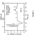
- Figure 5 depicts a z-axial optoacoustic image of absorbed laser fluence distribution in cock's comb.
- the transient stress profile induced by a 14-ns pulse at 532 nm in a cock's comb of a rooster was measured in vivo by an acoustic transducer.
- the laser beam was about 1 cm in diameter.
- Time "0" corresponds to a signal detected from the tissue surface. Distinct layers were observed in the cock's comb tissue. Alteration of the detected stress transient due to diffraction of acoustic waves generated in distributed capillary blood vessels (layer 2) yields a negative signal.
- the stress profile will have only positive components.
- Signals 1-6 were induced in blood vessels located at different depths in the tissue. Numbers 1-5 correspond to the acoustic transducer signals detected in layers with either enhanced density of small blood vessels (1 and 2) or in separated large blood vessels (3, 4 and 5).
- the layered structure of the cock's comb is clearly depicted (the layer with dense small dermal blood vessels that lies just below the epidermis, the layer of less vascular loose connective tissue, the comb core layer with arteries and veins that supply the more superficial vascular layers of the cockscomb). The depth of their location is measured correctly if compared with cockscomb histology. The lateral position can be found by scanning a focused laser beam along the tissue surface.
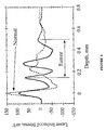
- Figure 6 shows a display of a typical profile of a stress wave induced by nanosecond laser pulses at 532 nm in tissues of a mouse with a small tumor beneath the skin. Signals detected from the volume with cancer and from the tissue with no cancer are presented for comparison. The difference between two presented signals indicate that breast tumor can be diagnosed with laser optoacoustic tomography system of the present invention in a mouse model in vivo .
- This is another example of laser optoacoustic tomography system of the present invention in the reflection embodiment performed in vivo .
- the object of study was a mouse with a cancer modeling a female's breast tumor grown inside the muscle of the mouse. The imaging experiment was performed twice with two different mice with similar tumor conditions. These embodiments presented demonstrated laser optoacoustic imaging in tissues by time-resolved detection of laser-induced stress transients.
Abstract
Description
Claims (8)
- An imaging system configured to reconstruct an image of high spatial resolution from pressure profiles optically-induced at a region of interest in a body, comprising:characterised in that the imaging system further includes means to deliver exogenous molecular probes or dyes to enhance contrast between pressure profiles of different structures at the region of interest and a computer with software for reconstruction of a three-dimensional image, a two-dimensional slice or an axial profile of the region of interest in the body from said detected pressure profiles and for analysis thereof.a pulsed laser source to produce said optically-induced pressure profiles using temporal stress confinement;a light delivery system for delivery of radiation from said pulsed laser source to the region of interest;at least one acoustic transducer with sufficient temporal resolution to detect optoacoustic signals representative of pressure profiles at said region of interest; andan electronic system for recording and processing said detected pressure profiles;
- The imaging system of claim 1, wherein said pulsed laser source has a spectral range of wavelengths from 600 nm to 1400 nm.
- The imaging system of claims 1 or 2, wherein said light delivery system comprises at least one separate optical fiber or laser beam to irradiate a large volume of said tissue.
- The imaging system of any one of the preceding claims, wherein said acoustic transducer is capable of detecting pressure profiles within a wide ultrasonic frequency range corresponding to dimensions of said region of interest or to a structure therein.
- The imaging system of claim 4, wherein said acoustic transducer is a piezoelectric transducer.
- The imaging system of any one of the preceding claims, wherein said acoustic transducer detects a pressure profile originating at a tissue depth of up to 12 cm.
- The imaging system of any one of the preceding claims, wherein said image reconstructed from temporally-resolved pressure profiles is an image of optical absorption coefficients in the region of interest.
- The imaging system of any one of the preceding claims, wherein the imaging system further comprises an endoscope having one or both of said light delivery system and said at least one acoustic transducer.
Applications Claiming Priority (3)
| Application Number | Priority Date | Filing Date | Title |
|---|---|---|---|
| US594758 | 1984-03-29 | ||
| US08/594,758 US5840023A (en) | 1996-01-31 | 1996-01-31 | Optoacoustic imaging for medical diagnosis |
| PCT/US1997/001815 WO1997027801A1 (en) | 1996-01-31 | 1997-01-31 | Laser opto-acoustic imaging system |
Publications (3)
| Publication Number | Publication Date |
|---|---|
| EP0920277A4 EP0920277A4 (en) | 1999-06-09 |
| EP0920277A1 EP0920277A1 (en) | 1999-06-09 |
| EP0920277B1 true EP0920277B1 (en) | 2005-09-14 |
Family
ID=24380282
Family Applications (1)
| Application Number | Title | Priority Date | Filing Date |
|---|---|---|---|
| EP97904228A Expired - Lifetime EP0920277B1 (en) | 1996-01-31 | 1997-01-31 | Laser opto-acoustic imaging system |
Country Status (7)
| Country | Link |
|---|---|
| US (1) | US5840023A (en) |
| EP (1) | EP0920277B1 (en) |
| JP (1) | JPH11514549A (en) |
| AU (1) | AU732799B2 (en) |
| CA (1) | CA2244732C (en) |
| DE (1) | DE69734203T2 (en) |
| WO (1) | WO1997027801A1 (en) |
Cited By (1)
| Publication number | Priority date | Publication date | Assignee | Title |
|---|---|---|---|---|
| DE102008017097A1 (en) | 2008-01-17 | 2009-07-30 | RUHR-UNIVERSITäT BOCHUM | Method for photoacoustic generation of illustration, involves emitting light pulse sequence by light source, where light pulse sequence has two electromagnetic pulses |
Families Citing this family (311)
| Publication number | Priority date | Publication date | Assignee | Title |
|---|---|---|---|---|
| US5585112A (en) | 1989-12-22 | 1996-12-17 | Imarx Pharmaceutical Corp. | Method of preparing gas and gaseous precursor-filled microspheres |
| US5776429A (en) | 1989-12-22 | 1998-07-07 | Imarx Pharmaceutical Corp. | Method of preparing gas-filled microspheres using a lyophilized lipids |
| US6088613A (en) | 1989-12-22 | 2000-07-11 | Imarx Pharmaceutical Corp. | Method of magnetic resonance focused surgical and therapeutic ultrasound |
| US5922304A (en) | 1989-12-22 | 1999-07-13 | Imarx Pharmaceutical Corp. | Gaseous precursor filled microspheres as magnetic resonance imaging contrast agents |
| US5542935A (en) | 1989-12-22 | 1996-08-06 | Imarx Pharmaceutical Corp. | Therapeutic delivery systems related applications |
| US6551576B1 (en) | 1989-12-22 | 2003-04-22 | Bristol-Myers Squibb Medical Imaging, Inc. | Container with multi-phase composition for use in diagnostic and therapeutic applications |
| US6146657A (en) | 1989-12-22 | 2000-11-14 | Imarx Pharmaceutical Corp. | Gas-filled lipid spheres for use in diagnostic and therapeutic applications |
| US5205290A (en) | 1991-04-05 | 1993-04-27 | Unger Evan C | Low density microspheres and their use as contrast agents for computed tomography |
| US5874062A (en) | 1991-04-05 | 1999-02-23 | Imarx Pharmaceutical Corp. | Methods of computed tomography using perfluorocarbon gaseous filled microspheres as contrast agents |
| US20080154257A1 (en) * | 2006-12-22 | 2008-06-26 | Shiva Sharareh | Real-time optoacoustic monitoring with electophysiologic catheters |
| US6743779B1 (en) | 1994-11-29 | 2004-06-01 | Imarx Pharmaceutical Corp. | Methods for delivering compounds into a cell |
| US5997898A (en) | 1995-06-06 | 1999-12-07 | Imarx Pharmaceutical Corp. | Stabilized compositions of fluorinated amphiphiles for methods of therapeutic delivery |
| US6033645A (en) | 1996-06-19 | 2000-03-07 | Unger; Evan C. | Methods for diagnostic imaging by regulating the administration rate of a contrast agent |
| US6521211B1 (en) | 1995-06-07 | 2003-02-18 | Bristol-Myers Squibb Medical Imaging, Inc. | Methods of imaging and treatment with targeted compositions |
| US6231834B1 (en) | 1995-06-07 | 2001-05-15 | Imarx Pharmaceutical Corp. | Methods for ultrasound imaging involving the use of a contrast agent and multiple images and processing of same |
| US6139819A (en) | 1995-06-07 | 2000-10-31 | Imarx Pharmaceutical Corp. | Targeted contrast agents for diagnostic and therapeutic use |
| US6405069B1 (en) * | 1996-01-31 | 2002-06-11 | Board Of Regents, The University Of Texas System | Time-resolved optoacoustic method and system for noninvasive monitoring of glucose |
| US6309352B1 (en) * | 1996-01-31 | 2001-10-30 | Board Of Regents, The University Of Texas System | Real time optoacoustic monitoring of changes in tissue properties |
| AU736301B2 (en) | 1996-05-01 | 2001-07-26 | Imarx Therapeutics, Inc. | Methods for delivering compounds into a cell |
| US6414139B1 (en) | 1996-09-03 | 2002-07-02 | Imarx Therapeutics, Inc. | Silicon amphiphilic compounds and the use thereof |
| WO1998010798A1 (en) | 1996-09-11 | 1998-03-19 | Imarx Pharmaceutical Corp. | Improved methods for diagnostic imaging using a contrast agent and a vasodilator |
| US5713356A (en) | 1996-10-04 | 1998-02-03 | Optosonics, Inc. | Photoacoustic breast scanner |
| US6745067B1 (en) * | 1998-09-14 | 2004-06-01 | Lucid, Inc. | System for marking the locations of imaged tissue with respect to the surface of the tissue |
| US6424852B1 (en) * | 1996-10-18 | 2002-07-23 | Lucid, Inc. | System for confocal imaging within dermal tissue |
| GB9704737D0 (en) * | 1997-03-07 | 1997-04-23 | Optel Instr Limited | Biological measurement system |
| US6537246B1 (en) | 1997-06-18 | 2003-03-25 | Imarx Therapeutics, Inc. | Oxygen delivery agents and uses for the same |
| US6143276A (en) | 1997-03-21 | 2000-11-07 | Imarx Pharmaceutical Corp. | Methods for delivering bioactive agents to regions of elevated temperatures |
| US6090800A (en) | 1997-05-06 | 2000-07-18 | Imarx Pharmaceutical Corp. | Lipid soluble steroid prodrugs |
| US6416740B1 (en) | 1997-05-13 | 2002-07-09 | Bristol-Myers Squibb Medical Imaging, Inc. | Acoustically active drug delivery systems |
| GB9712524D0 (en) * | 1997-06-16 | 1997-08-20 | Nycomed Imaging As | Method |
| US6548047B1 (en) | 1997-09-15 | 2003-04-15 | Bristol-Myers Squibb Medical Imaging, Inc. | Thermal preactivation of gaseous precursor filled compositions |
| US6091981A (en) * | 1997-09-16 | 2000-07-18 | Assurance Medical Inc. | Clinical tissue examination |
| US6063031A (en) * | 1997-10-14 | 2000-05-16 | Assurance Medical, Inc. | Diagnosis and treatment of tissue with instruments |
| US6123923A (en) | 1997-12-18 | 2000-09-26 | Imarx Pharmaceutical Corp. | Optoacoustic contrast agents and methods for their use |
| US20010003580A1 (en) | 1998-01-14 | 2001-06-14 | Poh K. Hui | Preparation of a lipid blend and a phospholipid suspension containing the lipid blend |
| US5977538A (en) * | 1998-05-11 | 1999-11-02 | Imarx Pharmaceutical Corp. | Optoacoustic imaging system |
| US6104942A (en) * | 1998-05-12 | 2000-08-15 | Optosonics, Inc. | Thermoacoustic tissue scanner |
| US6478735B1 (en) * | 1999-01-28 | 2002-11-12 | The United States Of America As Represented By The Administrator Of The National Aeronautics And Space Administration | Physiological feedback method and system |
| US6216025B1 (en) | 1999-02-02 | 2001-04-10 | Optosonics, Inc. | Thermoacoustic computed tomography scanner |
| GB9915082D0 (en) * | 1999-06-28 | 1999-08-25 | Univ London | Optical fibre probe |
| US6498942B1 (en) | 1999-08-06 | 2002-12-24 | The University Of Texas System | Optoacoustic monitoring of blood oxygenation |
| US6253621B1 (en) | 1999-08-31 | 2001-07-03 | Advanced Micro Devices | Micro-void detection |
| US6212421B1 (en) * | 1999-09-03 | 2001-04-03 | Lockheed Martin Energy Research Corp. | Method and apparatus of spectro-acoustically enhanced ultrasonic detection for diagnostics |
| US6277656B1 (en) | 1999-09-30 | 2001-08-21 | Rama R. Goruganthu | Substrate removal as a function of acoustic analysis |
| US6421811B1 (en) | 1999-09-30 | 2002-07-16 | Advanced Micro Devices, Inc. | Defect detection via acoustic analysis |
| US6430728B1 (en) | 1999-09-30 | 2002-08-06 | Advanced Micro Devices, Inc. | Acoustic 3D analysis of circuit structures |
| WO2001035881A1 (en) * | 1999-11-12 | 2001-05-25 | Thomas Bende | Non-contact photoacoustic spectroscopy for photoablation control |
| US6751490B2 (en) | 2000-03-01 | 2004-06-15 | The Board Of Regents Of The University Of Texas System | Continuous optoacoustic monitoring of hemoglobin concentration and hematocrit |
| GB0015250D0 (en) * | 2000-06-22 | 2000-08-16 | Payne Peter A | Ophthalmic uses of lasers |
| IL138073A0 (en) * | 2000-08-24 | 2001-10-31 | Glucon Inc | Photoacoustic assay and imaging system |
| US6669926B1 (en) | 2000-10-16 | 2003-12-30 | Mallinckrodt, Inc. | Hydrophilic light absorbing indole compounds for determination of physiological function in critically ill patients |
| US6673334B1 (en) * | 2000-10-16 | 2004-01-06 | Mallinkcrodt, Inc. | Light sensitive compounds for instant determination of organ function |
| AU2002230842A1 (en) | 2000-10-30 | 2002-05-15 | The General Hospital Corporation | Optical methods and systems for tissue analysis |
| US9295391B1 (en) | 2000-11-10 | 2016-03-29 | The General Hospital Corporation | Spectrally encoded miniature endoscopic imaging probe |
| US7179449B2 (en) | 2001-01-30 | 2007-02-20 | Barnes-Jewish Hospital | Enhanced ultrasound detection with temperature-dependent contrast agents |
| JP2002272738A (en) * | 2001-03-21 | 2002-09-24 | Fuji Photo Film Co Ltd | Ultrasonic diagnostic equipment |
| GB2408797B (en) | 2001-05-01 | 2006-09-20 | Gen Hospital Corp | Method and apparatus for determination of atherosclerotic plaque type by measurement of tissue optical properties |
| US6490470B1 (en) | 2001-06-19 | 2002-12-03 | Optosonics, Inc. | Thermoacoustic tissue scanner |
| US20050085725A1 (en) * | 2001-08-09 | 2005-04-21 | Ron Nagar | Photoacoustic assay and imaging system |
| US6980299B1 (en) | 2001-10-16 | 2005-12-27 | General Hospital Corporation | Systems and methods for imaging a sample |
| US7077565B2 (en) * | 2001-11-15 | 2006-07-18 | Glucon, Inc. | Method for measuring temperature of substances from measurement of absorption coefficients |
| EP1468245B1 (en) | 2002-01-11 | 2011-03-30 | The General Hospital Corporation | Apparatus for OCT imaging with axial line focus for improved resolution and depth of field |
| US7355716B2 (en) | 2002-01-24 | 2008-04-08 | The General Hospital Corporation | Apparatus and method for ranging and noise reduction of low coherence interferometry LCI and optical coherence tomography OCT signals by parallel detection of spectral bands |
| US7322972B2 (en) * | 2002-04-10 | 2008-01-29 | The Regents Of The University Of California | In vivo port wine stain, burn and melanin depth determination using a photoacoustic probe |
| AU2003238655A1 (en) * | 2002-06-25 | 2004-01-06 | Glucon Inc. | Method and apparatus for determining tissue viability |
| US7931596B2 (en) * | 2002-07-12 | 2011-04-26 | Iscience Interventional Corporation | Ultrasound interfacing device for tissue imaging |
| EP1526804B1 (en) | 2002-07-26 | 2009-03-11 | The Board of Regents, The University of Texas System | Continuous optoacoustic monitoring of hemoglobin concentration and hematocrit |
| US7761139B2 (en) | 2003-01-24 | 2010-07-20 | The General Hospital Corporation | System and method for identifying tissue using low-coherence interferometry |
| US8054468B2 (en) | 2003-01-24 | 2011-11-08 | The General Hospital Corporation | Apparatus and method for ranging and noise reduction of low coherence interferometry LCI and optical coherence tomography OCT signals by parallel detection of spectral bands |
| WO2004068405A2 (en) * | 2003-01-25 | 2004-08-12 | Oraevsky Alexander A | High contrast optoacoustical imaging using nanoparticles |
| JP4805142B2 (en) | 2003-03-31 | 2011-11-02 | ザ ジェネラル ホスピタル コーポレイション | Speckle reduction in optically interfering tomography by combining light of different angles with varying path length |
| WO2004096082A2 (en) * | 2003-04-24 | 2004-11-11 | The Board Of Regents Of The University Of Texas System | Noninvasive blood analysis by optical probing of the veins under the tongue |
| EP2280260B1 (en) | 2003-06-06 | 2017-03-08 | The General Hospital Corporation | Process and apparatus for a wavelength tuned light source |
| JP4406226B2 (en) * | 2003-07-02 | 2010-01-27 | 株式会社東芝 | Biological information video device |
| EP1685366B1 (en) | 2003-10-27 | 2011-06-15 | The General Hospital Corporation | Method and apparatus for performing optical imaging using frequency-domain interferometry |
| US7551293B2 (en) | 2003-11-28 | 2009-06-23 | The General Hospital Corporation | Method and apparatus for three-dimensional spectrally encoded imaging |
| US20050124897A1 (en) * | 2003-12-03 | 2005-06-09 | Scimed Life Systems, Inc. | Apparatus and methods for delivering acoustic energy to body tissue |
| JP4643153B2 (en) * | 2004-02-06 | 2011-03-02 | 株式会社東芝 | Non-invasive biological information imaging device |
| WO2005107592A1 (en) | 2004-05-06 | 2005-11-17 | Nippon Telegraph And Telephone Corporation | Component concentration measuring device and method of controlling component concentration measuring device |
| WO2005117534A2 (en) | 2004-05-29 | 2005-12-15 | The General Hospital Corporation | Process, system and software arrangement for a chromatic dispersion compensation using reflective layers in optical coherence tomography (oct) imaging |
| EP1771755B1 (en) | 2004-07-02 | 2016-09-21 | The General Hospital Corporation | Endoscopic imaging probe comprising dual clad fibre |
| AT414212B (en) * | 2004-07-20 | 2006-10-15 | Upper Austrian Res Gmbh | THERMOACUSTIC TOMOGRAPHY PROCESS AND THERMOACUSTIC TOMOGRAPH |
| KR101332222B1 (en) | 2004-08-06 | 2013-11-22 | 더 제너럴 하스피탈 코포레이션 | Process, system and software arrangement for determining at least one location in a sample using an optical coherence tomography |
| ES2379468T3 (en) | 2004-08-24 | 2012-04-26 | The General Hospital Corporation | Procedure, system and software configuration to determine the modulus of elasticity |
| KR20120062944A (en) | 2004-08-24 | 2012-06-14 | 더 제너럴 하스피탈 코포레이션 | Method and apparatus for imaging of vessel segments |
| KR101269455B1 (en) | 2004-09-10 | 2013-05-30 | 더 제너럴 하스피탈 코포레이션 | System and method for optical coherence imaging |
| EP2329759B1 (en) | 2004-09-29 | 2014-03-12 | The General Hospital Corporation | System and method for optical coherence imaging |
| WO2006050453A1 (en) | 2004-11-02 | 2006-05-11 | The General Hospital Corporation | Fiber-optic rotational device, optical system and method for imaging a sample |
| EP2278267A3 (en) | 2004-11-24 | 2011-06-29 | The General Hospital Corporation | Common-Path Interferometer for Endoscopic OCT |
| EP1816949A1 (en) | 2004-11-29 | 2007-08-15 | The General Hospital Corporation | Arrangements, devices, endoscopes, catheters and methods for performing optical imaging by simultaneously illuminating and detecting multiple points on a sample |
| EP1833361A1 (en) * | 2004-12-20 | 2007-09-19 | Koninklijke Philips Electronics N.V. | Investigation of body structures |
| IL166408A0 (en) * | 2005-01-20 | 2006-01-15 | Ultraview Ltd | Combined 2d pulse-echo ultrasound and optoacousticsignal for glaucoma treatment |
| US20060184042A1 (en) * | 2005-01-22 | 2006-08-17 | The Texas A&M University System | Method, system and apparatus for dark-field reflection-mode photoacoustic tomography |
| ATE451669T1 (en) | 2005-04-28 | 2009-12-15 | Gen Hospital Corp | EVALUATION OF IMAGE FEATURES OF AN ANATOMIC STRUCTURE IN OPTICAL COHERENCE TOMOGRAPHY IMAGES |
| US7859679B2 (en) | 2005-05-31 | 2010-12-28 | The General Hospital Corporation | System, method and arrangement which can use spectral encoding heterodyne interferometry techniques for imaging |
| JP5702049B2 (en) | 2005-06-01 | 2015-04-15 | ザ ジェネラル ホスピタル コーポレイション | Apparatus, method and system for performing phase resolved optical frequency domain imaging |
| US20070015992A1 (en) * | 2005-06-30 | 2007-01-18 | General Electric Company | System and method for optoacoustic imaging |
| ATE484727T1 (en) | 2005-08-09 | 2010-10-15 | Gen Hospital Corp | DEVICE AND METHOD FOR PERFORMING POLARIZATION-BASED QUADRATURE DEMODULATION IN OPTICAL COHERENCE TOMOGRAPHY |
| US7847949B2 (en) | 2005-09-29 | 2010-12-07 | The General Hospital Corporation | Method and apparatus for optical imaging via spectral encoding |
| EP1945094B1 (en) | 2005-10-14 | 2018-09-05 | The General Hospital Corporation | Spectral- and frequency- encoded fluorescence imaging |
| US7796270B2 (en) | 2006-01-10 | 2010-09-14 | The General Hospital Corporation | Systems and methods for generating data based on one or more spectrally-encoded endoscopy techniques |
| US20090227997A1 (en) * | 2006-01-19 | 2009-09-10 | The Regents Of The University Of Michigan | System and method for photoacoustic imaging and monitoring of laser therapy |
| DK1973466T3 (en) | 2006-01-19 | 2021-02-01 | Massachusetts Gen Hospital | BALLOON IMAGING CATHETER |
| US20090054763A1 (en) * | 2006-01-19 | 2009-02-26 | The Regents Of The University Of Michigan | System and method for spectroscopic photoacoustic tomography |
| US8145018B2 (en) | 2006-01-19 | 2012-03-27 | The General Hospital Corporation | Apparatus for obtaining information for a structure using spectrally-encoded endoscopy techniques and methods for producing one or more optical arrangements |
| US8135450B2 (en) * | 2006-01-20 | 2012-03-13 | Esenaliev Rinat O | Noninvasive glucose sensing methods and systems |
| EP2001362A4 (en) * | 2006-01-20 | 2010-10-06 | Seno Medical Instr Inc | Quantitative optoacoustic tomography with enhanced contrast |
| WO2007149602A2 (en) | 2006-02-01 | 2007-12-27 | The General Hospital Corporation | Methods and systems for providing electromagnetic radiation to at least one portion of a sample using conformal laser therapy procedures |
| JP2009537024A (en) | 2006-02-01 | 2009-10-22 | ザ ジェネラル ホスピタル コーポレイション | Apparatus for controlling at least one of at least two sites of at least one fiber |
| US7538859B2 (en) | 2006-02-01 | 2009-05-26 | The General Hospital Corporation | Methods and systems for monitoring and obtaining information of at least one portion of a sample using conformal laser therapy procedures, and providing electromagnetic radiation thereto |
| US9777053B2 (en) | 2006-02-08 | 2017-10-03 | The General Hospital Corporation | Methods, arrangements and systems for obtaining information associated with an anatomical sample using optical microscopy |
| US7982879B2 (en) | 2006-02-24 | 2011-07-19 | The General Hospital Corporation | Methods and systems for performing angle-resolved fourier-domain optical coherence tomography |
| US7750536B2 (en) | 2006-03-02 | 2010-07-06 | Visualsonics Inc. | High frequency ultrasonic transducer and matching layer comprising cyanoacrylate |
| EP2564769B1 (en) | 2006-04-05 | 2015-06-03 | The General Hospital Corporation | Apparatus for polarization-sensitive optical frequency domain imaging of a sample |
| JP2009536740A (en) | 2006-05-10 | 2009-10-15 | ザ ジェネラル ホスピタル コーポレイション | Process, configuration and system for providing frequency domain imaging of samples |
| WO2007133964A2 (en) * | 2006-05-12 | 2007-11-22 | The General Hospital Corporation | Processes, arrangements and systems for providing a fiber layer thickness map based on optical coherence tomography images |
| CN100456016C (en) * | 2006-05-30 | 2009-01-28 | 华南师范大学 | Multi-channel electronic parallel scanning photoacoustic real-time tomo graphic-imaging method and apparatus thereof |
| US20070291275A1 (en) * | 2006-06-16 | 2007-12-20 | Prescient Medical, Inc. | Side-viewing optical acoustic sensors and their use in intravascular diagnostic probes |
| CA2656504C (en) * | 2006-07-11 | 2013-02-26 | The Curators Of The University Of Missouri | Photo-acoustic detection device and method |
| US8501099B2 (en) * | 2006-07-11 | 2013-08-06 | The Curators Of The University Of Missouri | Photo-acoustic detection device and method |
| EP2054712B1 (en) | 2006-08-25 | 2015-10-07 | The General Hospital Corporation | Apparatus and methods for enhancing optical coherence tomography imaging using volumetric filtering techniques |
| US8219170B2 (en) | 2006-09-20 | 2012-07-10 | Nellcor Puritan Bennett Llc | System and method for practicing spectrophotometry using light emitting nanostructure devices |
| US8175671B2 (en) | 2006-09-22 | 2012-05-08 | Nellcor Puritan Bennett Llc | Medical sensor for reducing signal artifacts and technique for using the same |
| US8396527B2 (en) | 2006-09-22 | 2013-03-12 | Covidien Lp | Medical sensor for reducing signal artifacts and technique for using the same |
| US8190225B2 (en) | 2006-09-22 | 2012-05-29 | Nellcor Puritan Bennett Llc | Medical sensor for reducing signal artifacts and technique for using the same |
| US8838213B2 (en) | 2006-10-19 | 2014-09-16 | The General Hospital Corporation | Apparatus and method for obtaining and providing imaging information associated with at least one portion of a sample, and effecting such portion(s) |
| WO2008067438A2 (en) * | 2006-11-29 | 2008-06-05 | The Regents Of University Of Michigan | System and method for photoacoustic guided diffuse optical imaging |
| EP1935346A1 (en) | 2006-12-21 | 2008-06-25 | Stichting voor de Technische Wetenschappen | Imaging apparatus and method |
| US20080173093A1 (en) * | 2007-01-18 | 2008-07-24 | The Regents Of The University Of Michigan | System and method for photoacoustic tomography of joints |
| EP2104968A1 (en) | 2007-01-19 | 2009-09-30 | The General Hospital Corporation | Rotating disk reflection for fast wavelength scanning of dispersed broadband light |
| JP5507258B2 (en) | 2007-01-19 | 2014-05-28 | ザ ジェネラル ホスピタル コーポレイション | Apparatus and method for controlling measurement depth in optical frequency domain imaging |
| WO2008100386A2 (en) * | 2007-02-09 | 2008-08-21 | Board Of Regents, The University Of Texas System | Intravascular photoacoustic and ultrasound echo imaging |
| WO2008103982A2 (en) * | 2007-02-23 | 2008-08-28 | The Regents Of The University Of Michigan | System and method for monitoring photodynamic therapy |
| US20080228073A1 (en) * | 2007-03-12 | 2008-09-18 | Silverman Ronald H | System and method for optoacoustic imaging of peripheral tissues |
| WO2008115965A1 (en) * | 2007-03-19 | 2008-09-25 | The General Hospital Corporation | Apparatus and method for providing a noninvasive diagnosis of internal bleeding |
| EP2602651A3 (en) | 2007-03-23 | 2014-08-27 | The General Hospital Corporation | Methods, arrangements and apparatus for utilizing a wavelength-swept laser using angular scanning and dispersion procedures |
| WO2008121844A1 (en) | 2007-03-30 | 2008-10-09 | The General Hospital Corporation | System and method providing intracoronary laser speckle imaging for the detection of vulnerable plaque |
| US20080255433A1 (en) * | 2007-04-11 | 2008-10-16 | The Board Of Regents Of The University Of Texas Syatem | Optoacoustic monitoring of multiple parameters |
| US10231656B2 (en) | 2007-04-11 | 2019-03-19 | Noninvasix, Inc. | Systems and methods for measuring oxygenation |
| US10226206B2 (en) | 2007-04-11 | 2019-03-12 | The Board Of Regents Of The University Of Texas System | Systems and methods for measuring neonatal cerebral oxygenation |
| WO2008131082A1 (en) | 2007-04-17 | 2008-10-30 | The General Hospital Corporation | Apparatus and methods for measuring vibrations using spectrally-encoded endoscopy techniques |
| WO2008137637A2 (en) | 2007-05-04 | 2008-11-13 | The General Hospital Corporation | Methods, arrangements and systems for obtaining information associated with a sample using brillouin microscopy |
| JP4739363B2 (en) * | 2007-05-15 | 2011-08-03 | キヤノン株式会社 | Biological information imaging apparatus, biological information analysis method, and biological information imaging method |
| JP5595534B2 (en) * | 2007-05-15 | 2014-09-24 | キヤノン株式会社 | Biological information imaging apparatus, biological information analysis method, and biological information imaging method |
| US7952719B2 (en) | 2007-06-08 | 2011-05-31 | Prescient Medical, Inc. | Optical catheter configurations combining raman spectroscopy with optical fiber-based low coherence reflectometry |
| JP5349839B2 (en) * | 2007-06-22 | 2013-11-20 | キヤノン株式会社 | Biological information imaging device |
| WO2009014820A1 (en) * | 2007-07-20 | 2009-01-29 | Prescient Medical, Inc. | Wall-contacting intravascular ultrasound probe catheters |
| EP2173254A2 (en) | 2007-07-31 | 2010-04-14 | The General Hospital Corporation | Systems and methods for providing beam scan patterns for high speed doppler optical frequency domain imaging |
| US8361437B2 (en) | 2007-08-28 | 2013-01-29 | University Of Florida Research Foundation, Inc. | Multimodal nanoparticles for non-invasive bio-imaging |
| EP2191254B1 (en) | 2007-08-31 | 2017-07-19 | The General Hospital Corporation | System and method for self-interference fluorescence microscopy, and computer-accessible medium associated therewith |
| JP5201920B2 (en) * | 2007-09-12 | 2013-06-05 | キヤノン株式会社 | measuring device |
| JP5132228B2 (en) * | 2007-09-12 | 2013-01-30 | キヤノン株式会社 | Measuring method and measuring device |
| US20090076395A1 (en) * | 2007-09-19 | 2009-03-19 | Prescient Medical, Inc. | Optimized intravascular ultrasound probe catherers |
| WO2009045885A2 (en) * | 2007-10-02 | 2009-04-09 | Board Of Regents, The University Of Texas System | Real-time ultrasound monitoring of heat-induced tissue interactions |
| WO2009050632A1 (en) | 2007-10-16 | 2009-04-23 | Koninklijke Philips Electronics N.V. | Apparatus, systems and methods for production and integration of compact illumination schemes |
| JP5643101B2 (en) | 2007-10-25 | 2014-12-17 | ワシントン・ユニバーシティWashington University | Scattering medium imaging method, imaging apparatus, and imaging system |
| US7933021B2 (en) | 2007-10-30 | 2011-04-26 | The General Hospital Corporation | System and method for cladding mode detection |
| EP2231018A4 (en) * | 2007-12-12 | 2012-11-21 | Jeffrey J L Carson | Three-dimensional photoacoustic imager and methods for calibrating an imager |
| JP5698538B2 (en) * | 2007-12-28 | 2015-04-08 | コーニンクレッカ フィリップス エヌ ヴェ | Tissue resection device, tissue resection system, and program for monitoring the performance of tissue resection |
| US11123047B2 (en) | 2008-01-28 | 2021-09-21 | The General Hospital Corporation | Hybrid systems and methods for multi-modal acquisition of intravascular imaging data and counteracting the effects of signal absorption in blood |
| US9332942B2 (en) | 2008-01-28 | 2016-05-10 | The General Hospital Corporation | Systems, processes and computer-accessible medium for providing hybrid flourescence and optical coherence tomography imaging |
| JP5284129B2 (en) * | 2008-02-06 | 2013-09-11 | キヤノン株式会社 | Imaging apparatus and analysis method |
| EP2110076A1 (en) * | 2008-02-19 | 2009-10-21 | Helmholtz Zentrum München Deutsches Forschungszentrum für Gesundheit und Umwelt (GmbH) | Method and device for near-field dual-wave modality imaging |
| US9078617B2 (en) * | 2008-03-17 | 2015-07-14 | Or-Nim Medical Ltd. | Apparatus for non-invasive optical monitoring |
| WO2009137701A2 (en) | 2008-05-07 | 2009-11-12 | The General Hospital Corporation | System, method and computer-accessible medium for tracking vessel motion during three-dimensional coronary artery microscopy |
| EP2288948A4 (en) | 2008-06-20 | 2011-12-28 | Gen Hospital Corp | Fused fiber optic coupler arrangement and method for use thereof |
| JP4829934B2 (en) * | 2008-07-11 | 2011-12-07 | キヤノン株式会社 | Inspection device |
| US9254089B2 (en) | 2008-07-14 | 2016-02-09 | The General Hospital Corporation | Apparatus and methods for facilitating at least partial overlap of dispersed ration on at least one sample |
| CN102137618B (en) * | 2008-07-25 | 2015-06-17 | 健康与环境慕尼黑德国研究中心赫姆霍茨中心(有限公司) | Quantitative multi-spectral opto-acoustic tomography (MSOT) of tissue biomarkers |
| JP5419404B2 (en) * | 2008-09-04 | 2014-02-19 | キヤノン株式会社 | Photoacoustic device |
| JP5451014B2 (en) * | 2008-09-10 | 2014-03-26 | キヤノン株式会社 | Photoacoustic device |
| US8532751B2 (en) | 2008-09-30 | 2013-09-10 | Covidien Lp | Laser self-mixing sensors for biological sensing |
| US8433382B2 (en) | 2008-09-30 | 2013-04-30 | Covidien Lp | Transmission mode photon density wave system and method |
| US8386000B2 (en) | 2008-09-30 | 2013-02-26 | Covidien Lp | System and method for photon density wave pulse oximetry and pulse hemometry |
| US8142368B2 (en) * | 2008-10-02 | 2012-03-27 | Artann Laboratories Inc. | Method of characterization and differentiation of tissue |
| JP5305818B2 (en) * | 2008-10-03 | 2013-10-02 | キヤノン株式会社 | Biological information acquisition device |
| JP5279435B2 (en) * | 2008-10-03 | 2013-09-04 | キヤノン株式会社 | SUBJECT INFORMATION ACQUISITION DEVICE AND METHOD FOR CONTROLLING SUBJECT INFORMATION ACQUISITION DEVICE |
| JP5183406B2 (en) * | 2008-10-03 | 2013-04-17 | キヤノン株式会社 | Biological information processing apparatus and biological information processing method |
| JP5235586B2 (en) * | 2008-10-03 | 2013-07-10 | キヤノン株式会社 | Biological information processing apparatus and biological information processing method |
| JP2010088627A (en) * | 2008-10-07 | 2010-04-22 | Canon Inc | Apparatus and method for processing biological information |
| CN102215752A (en) * | 2008-10-13 | 2011-10-12 | 阿姆斯特丹大学学术医学中心 | Mammography-apparatus and method for screening the occurrence of malignant cells |
| US9528966B2 (en) * | 2008-10-23 | 2016-12-27 | Washington University | Reflection-mode photoacoustic tomography using a flexibly-supported cantilever beam |
| ES2957932T3 (en) | 2008-12-10 | 2024-01-30 | Massachusetts Gen Hospital | Systems, apparatus and procedures for extending the image depth range of optical coherence tomography using optical subsampling |
| JP5241465B2 (en) * | 2008-12-11 | 2013-07-17 | キヤノン株式会社 | Photoacoustic imaging apparatus and photoacoustic imaging method |
| US9351705B2 (en) | 2009-01-09 | 2016-05-31 | Washington University | Miniaturized photoacoustic imaging apparatus including a rotatable reflector |
| US8097864B2 (en) | 2009-01-26 | 2012-01-17 | The General Hospital Corporation | System, method and computer-accessible medium for providing wide-field superresolution microscopy |
| CA2749670A1 (en) | 2009-02-04 | 2010-08-12 | The General Hospital Corporation | Apparatus and method for utilization of a high-speed optical wavelength tuning source |
| WO2010105197A2 (en) | 2009-03-12 | 2010-09-16 | The General Hospital Corporation | Non-contact optical system, computer-accessible medium and method for measuring at least one mechanical property of tissue using coherent speckle techniques(s) |
| US8025406B2 (en) * | 2009-03-17 | 2011-09-27 | The Uwm Research Foundation, Inc. | Systems and methods for photoacoustic opthalmoscopy |
| AU2010239360A1 (en) * | 2009-04-20 | 2011-11-24 | The Curators Of The University Of Missouri | Photoacoustic detection of analytes in solid tissue and detection system |
| US9271654B2 (en) | 2009-06-29 | 2016-03-01 | Helmholtz Zentrum Munchen Deutsches Forschungszentrum Fur Gesundheit Und Umwelt (Gmbh) | Thermoacoustic imaging with quantitative extraction of absorption map |
| US11490826B2 (en) | 2009-07-14 | 2022-11-08 | The General Hospital Corporation | Apparatus, systems and methods for measuring flow and pressure within a vessel |
| WO2011012274A1 (en) | 2009-07-27 | 2011-02-03 | Helmholtz Zentrum München Deutsches Forschungszentrum Für Gesundheit Und Umwelt (Gmbh) | Imaging device and method for optoacoustic imaging of small animals |
| JP5525787B2 (en) * | 2009-09-14 | 2014-06-18 | 株式会社東芝 | Biological information video device |
| US8494604B2 (en) | 2009-09-21 | 2013-07-23 | Covidien Lp | Wavelength-division multiplexing in a multi-wavelength photon density wave system |
| US8788001B2 (en) | 2009-09-21 | 2014-07-22 | Covidien Lp | Time-division multiplexing in a multi-wavelength photon density wave system |
| US9057695B2 (en) * | 2009-09-24 | 2015-06-16 | Canon Kabushiki Kaisha | Apparatus and method for irradiating a scattering medium with a reconstructive wave |
| US8798704B2 (en) | 2009-09-24 | 2014-08-05 | Covidien Lp | Photoacoustic spectroscopy method and system to discern sepsis from shock |
| CN104856728B (en) * | 2009-10-29 | 2019-07-23 | 佳能株式会社 | Photo-acoustic device |
| CN102596049B (en) | 2009-10-29 | 2015-04-08 | 佳能株式会社 | Photo-acoustic device |
| JP5538855B2 (en) | 2009-12-11 | 2014-07-02 | キヤノン株式会社 | Photoacoustic apparatus and method for controlling the apparatus |
| JP5538856B2 (en) | 2009-12-11 | 2014-07-02 | キヤノン株式会社 | Photoacoustic device |
| EP2514364B1 (en) | 2009-12-17 | 2020-02-12 | Canon Kabushiki Kaisha | Measurement system, and image forming method and program |
| NL2005509C2 (en) | 2010-02-19 | 2011-08-23 | Academisch Medisch Ct Bij De Universiteit Van Amsterdam | Mammography-apparatus. |
| JP5448918B2 (en) * | 2010-02-24 | 2014-03-19 | キヤノン株式会社 | Biological information processing device |
| EP2542154B1 (en) | 2010-03-05 | 2020-09-09 | The General Hospital Corporation | Apparatus for providing electro-magnetic radiation to a sample |
| WO2011127428A2 (en) | 2010-04-09 | 2011-10-13 | Washington University | Quantification of optical absorption coefficients using acoustic spectra in photoacoustic tomography |
| JP5641773B2 (en) * | 2010-04-28 | 2014-12-17 | キヤノン株式会社 | measuring device |
| US9069130B2 (en) | 2010-05-03 | 2015-06-30 | The General Hospital Corporation | Apparatus, method and system for generating optical radiation from biological gain media |
| WO2011150069A2 (en) | 2010-05-25 | 2011-12-01 | The General Hospital Corporation | Apparatus, systems, methods and computer-accessible medium for spectral analysis of optical coherence tomography images |
| WO2011149972A2 (en) | 2010-05-25 | 2011-12-01 | The General Hospital Corporation | Systems, devices, methods, apparatus and computer-accessible media for providing optical imaging of structures and compositions |
| EP2575591A4 (en) | 2010-06-03 | 2017-09-13 | The General Hospital Corporation | Apparatus and method for devices for imaging structures in or at one or more luminal organs |
| JP5761935B2 (en) | 2010-07-22 | 2015-08-12 | キヤノン株式会社 | Subject information acquisition apparatus, subject information acquisition method, and subject information acquisition program |
| US8930145B2 (en) | 2010-07-28 | 2015-01-06 | Covidien Lp | Light focusing continuous wave photoacoustic spectroscopy and its applications to patient monitoring |
| US20130158383A1 (en) * | 2010-08-20 | 2013-06-20 | Purdue Research Foundation | Bond-selective vibrational photoacoustic imaging system and method |
| US9289191B2 (en) | 2011-10-12 | 2016-03-22 | Seno Medical Instruments, Inc. | System and method for acquiring optoacoustic data and producing parametric maps thereof |
| US8686335B2 (en) | 2011-12-31 | 2014-04-01 | Seno Medical Instruments, Inc. | System and method for adjusting the light output of an optoacoustic imaging system |
| US20130109950A1 (en) * | 2011-11-02 | 2013-05-02 | Seno Medical Instruments, Inc. | Handheld optoacoustic probe |
| WO2012058381A2 (en) | 2010-10-27 | 2012-05-03 | The General Hospital Corporation | Apparatus, systems and methods for measuring blood pressure within at least one vessel |
| US8817255B2 (en) | 2010-12-17 | 2014-08-26 | Canon Kabushiki Kaisha | Apparatus and method for irradiating a scattering medium |
| US8954130B2 (en) | 2010-12-17 | 2015-02-10 | Canon Kabushiki Kaisha | Apparatus and method for irradiating a medium |
| US8976433B2 (en) | 2010-12-17 | 2015-03-10 | Canon Kabushiki Kaisha | Apparatus and method for irradiating a scattering medium |
| US8521247B2 (en) | 2010-12-29 | 2013-08-27 | Covidien Lp | Certification apparatus and method for a medical device computer |
| US8997572B2 (en) | 2011-02-11 | 2015-04-07 | Washington University | Multi-focus optical-resolution photoacoustic microscopy with ultrasonic array detection |
| RU2486501C2 (en) * | 2011-02-28 | 2013-06-27 | Александр Алексеевич Карабутов | Laser optical-acoustic tomography method and apparatus for realising said method (versions) |
| US10390709B2 (en) * | 2011-03-14 | 2019-08-27 | Lawrence Livermore National Security, Llc | Non-contact optical system for detecting ultrasound waves from a surface |
| JP5917012B2 (en) * | 2011-04-11 | 2016-05-11 | キヤノン株式会社 | Laser apparatus and photoacoustic apparatus |
| JP6240064B2 (en) | 2011-04-29 | 2017-11-29 | ザ ジェネラル ホスピタル コーポレイション | Method for determining depth-resolved physical and / or optical properties of a scattering medium |
| EP2701624B1 (en) * | 2011-04-29 | 2017-11-22 | Board Of Regents, The University Of Texas System | Apparatus for optoacoustic guidance and confirmation of placement of indwelling medical apparatus |
| US9304490B2 (en) | 2011-05-27 | 2016-04-05 | Canon Kabushiki Kaisha | Apparatus and method for irradiating a medium |
| US9442095B2 (en) | 2011-06-15 | 2016-09-13 | Northwestern University | Optical coherence photoacoustic microscopy |
| US9330092B2 (en) | 2011-07-19 | 2016-05-03 | The General Hospital Corporation | Systems, methods, apparatus and computer-accessible-medium for providing polarization-mode dispersion compensation in optical coherence tomography |
| JP5917037B2 (en) | 2011-07-29 | 2016-05-11 | キヤノン株式会社 | Subject information acquisition apparatus and subject information acquisition method |
| EP3835718B1 (en) | 2011-08-25 | 2023-07-26 | The General Hospital Corporation | Apparatus for providing micro-optical coherence tomography inside a respiratory system |
| US9341783B2 (en) | 2011-10-18 | 2016-05-17 | The General Hospital Corporation | Apparatus and methods for producing and/or providing recirculating optical delay(s) |
| US9055869B2 (en) | 2011-10-28 | 2015-06-16 | Covidien Lp | Methods and systems for photoacoustic signal processing |
| US9445786B2 (en) | 2011-11-02 | 2016-09-20 | Seno Medical Instruments, Inc. | Interframe energy normalization in an optoacoustic imaging system |
| US9730587B2 (en) * | 2011-11-02 | 2017-08-15 | Seno Medical Instruments, Inc. | Diagnostic simulator |
| US9743839B2 (en) | 2011-11-02 | 2017-08-29 | Seno Medical Instruments, Inc. | Playback mode in an optoacoustic imaging system |
| US11287309B2 (en) | 2011-11-02 | 2022-03-29 | Seno Medical Instruments, Inc. | Optoacoustic component utilization tracking |
| US20130289381A1 (en) | 2011-11-02 | 2013-10-31 | Seno Medical Instruments, Inc. | Dual modality imaging system for coregistered functional and anatomical mapping |
| US10433732B2 (en) | 2011-11-02 | 2019-10-08 | Seno Medical Instruments, Inc. | Optoacoustic imaging system having handheld probe utilizing optically reflective material |
| US9757092B2 (en) | 2011-11-02 | 2017-09-12 | Seno Medical Instruments, Inc. | Method for dual modality optoacoustic imaging |
| US9733119B2 (en) | 2011-11-02 | 2017-08-15 | Seno Medical Instruments, Inc. | Optoacoustic component utilization tracking |
| US20130338475A1 (en) | 2012-06-13 | 2013-12-19 | Seno Medical Instruments, Inc. | Optoacoustic imaging system with fiber optic cable |
| US11191435B2 (en) | 2013-01-22 | 2021-12-07 | Seno Medical Instruments, Inc. | Probe with optoacoustic isolator |
| US20130116538A1 (en) | 2011-11-02 | 2013-05-09 | Seno Medical Instruments, Inc. | Optoacoustic imaging systems and methods with enhanced safety |
| US9814394B2 (en) | 2011-11-02 | 2017-11-14 | Seno Medical Instruments, Inc. | Noise suppression in an optoacoustic system |
| US20140005544A1 (en) | 2011-11-02 | 2014-01-02 | Seno Medical Instruments, Inc. | System and method for providing selective channel sensitivity in an optoacoustic imaging system |
| JP5936330B2 (en) | 2011-11-04 | 2016-06-22 | キヤノン株式会社 | Acoustic wave measuring device and control method of acoustic wave measuring device |
| US8886294B2 (en) | 2011-11-30 | 2014-11-11 | Covidien Lp | Methods and systems for photoacoustic monitoring using indicator dilution |
| US9186068B2 (en) | 2011-12-05 | 2015-11-17 | Covidien Lp | Methods and systems for photoacoustic monitoring using hypertonic and isotonic indicator dilutions |
| US9131852B2 (en) | 2011-12-05 | 2015-09-15 | Covidien Lp | Methods and systems for photoacoustic monitoring using indicator dilution |
| EP2806803B1 (en) * | 2012-01-23 | 2019-03-13 | Tomowave Laboratories, Inc. | Laser optoacoustic ultrasonic imaging system (louis) and methods of use |
| JP2015509443A (en) | 2012-03-09 | 2015-03-30 | セノ メディカル インストルメンツ,インク. | Statistical mapping in photoacoustic imaging systems |
| WO2013148306A1 (en) | 2012-03-30 | 2013-10-03 | The General Hospital Corporation | Imaging system, method and distal attachment for multidirectional field of view endoscopy |
| US9833146B2 (en) | 2012-04-17 | 2017-12-05 | Covidien Lp | Surgical system and method of use of the same |
| US8885155B2 (en) | 2012-04-30 | 2014-11-11 | Covidien Lp | Combined light source photoacoustic system |
| KR101974580B1 (en) | 2012-05-03 | 2019-05-02 | 삼성전자주식회사 | The laser-induced ultrasonic wave apparatus and the method of generating a image using the same |
| US11490797B2 (en) | 2012-05-21 | 2022-11-08 | The General Hospital Corporation | Apparatus, device and method for capsule microscopy |
| JP5984547B2 (en) | 2012-07-17 | 2016-09-06 | キヤノン株式会社 | Subject information acquisition apparatus and control method thereof |
| JP6227652B2 (en) | 2012-08-22 | 2017-11-08 | ザ ジェネラル ホスピタル コーポレイション | System, method, and computer-accessible medium for fabricating a miniature endoscope using soft lithography |
| US11020006B2 (en) | 2012-10-18 | 2021-06-01 | California Institute Of Technology | Transcranial photoacoustic/thermoacoustic tomography brain imaging informed by adjunct image data |
| JP6080485B2 (en) * | 2012-10-18 | 2017-02-15 | キヤノン株式会社 | SUBJECT INFORMATION ACQUISITION DEVICE AND METHOD FOR CONTROLLING SUBJECT INFORMATION ACQUISITION DEVICE |
| EP2742854B1 (en) | 2012-12-11 | 2021-03-10 | iThera Medical GmbH | Handheld device and method for tomographic optoacoustic imaging of an object |
| DE102012223651A1 (en) * | 2012-12-18 | 2014-06-18 | Carl Zeiss Ag | A resection imaging method and apparatus for performing such an imaging method |
| JP6112861B2 (en) | 2012-12-28 | 2017-04-12 | キヤノン株式会社 | SUBJECT INFORMATION ACQUISITION DEVICE, SIGNAL PROCESSING DEVICE, AND DISPLAY DEVICE |
| EP2754388B1 (en) | 2013-01-15 | 2020-09-09 | Helmholtz Zentrum München Deutsches Forschungszentrum für Gesundheit und Umwelt GmbH | System and method for quality-enhanced high-rate optoacoustic imaging of an object |
| JP6192297B2 (en) | 2013-01-16 | 2017-09-06 | キヤノン株式会社 | SUBJECT INFORMATION ACQUISITION DEVICE, DISPLAY CONTROL METHOD, AND PROGRAM |
| WO2014120791A1 (en) | 2013-01-29 | 2014-08-07 | The General Hospital Corporation | Apparatus, systems and methods for providing information regarding the aortic valve |
| WO2014121082A1 (en) | 2013-02-01 | 2014-08-07 | The General Hospital Corporation | Objective lens arrangement for confocal endomicroscopy |
| US9398893B2 (en) | 2013-03-15 | 2016-07-26 | Seno Medical Instruments, Inc. | System and method for diagnostic vector classification support |
| US10478072B2 (en) | 2013-03-15 | 2019-11-19 | The General Hospital Corporation | Methods and system for characterizing an object |
| US9380981B2 (en) | 2013-03-15 | 2016-07-05 | Covidien Lp | Photoacoustic monitoring technique with noise reduction |
| JP5575293B2 (en) * | 2013-03-22 | 2014-08-20 | キヤノン株式会社 | Subject information acquisition apparatus and subject information acquisition method |
| US20160051149A1 (en) * | 2013-04-22 | 2016-02-25 | The Curators Of The University Of Missouri | Photoacoustic Probe for Burn Injury Diagnosis |
| EP2997354A4 (en) | 2013-05-13 | 2017-01-18 | The General Hospital Corporation | Detecting self-interefering fluorescence phase and amplitude |
| US11452433B2 (en) | 2013-07-19 | 2022-09-27 | The General Hospital Corporation | Imaging apparatus and method which utilizes multidirectional field of view endoscopy |
| EP3021735A4 (en) | 2013-07-19 | 2017-04-19 | The General Hospital Corporation | Determining eye motion by imaging retina. with feedback |
| EP3025173B1 (en) | 2013-07-26 | 2021-07-07 | The General Hospital Corporation | Apparatus with a laser arrangement utilizing optical dispersion for applications in fourier-domain optical coherence tomography |
| EP3054853A4 (en) | 2013-10-11 | 2017-05-17 | Seno Medical Instruments, Inc. | Systems and methods for component separation in medical imaging |
| US11137375B2 (en) | 2013-11-19 | 2021-10-05 | California Institute Of Technology | Systems and methods of grueneisen-relaxation photoacoustic microscopy and photoacoustic wavefront shaping |
| JP2016537136A (en) | 2013-11-22 | 2016-12-01 | マサチューセッツ インスティテュート オブ テクノロジー | System and method for non-contact ultrasound |
| WO2015105870A1 (en) | 2014-01-08 | 2015-07-16 | The General Hospital Corporation | Method and apparatus for microscopic imaging |
| US10736494B2 (en) | 2014-01-31 | 2020-08-11 | The General Hospital Corporation | System and method for facilitating manual and/or automatic volumetric imaging with real-time tension or force feedback using a tethered imaging device |
| EP3099242B1 (en) | 2014-01-31 | 2021-05-19 | Tomowave Laboratories, Inc. | Optoacoustic image mapping of tissue temperature |
| WO2015131098A1 (en) | 2014-02-27 | 2015-09-03 | Seno Medical Instruments, Inc. | Probe adapted to control blood flow through vessels during imaging and method of use of same |
| EP3116405A4 (en) | 2014-03-12 | 2018-01-03 | Fujifilm Sonosite, Inc. | High frequency ultrasound transducer having an ultrasonic lens with integral central matching layer |
| US10228556B2 (en) | 2014-04-04 | 2019-03-12 | The General Hospital Corporation | Apparatus and method for controlling propagation and/or transmission of electromagnetic radiation in flexible waveguide(s) |
| US10238369B2 (en) | 2014-06-10 | 2019-03-26 | The Johns Hopkins University | Real time ultrasound thermal dose monitoring system for tumor ablation therapy |
| EP3166494A4 (en) | 2014-07-08 | 2018-03-21 | The Board of Regents of The University of Texas System | Systems and methods for measuring fetal cerebral oxygenation |
| KR102513779B1 (en) | 2014-07-25 | 2023-03-24 | 더 제너럴 하스피탈 코포레이션 | Apparatus, devices and methods for in vivo imaging and diagnosis |
| EP3212065B1 (en) | 2014-10-30 | 2022-03-30 | Seno Medical Instruments, Inc. | Opto-acoustic imaging system with detection of relative orientation of light source and acoustic receiver using acoustic waves |
| WO2016149107A1 (en) | 2015-03-14 | 2016-09-22 | Board Of Regents | Systems and methods for measuring neonatal cerebral oxygenation |
| US10898166B2 (en) | 2015-05-14 | 2021-01-26 | Endra Life Sciences Inc. | Systems and methods for imaging biological tissue structures |
| US10028662B2 (en) | 2015-05-14 | 2018-07-24 | Endra Life Sciences Inc. | Systems and methods for imaging biological tissue structures |
| WO2017160858A1 (en) | 2016-03-14 | 2017-09-21 | Massachusetts Institute Of Technology | System and method for non-contact ultrasound with enhanced safety |
| US11672426B2 (en) | 2017-05-10 | 2023-06-13 | California Institute Of Technology | Snapshot photoacoustic photography using an ergodic relay |
| NL2019124B1 (en) | 2017-06-27 | 2019-01-07 | Sigmascreening B V | Mammography apparatus |
| NL2019834B1 (en) | 2017-10-31 | 2019-05-08 | Sigmascreening B V | Mammography apparatus and method of pressurizing a breast |
| US10806513B2 (en) * | 2018-01-16 | 2020-10-20 | Bin Rao | Methods and apparatus for optimizing selective photothermolysis |
| WO2019152045A1 (en) * | 2018-02-02 | 2019-08-08 | Xinova, LLC | Real-time treatment adjustment based on dosimetric data |
| EP3761851A1 (en) | 2018-03-09 | 2021-01-13 | Technische Universität München | Sensor for tissue measurements |
| US10799292B2 (en) * | 2018-05-04 | 2020-10-13 | Bin Rao | High power tunable optical parametric oscillator for selective photothermolysis laser surgeries |
| IL261039B (en) | 2018-08-07 | 2020-10-29 | Elbit Systems Land & C4I Ltd | Two-stage photo-acoustic excitation sensing systems and methods |
| WO2020037082A1 (en) | 2018-08-14 | 2020-02-20 | California Institute Of Technology | Multifocal photoacoustic microscopy through an ergodic relay |
| EP3847453A4 (en) | 2018-09-04 | 2022-06-22 | California Institute of Technology | Enhanced-resolution infrared photoacoustic microscopy and spectroscopy |
| US11369280B2 (en) | 2019-03-01 | 2022-06-28 | California Institute Of Technology | Velocity-matched ultrasonic tagging in photoacoustic flowgraphy |
| US11701035B2 (en) | 2019-10-28 | 2023-07-18 | Bruce M. Landy | Noninvasive blood glucose detector and method using IR |
| JP2023536415A (en) * | 2020-07-21 | 2023-08-25 | ジャイラス エーシーエムアイ インク ディー/ビー/エー オリンパス サージカル テクノロジーズ アメリカ | Laser therapy using acoustic feedback |
| CN114469010A (en) * | 2022-04-19 | 2022-05-13 | 常州英诺激光科技有限公司 | Photoacoustic blood oxygen measuring device and method based on laser energy compensation technology |
Family Cites Families (22)
| Publication number | Priority date | Publication date | Assignee | Title |
|---|---|---|---|---|
| US4028933A (en) * | 1974-02-15 | 1977-06-14 | The Board Of Trustees Of Leland Stanford Junior University | Acoustic microscope |
| FR2405485A1 (en) * | 1977-10-07 | 1979-05-04 | Thomson Csf | ULTRASONIC VISUALIZATION SYSTEM OF A THIN SECTION OF A BODY |
| US4430897A (en) * | 1981-05-14 | 1984-02-14 | The Board Of Trustees Of The Leland Stanford University | Acoustic microscope and method |
| US4727420A (en) * | 1984-10-16 | 1988-02-23 | Fuji Photo Film Co., Ltd. | Acoustooptic image formation |
| US4710030A (en) * | 1985-05-17 | 1987-12-01 | Bw Brown University Research Foundation | Optical generator and detector of stress pulses |
| NO880891L (en) * | 1987-03-03 | 1988-09-05 | Elizabeth May Dowling | PROCEDURE AND APPARATUS FOR MEASURING OR DETECTING THE CONCENTRATION OF A SUBSTANCE. |
| EP0362307B1 (en) * | 1988-02-19 | 1992-12-09 | OEHLER, Oscar, Dr. | Ultrasonic temperature measurement and applications in optical spectroscopy and calorimetry |
| JP2685482B2 (en) * | 1988-04-08 | 1997-12-03 | 株式会社日立製作所 | Method and apparatus for analyzing particulate matter |
| DE3842916C1 (en) * | 1988-12-21 | 1990-02-01 | Messerschmitt-Boelkow-Blohm Gmbh, 8012 Ottobrunn, De | |
| US4950897A (en) * | 1989-01-04 | 1990-08-21 | University Of Toronto Innovations Foundation | Thermal wave sub-surface defect imaging and tomography apparatus |
| US5421337A (en) * | 1989-04-14 | 1995-06-06 | Massachusetts Institute Of Technology | Spectral diagnosis of diseased tissue |
| US5136172A (en) * | 1989-08-16 | 1992-08-04 | Hitachi, Ltd. | Method and apparatus for detecting photoacoustic signal |
| US5254112A (en) * | 1990-10-29 | 1993-10-19 | C. R. Bard, Inc. | Device for use in laser angioplasty |
| DE4128744C1 (en) * | 1991-08-29 | 1993-04-22 | Siemens Ag, 8000 Muenchen, De | |
| US5269309A (en) * | 1991-12-11 | 1993-12-14 | Fort J Robert | Synthetic aperture ultrasound imaging system |
| US5398685A (en) * | 1992-01-10 | 1995-03-21 | Wilk; Peter J. | Endoscopic diagnostic system and associated method |
| FR2712697B1 (en) * | 1993-11-19 | 1995-12-15 | Commissariat Energie Atomique | Elementary analysis method by optical emission spectrometry on plasma produced by laser in the presence of argon. |
| DE4400674C2 (en) * | 1994-01-12 | 1995-10-26 | Siemens Ag | Photoacoustic sensor |
| US5602894A (en) * | 1994-08-04 | 1997-02-11 | Bardash; Michael J. | Three-dimensional imaging system using laser generated ultrashort x-ray pulses |
| FR2728452B1 (en) * | 1994-12-22 | 1997-07-25 | Dior Christian Parfums | DEVICE FOR PHOTOACOUSTIC ANALYSIS OF SKIN IN SITU |
| US5582578A (en) * | 1995-08-01 | 1996-12-10 | Duke University | Method for the comminution of concretions |
| US5615675A (en) * | 1996-04-19 | 1997-04-01 | Regents Of The University Of Michigan | Method and system for 3-D acoustic microscopy using short pulse excitation and 3-D acoustic microscope for use therein |
-
1996
- 1996-01-31 US US08/594,758 patent/US5840023A/en not_active Expired - Lifetime
-
1997
- 1997-01-31 EP EP97904228A patent/EP0920277B1/en not_active Expired - Lifetime
- 1997-01-31 DE DE69734203T patent/DE69734203T2/en not_active Expired - Lifetime
- 1997-01-31 WO PCT/US1997/001815 patent/WO1997027801A1/en active IP Right Grant
- 1997-01-31 JP JP9519218A patent/JPH11514549A/en not_active Ceased
- 1997-01-31 AU AU18570/97A patent/AU732799B2/en not_active Expired
- 1997-01-31 CA CA2244732A patent/CA2244732C/en not_active Expired - Lifetime
Non-Patent Citations (2)
| Title |
|---|
| KARABUTOV ET AL: "Time-resolved optoacoustic tomography of inhomogeneous media", PROC. SPIE, vol. 2389, 1995 * |
| KRUGER ET AL: "Photoacoustic ultrasound reconstruction tomography", MEDICAL PHYSICS, vol. 22, no. 10, October 1995 (1995-10-01), pages 1605 - 1609, XP000543040, DOI: doi:10.1118/1.597429 * |
Cited By (1)
| Publication number | Priority date | Publication date | Assignee | Title |
|---|---|---|---|---|
| DE102008017097A1 (en) | 2008-01-17 | 2009-07-30 | RUHR-UNIVERSITäT BOCHUM | Method for photoacoustic generation of illustration, involves emitting light pulse sequence by light source, where light pulse sequence has two electromagnetic pulses |
Also Published As
| Publication number | Publication date |
|---|---|
| EP0920277A4 (en) | 1999-06-09 |
| US5840023A (en) | 1998-11-24 |
| DE69734203D1 (en) | 2005-10-20 |
| CA2244732A1 (en) | 1997-08-07 |
| AU732799B2 (en) | 2001-05-03 |
| WO1997027801A1 (en) | 1997-08-07 |
| DE69734203T2 (en) | 2006-07-13 |
| CA2244732C (en) | 2010-04-20 |
| JPH11514549A (en) | 1999-12-14 |
| AU1857097A (en) | 1997-08-22 |
| EP0920277A1 (en) | 1999-06-09 |
Similar Documents
| Publication | Publication Date | Title |
|---|---|---|
| EP0920277B1 (en) | Laser opto-acoustic imaging system | |
| JP6732830B2 (en) | Dual modality image processing system for simultaneous functional and anatomical display mapping | |
| Oraevsky et al. | Laser optic-acoustic tomography for medical diagnostics: principles | |
| Kolkman et al. | In vivo photoacoustic imaging of blood vessels using an extreme-narrow aperture sensor | |
| Emelianov et al. | Photoacoustics for molecular imaging and therapy | |
| Kostli et al. | Optoacoustic imaging using a three-dimensional reconstruction algorithm | |
| Esenaliev et al. | Sensitivity of laser opto-acoustic imaging in detection of small deeply embedded tumors | |
| Piras et al. | Photoacoustic imaging of the breast using the twente photoacoustic mammoscope: present status and future perspectives | |
| Andreev et al. | Optoacoustic tomography of breast cancer with arc-array transducer | |
| US20060184042A1 (en) | Method, system and apparatus for dark-field reflection-mode photoacoustic tomography | |
| EP2553425B1 (en) | Photoacoustic imaging apparatus and photoacoustic imaging method | |
| CN100446730C (en) | Photoacoustic imaging and chromatographic imaging method based on acoustic lens and apparatus thereof | |
| EP2074418B1 (en) | Ultrasonic determination of optical absorption coefficients | |
| Payne et al. | Optoacoustic tomography using time-resolved interferometric detection of surface displacement | |
| Karabutov et al. | Imaging of layered structures in biological tissues with optoacoustic front surface transducer | |
| Bost et al. | Optoacoustic imaging of subcutaneous microvasculature with a class one laser | |
| De Montigny | Photoacoustic tomography: principles and applications | |
| Beard et al. | 2D line-scan photoacoustic imaging of absorbers in a scattering tissue phantom | |
| Ermilov et al. | Detection and noninvasive diagnostics of breast cancer with 2-color laser optoacoustic imaging system | |
| Karabutov et al. | Optoacoustic images of early cancer in forward and backward modes | |
| Lashkari | Photoacoustic imaging using chirp technique: comparison with pulsed laser photoacoustics | |
| Salih et al. | Evaluation of Cholesterol Thickness of Blood Vessels Using Photoacoustic Technology | |
| Beard et al. | 3D photoacoustic scanner based on an optical ultrasound-mapping system for imaging superficial vascular anatomy in vivo | |
| Kolkman et al. | Photoacoustic monitoring and imaging of blood vessel size in vivo | |
| Manohar et al. | Speed-of-sound imaging in a photoacoustic imager |
Legal Events
| Date | Code | Title | Description |
|---|---|---|---|
| PUAI | Public reference made under article 153(3) epc to a published international application that has entered the european phase |
Free format text: ORIGINAL CODE: 0009012 |
|
| 17P | Request for examination filed |
Effective date: 19980925 |
|
| A4 | Supplementary search report drawn up and despatched |
Effective date: 19990223 |
|
| AK | Designated contracting states |
Kind code of ref document: A4 Designated state(s): CH DE ES FR GB IT LI NL Kind code of ref document: A1 Designated state(s): CH DE ES FR GB IT LI NL |
|
| 17Q | First examination report despatched |
Effective date: 20020912 |
|
| GRAP | Despatch of communication of intention to grant a patent |
Free format text: ORIGINAL CODE: EPIDOSNIGR1 |
|
| GRAS | Grant fee paid |
Free format text: ORIGINAL CODE: EPIDOSNIGR3 |
|
| GRAA | (expected) grant |
Free format text: ORIGINAL CODE: 0009210 |
|
| AK | Designated contracting states |
Kind code of ref document: B1 Designated state(s): CH DE ES FR GB IT LI NL |
|
| PG25 | Lapsed in a contracting state [announced via postgrant information from national office to epo] |
Ref country code: LI Free format text: LAPSE BECAUSE OF FAILURE TO SUBMIT A TRANSLATION OF THE DESCRIPTION OR TO PAY THE FEE WITHIN THE PRESCRIBED TIME-LIMIT Effective date: 20050914 Ref country code: CH Free format text: LAPSE BECAUSE OF FAILURE TO SUBMIT A TRANSLATION OF THE DESCRIPTION OR TO PAY THE FEE WITHIN THE PRESCRIBED TIME-LIMIT Effective date: 20050914 |
|
| REG | Reference to a national code |
Ref country code: GB Ref legal event code: FG4D |
|
| REG | Reference to a national code |
Ref country code: CH Ref legal event code: EP |
|
| REF | Corresponds to: |
Ref document number: 69734203 Country of ref document: DE Date of ref document: 20051020 Kind code of ref document: P |
|
| PG25 | Lapsed in a contracting state [announced via postgrant information from national office to epo] |
Ref country code: ES Free format text: LAPSE BECAUSE OF FAILURE TO SUBMIT A TRANSLATION OF THE DESCRIPTION OR TO PAY THE FEE WITHIN THE PRESCRIBED TIME-LIMIT Effective date: 20051225 |
|
| REG | Reference to a national code |
Ref country code: CH Ref legal event code: PL |
|
| ET | Fr: translation filed | ||
| PLBE | No opposition filed within time limit |
Free format text: ORIGINAL CODE: 0009261 |
|
| STAA | Information on the status of an ep patent application or granted ep patent |
Free format text: STATUS: NO OPPOSITION FILED WITHIN TIME LIMIT |
|
| 26N | No opposition filed |
Effective date: 20060615 |
|
| NLS | Nl: assignments of ep-patents |
Owner name: SENO MEDICAL INSTRUMENTS, INC. Effective date: 20080114 Owner name: LOIS IMAGING, L.C. Effective date: 20080114 Owner name: LASERSONIX TECHNOLOGIES INC. Effective date: 20080114 Owner name: ALEXANDER A. ORAEVSKY Effective date: 20080114 Owner name: STEVEN L. JACQUES Effective date: 20080114 Owner name: RINAT O. ESENALIEV Effective date: 20080114 |
|
| REG | Reference to a national code |
Ref country code: FR Ref legal event code: TP |
|
| PG25 | Lapsed in a contracting state [announced via postgrant information from national office to epo] |
Ref country code: IT Free format text: LAPSE BECAUSE OF NON-PAYMENT OF DUE FEES Effective date: 20100131 |
|
| PGRI | Patent reinstated in contracting state [announced from national office to epo] |
Ref country code: IT Effective date: 20110616 |
|
| REG | Reference to a national code |
Ref country code: FR Ref legal event code: PLFP Year of fee payment: 20 |
|
| PGFP | Annual fee paid to national office [announced via postgrant information from national office to epo] |
Ref country code: FR Payment date: 20151208 Year of fee payment: 20 |
|
| PGFP | Annual fee paid to national office [announced via postgrant information from national office to epo] |
Ref country code: NL Payment date: 20160111 Year of fee payment: 20 |
|
| PGFP | Annual fee paid to national office [announced via postgrant information from national office to epo] |
Ref country code: IT Payment date: 20160127 Year of fee payment: 20 Ref country code: DE Payment date: 20160127 Year of fee payment: 20 |
|
| PGFP | Annual fee paid to national office [announced via postgrant information from national office to epo] |
Ref country code: GB Payment date: 20160127 Year of fee payment: 20 |
|
| REG | Reference to a national code |
Ref country code: DE Ref legal event code: R071 Ref document number: 69734203 Country of ref document: DE |
|
| REG | Reference to a national code |
Ref country code: NL Ref legal event code: MK Effective date: 20170130 |
|
| REG | Reference to a national code |
Ref country code: GB Ref legal event code: PE20 Expiry date: 20170130 |
|
| PG25 | Lapsed in a contracting state [announced via postgrant information from national office to epo] |
Ref country code: GB Free format text: LAPSE BECAUSE OF EXPIRATION OF PROTECTION Effective date: 20170130 |